midwivesrevision@gmail.com

PELVIC ASSESSMENT MIDWIVES REVISION

Pelvic Assessment During Labor

A mother who is pregnant for the second time (Gravida 2, Para 1+0) reports with labor pains. Your task is to perform an internal pelvic assessment and create a plan for the mode of delivery.

Pelvic Assessment

Pelvic assessment is a process to determine whether a mother’s pelvis is wide enough for a baby to pass through safely during delivery.

Methods of Pelvic Assessment

  1. External Pelvic Assessment: This is a non-invasive assessment that can be performed by a midwife or healthcare provider. It involves observing the woman’s physical characteristics and taking a detailed history.
  2. Internal Pelvic Assessment: This is a more invasive assessment that requires a vaginal examination. It is typically performed by a doctor, especially during labor.

External Pelvic Assessment

The woman is observed/as she moves towards the HCW/ midwife, note the stature, gait and shape of the abdomen. Any mother with a pendulous abdomen is suspected of having a contracted pelvis.

1. Observation:
Observe the woman as she approaches. Pay attention to her;

  • Stature: Observe the woman’s height and build. A woman with a shorter stature might have a smaller pelvis.
  • Gait: Observe how the woman walks. A waddling gait can indicate a wider pelvis.
  • Abdomen: Observe the shape of the abdomen. A pendulous abdomen (protruding belly) can suggest a contracted pelvis

2. History Taking:

  • Social History:
      • Age:

  • Under 18 years may indicate an immature pelvis with smaller diameters.
  • Over 30 years may suggest that the pelvic joints are less flexible due to ossification, making labor more difficult.
      • Tribe: Some tribes are known to have smaller or larger pelvises, which can influence delivery outcomes.

  • Medical History:
    Ask if the mother has had diseases like poliomyelitis or rickets, which can affect the pelvis’s shape and size.
  • Surgical History:
    Inquire about any accidents or surgeries involving the spine, pelvis, or lower limbs, as these may lead to a contracted pelvis. 
  • Past Obstetrical History:
  1. This is especially important for mothers who have been pregnant before (multigravida).
  2. Ask about previous deliveries: Were they normal or assisted?
  3. Ask about the condition of babies at birth: This can help rule out obstructed labor.
  4. Ask about the baby’s health: This can help rule out mental retardation, which could be a result of abnormal labor.
  5. Ask about the baby’s birth weight: This gives an idea of the size of baby that can pass through the pelvis without complications.

3. General Examination:

  • Shoe Size: A woman wearing a smaller shoe size (size 4 or less) might have a smaller pelvis.
  • Size of Hands and Feet: Smaller hands and feet can indicate a smaller pelvis.
  • Height: A woman shorter than 152 cm might have a smaller pelvis that may not allow an average-sized baby to pass through.
Internal Pelvic Assessment MIDWIVES REVISION

Internal Pelvic Assessment

Internal pelvic assessment is usually done around 36 weeks of pregnancy for first-time mothers (primigravida) or by a midwife during labor. This assessment helps determine if the pelvis can accommodate the baby during delivery.

Scenario

A mother who is pregnant for the third time (Gravida 3, Para 1) arrives with labor pains. Your task is to perform a pelvic assessment to evaluate pelvic capacity.

Objectives

  1. Prepare the necessary equipment for an internal pelvic assessment.
  2. Conduct the internal pelvic assessment for the mother in labor.

Requirements

  • A pack containing

  • Two receivers

  • A gallipot of sterile swabs

  • Clean pad, Antiseptic lotion, Sterile gloves, Sterile bowl for lotion, Clean gloves, Lubricant, Mackintosh and draw sheet. 

  • At the bedside

  • Screen

  • Hand washing equipment 

  • Bedpan

NOTE: Measure the length of your fingers from the curve of the thumb to the middle finger, to measure the diagonal conjugate. 

Procedure

Step

Action

Rationale

1

Explain the procedure to the mother using soft skills.

To ensure the mother understands and feels comfortable.

2

Ask the mother to empty her bladder and provide privacy by screening the bed.

To allow accurate assessment and maintain the mother’s privacy.

3

Put on clean gloves.

To maintain hygiene.

4

Assist the mother into the dorsal position.

To allow proper access for the examination.

5

Place the mackintosh and draw sheet under her buttocks.

To provide a clean field.

6

Drape the mother.

To create a sterile area for the procedure.

7

Remove gloves, wash hands, and put on sterile gloves.

To prevent infection.

8

Observe the vulva.

To rule out any abnormalities.

9

Swab the vulva.

To prevent infection.

10

Lubricate the index and middle fingers of your dominant hand and insert them into the vagina, reaching under the symphysis pubis to feel for the sacral promontory.

This must not be prominent /tipped as this will reduce the Anteroposterior diameter of

the pelvic brim. 

To measure the diagonal conjugate.

11

Examine the sacral hollow, ensuring it is well curved, to allow proper rotation off the fetal head.

To check if internal rotation of the fetal head is possible.

12

Feel the left and right greater sciatic notches. They should be wide and round.

To assess the transverse diameter of the pelvic outlet.

13

Feel for the ischial spines; they should be blunt and round, not sharp, not to reduce the diameters of the outlet. If prominent, it can cause obstructed labour. 

Prominent spines can obstruct labor.

14

Examine the subpubic arch; it should accommodate two fingers with some space left.

If the space is less, it may reduce the pelvic outlet diameter.

15

Place four knuckles between the ischial tuberosities.

To measure the intertuberous diameter.

16

Clean the vulva, make the mother comfortable, and provide feedback.

To ensure the mother knows her status.

17

Clear the surroundings and record findings.

For follow-up and documentation.

Note:
During labour, while performing pelvic assessment, also assess the station of the fetus. Stations indicate how far the fetus has descended into the pelvis and can be felt during a vaginal examination, especially at stations -3, -2, and -1.

Station Table:

Station

Measurement from the Ischial Spine

Part of the True Pelvis

-3

-5 cm

Pelvic inlet or brim

-2

-3.3 cm

-1

-1.6 cm

0

0

Ischial spine

+1

+1.6 cm

Pelvic outlet

+2

+3.3 cm

+3

+5 cm

  • The table represents fetal station measurements during labor, which describe the position of the fetus’s presenting part (usually the head) in relation to the maternal ischial spines.

  • The ischial spines are bony protrusions in the pelvis and serve as a key landmark in determining the station.

Stations and their Significance:

Station 0: When the fetal head is at the level of the ischial spines, it is said to be at “0 station.” This is considered the midpoint, meaning the fetal head has engaged in the pelvis but hasn’t descended past the spines.

Negative Stations (-3 to -1): When the fetal head is above the ischial spines, it is in a negative station. The numbers reflect the distance in centimetres above the spines. For example:

  • -3 Station: The head is 5 cm above the ischial spines, closer to the pelvic inlet.
  • -2 Station: The head is 3.3 cm above the ischial spines, indicating descent but not yet engaged.

Positive Stations (+1 to +3): When the fetal head is below the ischial spines, it is in a positive station. The numbers reflect the distance in centimetres below the spines:

  • +1 Station: The head is 1.6 cm below the ischial spines.
  • +3 Station: The head is 5 cm below the ischial spines, nearing the pelvic outlet, indicating significant descent and progress toward delivery.
PELVIC ASSESSMENT CHECKLIST

PELVIC ASSESSMENT Read More »

female pelvis midwives revision

Introduction To Obstetric Anatomy

This field focuses on the anatomical structures involved in pregnancy, labor, and the postpartum period. Key areas of study include the pelvis, pelvic floor, female reproductive system, female breast, male reproductive system, embryology, fetal skull, and the female urinary system.

Definition of Terms

  • Anatomy: The study of the structures of the body.
  • Physiology: The study of how the body functions.
  • Obstetrics: A branch of medicine that focuses on pregnancy, childbirth, and the postpartum period (puerperium).

The Female Pelvis

The pelvis, or pelvic girdle, is a bony structure that forms a canal through which a fetus passes during birth.

Location: The pelvis is positioned between the movable vertebral column, which it supports, and the lower limbs, upon which it rests. It connects to the fifth lumbar vertebra above and the head of the femur (thigh bone) in the acetabulum (hip socket) below.

Shape: The pelvis resembles a bony basin.

Size: It is the largest bony structure in the body, with size varying based on individual age and body size.

Structure: The pelvis consists of the following components:

  1. Bones
  2. Joints
  3. Ligaments
bones of the pelvis midwives revision uganda

Bones of the Pelvis

1. The Innominate Bones:

These are two large bones on either side of the sacrum, where the femur bones connect. Each innominate bone is made up of three parts that meet at a cup-shaped depression known as the acetabulum:

Innominate Bones midwives revision

Ilium:
The largest and flared-out part of the innominate bone. It articulates with the alae (wings) of the sacrum and forms the upper two-fifths of the acetabulum.

  • Iliac Crest: The upper border of the ilium.
  • Anterior Superior Iliac Spine: The point where the iliac crest ends at the front.
  • Anterior Inferior Iliac Spine: Located about 2.5 cm below the anterior superior iliac spine.
  • Posterior Superior Iliac Spine: The point where the iliac crest ends at the back.
  • Posterior Inferior Iliac Spine: Located about 2.5 cm below the posterior superior iliac spine. This marks the upper border of the greater sciatic notch, where the sciatic nerves pass.

Ischium:
The lowest part of the innominate bone, forming the lower two-fifths of the acetabulum. 

  • Ischial Tuberosity: The body of the ischium, where the body rests.
  • Ischial Spine: Located about 2.5 cm above the ischial tuberosity. It divides the lesser and greater sciatic notches.

Pubis:
The smallest part, forming the lowest fifth of the acetabulum. It includes the superior ramus, body, and inferior ramus. The two pubic bones join at the symphysis pubis.

  • Superior Ramus: The upper part of the pubis.
  • Body: The main part of the pubis.
  • Inferior Ramus: The lower part of the pubis.
  • Symphysis Pubis: The right and left pubic bones fuse together with a pad of cartilage at the symphysis pubis.
  • Obturator Foramen: The space surrounded by the inferior and superior pubic rami.
 2. The Sacrum:

A wedge-shaped structure made up of five fused sacral vertebrae, with foramina (holes) through which blood vessels, nerves, and lymphatics pass. The sacrum is smooth inside to protect organs and rough outside for muscle attachment.
Parts of the Sacrum:

  • Sacral Promontory: The upper border of the first sacral vertebra, projecting forward over the hollow of the sacrum.
  • Hollow of Sacrum: The smooth, concave anterior surface.
  • Alae of Sacrum: Wing-like extensions on each side of the first sacral vertebra.
  • Sacral Canal: Runs through the center of the bone and opens at the level of the fifth sacral vertebra. It provides a passage for the spinal cord and spinal nerves. At the level of the second and third sacral vertebrae, the nerves spread out to form the Cauda equina (horse’s tail).
sacrum_coccyx_ midwives revision
3. The Coccyx:

A vestigial tailbone, made up of four fused coccygeal vertebrae. It is triangular in shape and articulates with the sacrum at the sacro-coccygeal joint.

The-pelvis-and-its-joints midwives revision

Pelvic Joints

1. Sacroiliac Joints:
The strongest joints in the body, located between the first two sacral vertebrae and the ilium. These joints allow limited movement and are supported by ligaments.

  • Location: Between the first two bodies of the sacral vertebrae and the upper surface of the ilium.
  • Function: Strongest joints in the body, allowing limited movement. They are surrounded and supported by ligaments.

2. Symphysis Pubis:
A pad of cartilage between the two pubic bones, forming a cartilaginous joint that unites the pubic rami.

  • Definition: A pad of cartilage lying between the two bodies of the pubic bone.
  • Function: A cartilaginous joint uniting the two rami of the pubic bone.
  • Size: Approximately 4 cm in length, with supporting ligaments around it. Clinical Note:
    During pregnancy, the hormone progesterone relaxes and softens the smooth muscles of these joints, causing backache and pain at the symphysis.

3. Sacrococcygeal Joint:
This joint allows the coccyx to bend backward during labor, providing more room for the fetal head to pass through the birth canal.

  • Location: Where the base of the coccyx articulates with the tip of the sacrum.
  • Function: Allows a bend backwards during labor, providing more room for the fetal head as it passes through the birth canal.
Pelvic Ligaments midwives revision

Pelvic Ligaments

These strong fibrous bands strengthen the pelvic joints and support the pelvic organs. They include:

  1. Sacro-Iliac Ligaments: Strengthen the sacroiliac joint.
  2. Sacro-Tuberous Ligaments: Stretch from the lower sacrum to the ischial tuberosities.
  3. Sacro-Spinous Ligaments: Extend from the lower sacrum to the ischial spines, forming the posterior wall of the pelvic outlet.
  4. Inter-Pubic Ligament: Strengthens the pubic bones.
  5. Inguinal Ligaments: Extend between the anterior superior iliac spine and the pubic body.
  6. Lacuna Ligaments: Lie beneath the inguinal ligaments.
  7. Sacro-Coccygeal Ligament: Strengthens the sacro-coccygeal joint.
  8. Obturator Ligaments: Cover the obturator foramen, allowing the passage of blood vessels, nerves, and lymphatics.
Greater-and-Lesser-Pelvis-Divided-by-the-Pelvic-Brim midwives revision

Divisions of the Pelvis

The pelvis is divided into:

1. The Lesser or True Pelvis (Pelvis Minor): The lower part of the pelvis, crucial in childbirth.

  • Location: The bottom part of the false pelvis.
  • Importance: It is for childbirth as it forms the birth canal.
  • Components:
  1. Brim (Inlet): The ring-shaped bone separating the false pelvis from the true pelvis.
  2. Cavity: The space between the brim and the outlet.
  3. Outlet: The lowest part of the true pelvis.

2. The Greater or False Pelvis (Pelvis Major): The upper part, less important in midwifery.

  • Location: The part above the pelvic brim.
  • Importance: Less significant in midwifery.

The True Pelvis

Brim/Inlet: A ring-shaped bone separating the false pelvis from the true pelvis.

  • Importance: The site where the engagement of the fetal head takes place.
  • Shape: Round, except where the sacral promontory projects into it.
  • Circumference: Approximately 36 cm, allowing a fetal head with a circumference of 35 cm to pass.

Landmarks:

  1. Promontory of the sacrum
  2. Alae of the sacrum
  3. Sacroiliac joint
  4. Ilio-pectineal eminence
  5. Superior pubic ramus
  6. Upper inner border of the body of the pubic bone
  7. Upper border of the symphysis pubis
  8. Ilio-pectineal line
pelvic inlett and pelvic outlet midwives revision (1)
Important Landmarks:
  1. Sacral Promontory: If prominent, it can reduce the antero-posterior diameter, obstructing labor.
  2. Ilio-pectineal Eminence: Important for determining the fetal head’s position.  Important because it’s the point where the denominator (the presenting part of the fetus) faces during labor.
  3. Symphysis Pubis: If narrow, it reduces the antero-posterior diameter.
  4. Engagement: The oblique diameter is crucial for the engagement of the fetal head.

Diameters of the Pelvic Brim/Inlet:(11-12-13)

  1. Antero-Posterior Diameter: Measures 11 cm from the sacral promontory to the upper inner border of the symphysis pubis. Three conjugates are involved: anatomical (12 cm), obstetrical, and diagonal (12-13 cm, subtracting 1-2 cm for tissues).
  2. Oblique Diameters: Right and left, measuring 12 cm from the sacro-iliac joint to the ilio-pectineal eminence.
  3. Transverse Diameter: The largest diameter on the brim, measuring 13 cm from the ilio-pectineal line.
pelvic-dimensions pelvic diameters midwives revision

The Pelvic Outlet

  • Definition: The lowest part of the true pelvis.
  • Significance: Forms the narrow pelvic strait through which the fetus must pass.
    The pelvic outlet is the narrowest part of the pelvis, through which the fetus must pass.
  • Shape: Diamond-shaped.
  • Circumference: 36 cm.
Borders-of-the-Pelvic-Outlet pelvic outlet midwives revision
Measurements of the outlet:
  1. Antero-posterior Diameter: Measured from the lower border of the symphysis pubis to the lower border of the sacrum (13 cm).
  2. Oblique Diameter: Difficult to measure accurately due to the stretching of the sacro-tuberous ligaments by the fetal head. It’s accepted to lie parallel to the oblique diameter of the brim and cavity and should be at least 12 cm.
  3. Transverse Diameter: Measured between the ischial spines (11 cm).
Important Landmarks of the Pelvic Outlet:
  • Coccyx: Important because it tilts (extends) backwards during labor to give more room for the passing fetus.
  • Sub-pubic Arch: Should accommodate at least two fingers and leave space for the passage of the baby.
  • Ischial Spines: Should be round. If prominent, they reduce the transverse diameter and obstruct labor.
  • Sacro-spinous Ligament: Should be soft and stretch outwards for the baby to pass.

Functions of the Pelvis:

  1. Birth Canal: Provides the passage through which the fetus must pass to be born.
  2. Protection: Contains and protects internal reproductive organs such as the bladder, uterus, and vagina.
  3. Weight Transmission: Transmits the weight of the trunk to the legs, acting as a bridge between the femurs.
  4. Movement: The primary function of the pelvic girdle is to allow movement of the body.
  5. Sitting and Kneeling: Permits a person to sit or kneel.
  6. Nerve Transmission: The sacrum transmits the cauda equina (the continuation of the nerve roots in the lumbar and sacral region) to and conveys nerves to various parts of the pelvis.
types of pelvis midwives revision

Types of Pelvis

There are four main types of pelvis, each with distinct characteristics:

Gynaecoid Pelvis (Normal Female Pelvis)

The normal female pelvis for childbirth. Found in women of average weight and height with a shoe size 4 or larger.

Characteristics:

  • Brim: Round, except where the sacral promontory protrudes slightly inward.
  • Cavity: Generous fore pelvis (the part in front of the transverse diameter), shallow cavity, broad, and well-curved sacrum.
  • Outlet: Blunt ischial spines, well-rounded sciatic notches, and a subpubic angle of 90 degrees.

Effects on Labour:

  • The rounded shape is favourable for childbirth. The fetus presents with its head in the occipito anterior position, which aligns with the rounded part of the pelvis, facilitating a smoother labor.
  • Favorable for the fetus at the start of labor because the pelvis is well-rounded anteriorly, allowing the fetus to present with the most rounded part of its head (occipital anterior).

Android Pelvis (Male-like Pelvis)

Resembles a male pelvis. Found in short and heavily built women.

Characteristics

  • Brim: Heart or triangular-shaped.
  • Cavity: Narrow fore pelvis, deep cavity, and straight sacrum.
  • Outlet: Prominent ischial spines, narrow sciatic notches, and a subpubic angle less than 90 degrees.

Effects on Labour:

  • Brim: Favours a posterior position of the occiput (fetus presents with the occiput lying posteriorly).
  • Outlet: May become obstructed at the outlet due to prominent ischial spines reducing the transverse diameter (Deep transverse arrest). Emergency Caesarean section is often necessary.

Platypelloid Pelvis (Flat Pelvis)

A flat pelvis characterized by a kidney/bean-shaped brim and a short anteroposterior diameter.

Characteristics

  • Brim: Kidney or bean-shaped with a short anteroposterior diameter.
  • Cavity: Wide transverse diameter, flat sacrum, and shallow cavity.
  • Outlet: Blunt ischial spines, wide sciatic notches, and a subpubic angle greater than 90 degrees.

Effects on Labour:

The fetus’s head usually engages in the transverse diameter. However, due to the narrow anteroposterior diameter, the head may require tilting (asynclitism) to pass through, sometimes leading to face presentation or requiring a Caesarean section if the head remains high.

  • Engagement: The head must engage with the sagittal suture in the transverse diameter.
  • Descent: Descent through the cavity is usually without difficulty.
  • Asynclitism: Lateral tilting of the head is necessary to allow the bi-parietal diameter to pass the narrowest anteroposterior diameter of the brim.
  • Presentation: Can result in face presentation.
  • Contracted Brim: If the brim is severely contracted, the fetal head remains floating high above it, requiring a Cesarean section.

Anthropoid Pelvis

Found in tall women with narrow shoulders.

Characteristics

  • Brim: Long oval shape, with a longer anteroposterior diameter than the transverse diameter.
  • Cavity: Long, deep sacrum with side walls that diverge.
  • Outlet: Less prominent ischial spines, very wide sciatic notch, and a subpubic angle greater than 90 degrees.

Effects on Labour:
Labour is usually not problematic, but the fetus often remains in a posterior position, leading to delivery with the face towards the pubis instead of the perineum.

  • Labor: Usually does not present any difficulties.
  • Position: Direct occipito posterior position is often a feature, and the position adopted for engagement may persist up to delivery.
  • Delivery: The fetus passes through the pelvis remaining in the same position and so delivers face to pubis instead of face to perineum.

Summary of Pelvic Types

Features

Gynaecoid

Android

Anthropoid

Platypelloid

Brim

Rounded

Heart-shaped

Long oval

Kidney-shaped

Fore pelvis

Generous

Narrow

Narrowed

Wide

Side walls

Straight

Convergent

Divergent

Divergent

Ischial spines

Blunt

Prominent

Blunt

Blunt

Sciatic notch

Rounded

Narrow

Wide

Wide

Subpubic angle

90°

<90°

>90°

>90°

Incidence

50%

20%

25%

5%

Other Pelvic Variations

These variations can result from developmental anomalies, dietary deficiencies, injuries, or diseases. They often lead to a contracted pelvis, where one or more diameters are reduced, complicating the normal labor process.

1. Developmental Anomalies.

  • Robert’s Pelvis: The sacrum’s wings are underdeveloped or absent, causing contraction in all diameters, requiring a Caesarean section.
  • Naegele’s Pelvis: The sacrum has only one wing due to congenital abnormalities or disease, a true Naegele’s pelvis may occur in a woman who has walked with a limp for many years, also requiring a Caesarean section.
  • Assimilation Pelvis: Can be high (six vertebrae in the sacrum) or low (four vertebrae in the sacrum). Normal is 5.
  1. High Assimilation Pelvis: The sacrum consists of six vertebrae.
  2. Low Assimilation Pelvis: The sacrum consists of four vertebrae.
  • Justominor Pelvis: A smaller version of the gynaecoid pelvis, common in petite women, with proportional reductions in all measurements. A Gynaecoid type of pelvis where all measurements are reduced but in correct proportions. Common in petite women of small stature. Effects on Labor:
  1. Small Baby: Can be delivered vaginally with little or no problems.
  2. Large Baby: May require Cesarean section.

2. Dietary Deficiencies.

  • Rachitic Pelvis: Deformity due to rickets, which affects the person in early childhood due to lack of vitamin D and calcium.
  • Osteomalacic Pelvis: An extreme deformity due to osteomalacia from dietary deficiencies, caused by a deficient diet and lack of vitamin D, leading to softening of the bones and a Y-shaped pelvic brim.

3. Injuries and Diseases.

  • Asymmetrical Pelvis: May result from congenital hip dislocation or polio, causing distortion on one side.

Main Differences Between Male and Female Pelvis

Feature

Male

Female

General structure

Thick and heavy

Thin and light

Muscle attachments

Well marked

Poorly marked

False pelvis

Deep

Shallow

True pelvis

Narrow and deep

Wide and shallow

Superior pelvic aperture

Heart-shaped

Oval or rounded

Inferior pelvic aperture

Comparatively small

Comparatively large

Subpubic angle

Narrow

Wide

Obturator foramen

Round

Oval

Acetabulum

Large

Small

Revision Questions

  • Describe the four bones of the gynaecoid pelvis.
  • Describe the innominate bones.
  • List four diameters of the pelvic brim.
  • Outline three important landmarks of the pelvic brim.
  • Describe the three parts of the true pelvis in structure.
  • List four joints of the pelvis.
  • Outline six ligaments of the pelvis.
  • Describe the main four types of pelvis.
  • A prime gravida at 36 weeks of gestation comes for antenatal care. How would you assess her pelvis?
  • Explain five causes of a contracted pelvis.

Female Pelvis Read More »

FEMALE EXTERNAL GENITAL ORGANS

OVERVIEW OF ANATOMY AND PHYSIOLOGY OF THE FEMALE REPRODUCTIVE SYSTEM

EXTERNAL GENITALIA

Female external genitalia (the vulva) include the mons pubis, labia majora, labia minora, clitoris, vestibule, the greater vestibular glands (Bartholin’s glands) and bulbs of the vestibule

female

Mons Pubis: The mons pubis is a rounded, fatty region located over the pubic bone. It becomes covered with hair after puberty and acts as a cushion during sexual intercourse.

Labia Majora (‘greater lips’): These are two prominent, fatty skin folds that extend from the mons pubis to the perineum. They protect the delicate structures within and typically become thinner with age or after childbirth.

Labia Minora (‘lesser lips’): These are smaller, thinner, and more pigmented skin folds situated inside the labia majora. They encircle the vaginal and urethral openings and contain numerous sweat and oil glands. The labia minora are composed of erectile tissue, which becomes engorged during sexual arousal, and they are highly sensitive to touch.  Anteriorly, each labium minus divides into two parts: the upper layer passes above the clitoris to form along with its fellow fold, the prepuce, which overhangs the clitoris. The prepuce is a retractable piece of skin which surrounds and protects the clitoris. The lower layer passes below the clitoris to form with its fellow the frenulum of the clitoris.

Clitoris: This is a highly sensitive and erectile organ located at the top of the vulva, partially hidden beneath the upper junction of the labia minora. It is analogous to the male penis and is a central focus of sexual response, becoming swollen with blood and sensitive to stimulation during sexual arousal. The clitoris is a small rudimentary sexual organ corresponding to the male penis. The visible knob-like portion is located near the anterior junction of the labia minora, above the opening of the urethra and vagina. Unlike the penis, the clitoris does not contain the distal portion of the urethra and functions solely to induce the orgasm during sexual intercourse.

Vestibule: The vestibule is a space or cleft enclosed by the labia minora. It contains the openings to the urethra (the tube that allows urine to exit the body) and the vagina.

Vaginal Opening (Introitus): This is the entrance to the vagina, occupies the posterior two-thirds of the vestibule. In many women, this opening is partially closed by a membrane called the hymen. The orifice is partially closed by the hymen, a thin membrane that tears during sexual intercourse. The remaining tags of hymen are known as the ‘carunculae myrtiformes’ because they are thought to resemble myrtle berries.

The urethral orifice: This lies 2.5 cm posterior to the clitoris and immediately in front of the vaginal orifice. On either side lie the openings of the Skene’s ducts, two small blind-ended tubules 0.5 cm long running within the urethral wall.

The greater vestibular glands (Bartholin’s glands) are two small glands that open on either side of the vaginal orifice and lie in the posterior part of the labia majora. They secrete mucus, which lubricates the vaginal opening. The duct may occasionally become blocked, which can cause the secretions from the gland to accommodate within it and form a cyst.

Blood supply: The blood supply comes from the internal and the external pudendal arteries. The blood drains through corresponding veins.

Lymphatic drainage: Lymphatic drainage is mainly via the inguinal glands.

Innervation: The nerve supply is derived from branches of the pudendal nerve.

Functions of the Vulva

  • Protection: The labia majora act as a protective barrier for the internal reproductive organs, helping to shield them from injury and infection.
  • Sexual Arousal: The clitoris and the highly sensitive nerve endings in the labia minora play a crucial role in sexual arousal and pleasure.
  • Reproduction: The vaginal opening allows for sexual intercourse and serves as the birth canal during childbirth.
  • Urination: The urethral opening within the vestibule allows for the passage of urine from the bladder to the outside of the body.
  • Secretion: The vulva contains numerous sweat and oil glands that secrete fluids to keep the area moist and lubricated.
  • Childbirth: During childbirth, the vulva and vaginal opening stretch to accommodate the passage of the baby.
Scenario for Practical

Mother x has come for a postnatal examination. You are required to do vulva swabbing on her.
Task; Perform Vulva Swabbing
Objectives.
1. Set requirements for Vulva swabbing.
2. Perform the Vulva swabbing procedure. 

Requirements

Top shelf

Bottom shelf

Bedside

– A pack containing

– Bed pan

– Screens

– 2 Bowls

– Mackintosh

– Hand washing equipment.

– Receivers

– Sanitary pad


– 1 drum of swabs


– 1 drum of drapes


Procedure

Steps

Action

Rationale

1.

Apply soft skills.

To maintain relationship.

2.

Offer a bed pan if necessary

For comfort and accurate procedure

3.

Position the patient in a dorsal position and cover the trunk.

To perform the procedure

4.

Place mackintosh and under the mother’s buttocks.

To protect the bedding.

5.

Assemble the equipment on the top shelf.

To save time.

6.

Wash hands and put on sterile gloves.

To prevent cross infection.

7.

Drape the thighs.

To provide a sterile area.

8.

Inspect the Vulva for any discharge and abnormality

For appropriate interventions.

9.

Place 5 swabs in the dominant bowl and leave a swab in the hand for drying the mother.

To prevent contamination.

10.

For each part in the following order;
Left labia majora
Left labia minora
Right labia majora
Right labia minora
The vestibule including the vaginal orifice.

To prevent infection.

11.

Dry the vulva and apply a sanitary pad as required.

For promotion of hygiene

12.

Turn the patient on the left side. Dry the perineum.

To prevent irritation.

13.

Leave the mother in a comfortable position.

To promote hygiene.

14.

Clear away the equipment and wash hands.


15.

Document the findings

For proper follow up.

INTERNAL GENITALIA

The internal reproductive system comprises the vagina, cervix, uterus, fallopian tubes, and ovaries, all situated within the pelvic region.

INTERNAL GENITALIA midwives revision

The vagina is a fibro-muscular tube extending from the vulva’s vestibule to the cervix

The vagina is a fibro-muscular tube which is part of the internal organs of the reproductive system. It extends from the vestibule below to the cervix above, running in an upward and backward direction. The upper end of the vagina is called the vault.

Approximately 10 cm in length, it can extend further during childbirth. The vaginal mucous membranes secrete fluids that cleanse and maintain an acidic environment. The hymen may cover the vaginal opening, breaking during the first penetrative sexual encounter.

Shape:

  • The vagina is a potential tube.
  • Its walls are in close contact but can be separated during intercourse, vaginal examination, and childbirth.

Size:

The posterior wall is longer and measures 10cm, but the anterior wall measures 7.5cm because the uterus enters it at right angles and then bends forward, thus encroaching on the anterior wall.

Gross Structure:

The vagina has four fornices: 

  • The posterior fornix, which is the deepest.
  • The anterior fornix, which is fairly deep.
  • The lateral fornices( left and right), which are shallow.

Microscopic Structure of the Vagina:

  1. Mucosa: Composed of stratified squamous non-keratinized epithelium which falls into folds known as rugae. These give the vagina an ability to stretch when needed.
  2. Vascular Connective Tissue: Found beneath the epithelium and contains blood vessels, lymph vessels, and nerves.
  3. Muscular Coat: A thin but strong layer (smooth muscle) composed of inner circular and outer longitudinal fibres.
  4. Fascia/Adventitia: This forms the outer protective coat and is continuous with the pelvic fascia.
Contents of the Vagina midwives revision

Contents of the Vagina:

The vagina itself does not contain any glands but is kept moist by mucus and a transudation from underlying blood vessels through the epithelium and Bartholin’s secretions. The vaginal media is acidic (pH 4.5), made possible by the presence of Doderlein’s bacilli which produce lactic acid after the action of glycogen. These are normal lactobacilli that help to prevent infection. The acidic media helps prevent infection.

Lymphatic Drainage: Into inguinal and sacral glands.

Nerve Supply:

By nerves derived from the pelvic plexus.
The vaginal nerves follow the vaginal arteries to supply the vaginal walls and the erectile tissue of the vulva.

Relations to the Vagina:

  1. Laterally: Pubococcygeus muscle below and pelvic fascia above.
  2. Inferiorly: Vulva.
  3. Superiorly: Cervix.
  4. Anteriorly: Upper half of the bladder, lower half of the urethra.
  5. Posteriorly: Upper third – pouch of Douglas; Middle third – rectum; Lower third – perineal body.

Functions of the Vagina:

  • Exit for the menstrual flow.
  • Entrance for spermatozoa.
  • Exit for products of conception.
  • Supports the uterus.
  • Prevents ascending infections.
  • Receives the penis and sperm during sexual intercourse.
  • Provides the pathway for the foetus during vaginal delivery.

Revision Questions:

  1. List two contents of the vagina.
  2. List four fornices of the vagina.
  3. Describe the microscopic structure of the vagina.
  4. List five organs that are related to the vagina.
  5. Outline five functions of the vagina.
Clinical procedure

A mother reports to the labour ward with labour like pains. You are required to do a vaginal examination to confirm labour.

TASK: CARRY OUT VAGINAL EXAMINATION

Objectives

  • To carry out Vaginal examination to mother in labour.

Requirement 

Procedure 

Step

Action

Rationale

1.

Welcome and explain the procedure to the mother. (Apply soft skills)

To allay anxiety and promote corporation

2.

Request mother to empty the bladder.

For comfort and easy examination.

3.

Put on clean gloves.

To protect self.

4.

Assist mother into dorsal position.

Visualization of the parts.

5.

Place a mackintosh and draw sheet under the buttocks.

Protection of beddings.

6.

Remove gloves, wash hands and dry them and Put on sterile gloves.


7.

Observe external genitalia for;
* Varicose veins, Oedema, Warts or sores.
* Scars from previous episiotomy, tear or excision
* Discharge or bleeding.
* Colour and odour of discharge or amniotic fluid if membranes ruptured.

To detect abnormalities.

8.

Swab the vulva.

To prevent ascending infection.

9.

Lubricate the index and middle insert them into the vagina.

To assess the state of the vagina.

10.

Feel the vaginal wall with scars, and any abnormality.

To exclude abnormalities.

11.

Locate the cervical as for;
* Effacement.
* Dilatation.
* Fore waters.

To assess the state of the cervix and membranes.

12.

Feel for the vault, sutures and fontanels, Position, Caput and moulding.

To determine the degree of moulding.

13.

Clean the mother. Leave her comfortable and provide a clean pad.

To provide comfort.

14.

Thank and explain findings to her.


15.

Clear away, remove gloves and document findings.

For continuity of care.

Cervix

The cervix, the most inferior part of the uterus, extends into the vaginal canal. It connects the uterus to the vagina, facilitating the passage of menstrual contents, sperm, and the baby during childbirth.

It makes up 1/3 of the uterus from the isthmus above to the vagina below. It is also known as the neck of the uterus.

 The cervix has two main portions: 

  • The ectocervix (visible during gynecologic examination) and 
  • The endocervix (a tunnel through the cervix leading to the uterus).
  • During Childbirth: The cervix undergoes changes, becoming soft and dilating to accommodate the fetus. Cervical dilation is indicative of labor initiation.

Situation:
It is situated in the true pelvis.

Shape:
It is cylindrical in shape, and the canal is spindle-shaped.

Size:
It measures 2.5cm to 3.5cm before pregnancy and 3.5cm to 4cm in women with parity.

Gross Structure of the cervix:
The cervix consists of the following parts:

  1. Supra-vaginal portion: The part above the vagina.
  2. Infra-vaginal portion: Found in the vault of the vagina, and enters it at a right angle provided that the uterus is anteverted and anteflexed.
  3. Internal os/endocervix: Which opens into the cavity of the uterus.
  4. The ectocervix or exocervix: The outer part of the cervix that can be seen during a speculum examination. It has an external os which opens into the vagina.
  5. Endocervical canal: The part between the external and internal os. The overlapping border between the endocervix and ectocervix is called the transformation zone.

Microscopic Structure of the Cervix:
The cervix consists of the following layers of tissue:

  • An inner lining of the endometrium: Arranged in a pattern of crypts (folds) giving it a tree-like appearance called arborvitae. These folds prevent sperms from flowing back into the vagina. The crypts contain endocervical glands that are lined by columnar epithelium that secretes cervical mucus.
  • The endometrium: Made up of endocervical glands which are sub columnar basal cells, rasmus glands, mucus-secreting cells, and ciliated columnar cells. The endometrium is not the same as that of the uterus because it does not slough/shed during menstruation.
  • A middle layer of muscular tissue: Arranged into circular and longitudinal fibres. The circular fibres help in dilatation of the cervical os during labour.
  • An outer layer of peritoneum: Covering that part of the cervix which lies anteriorly and posteriorly from where it is reflected up over the bladder.

Blood Supply: By uterine arteries.

Venous Drainage: Uterine veins.

Lymphatic Drainage: Into the internal iliac and sacral glands.

Nerve Supply: By sympathetic and parasympathetic nerves from the Lee-Franken Hauser plexus.

Supports:

  • Cardinal ligaments (transverse cervical ligaments): Extending from the lateral walls of the pelvis.
  • Pubo cervical ligament: Running forward from the cervix to the pubic bone.
  • Utero sacral ligament: Extending from the cervix, passing backwards to the sacrum.

Relations to the Cervix:

  • Anteriorly: By the utero-vesicle pouch and bladder.
  • Posteriorly: The rectal uterine pouch or pouch of Douglas and rectum.
  • Laterally: The broad ureters and uterine arteries.

Functions of the Cervix:

  1. Limits microbial access to the uterus: By the mucus and during pregnancy it is sealed by the operculum.
  2. It dilates and withdraws during labour: To enable vaginal delivery of the fetus and placenta.
  3. The tree of life “arborvitae” prevents sperms deposited during sexual intercourse from flowing back: Due to the crypts and cervical mucus.
  4. It is an exit to the menstrual flow.
  5. The cervical glands provide nutrition to the sperms.
  6. Produces fertile mucus that eases movement of the sperms.

Revision Questions:

  1. Explain two functions of the arborvitae.
  2. State two functions of the cervix.
  3. Outline four reasons why the cervix is examined.

Scenario for Practical Procedure:

A 35-year-old mother reports to a gynaecological clinic with a history of dyspareunia. 

Task: Performing visual inspection with acetic acid.

Objectives:

  • To observe any changes in the squamous columnar junction with application of acetic acid.

Requirements:

As for internal pelvic assessment, but in addition, a Cusco speculum and a sponge holding forceps are important in the procedure.

Procedure

Steps

Action

Rationale

1

Welcome and explain the procedure to the mother. (Soft skills apply)

To allay anxiety and Promote corporation.

2

Request mother to empty the bladder.

For comfort.

3

Put on clean gloves and assist mother into dorsal position

Visualization of the parts.

4

Place a mackintosh and draw sheet under the buttocks.

Protection of beddings.

5

Remove the clean gloves, wash hands and dry them then Put on sterile gloves.

To prevent infections

6

Do inspection of the genitalia for:
>> Varicose veins, Oedema, Warts or sores.
>> Discharge or bleeding.

To detect abnormalities.

7

Swab the vulva.

To prevent ascending infection.

8

Lubricate the cusco’s speculum, insert it into the vagina and lock it.

To view the cervix.

9

Inspect the cervix for discharge, blood, sores or new growth.

For proper management.

10

Clean the cervix gently with cotton using a sponge holding forcep.


11

Apply acetic acid on the cervix and observe. In case of pap smear, obtain the specimen of cervical mucus.

To detect changes in the squamous epithelial junction.

12

Release the screw of Cuscos Speculum and let it out.


13

Clean the mother and make her comfortable.

To prevent infections

14

Clear away and tell the mother findings.


15

Remove gloves and wash hands.


16

Document findings.


Note.

  1. If Pap smear is to be done, follow the same steps but obtain a specimen of cervical discharge for examination,
  2. The epithelium of the cervix undergoes squamous metaplasia at the transformation zone and can form endocervical ectropion and cancer. 

Click here for Uterus, Fallopian tubes and Ovaries notes

FEMALE EXTERNAL GENITAL ORGANS Read More »

FETAL SKULL

THE FETAL SKULL

The fetal skull is a bony compartment forming the head, containing the vital brain, which is susceptible to injury during delivery

The fetal head’s presentation during normal labor is important; its successful delivery facilitates the delivery of the rest of the body.

Fetal skull is to some extent compressible and made mainly of thin pliable tabular (flat) bones forming the vault. This is anchored to the rigid and incompressible bones at the base of the skull.

 Divisions of the Fetal Head:

  • Face: Composed of 14 bones developing from cartilage. These bones are nearly fully ossified at birth, fused, and firm, protecting the brain. The face extends from the orbital ridges to the chin-neck junction.
  • Base: Firmly united bones protecting vital centers. Five in number, they develop from cartilage and are fully ossified at birth.
  • Vault: The area above an imaginary line from the nape of the neck to the orbital ridges. This is the largest part of the head and typically the first to pass through the birth canal. These bones develop from membranes.

Fetal skull showing different regions and landmarks of obstetrical significance

fetal skull nurses revision

    Sinciput is the area lying in front of the anterior fontanel and corresponds to the area of brow and the occiput is limited to the occipital bone.
    Flat bones of the vault are united together by non-ossified membranes attached to the margins of the bones. These are called sutures and fontanels. Of the many sutures and fontanels, the following are
of obstetric significance.

Bones of the Vault of the Fetal Skull:

The bony structure of the vault originates within a membrane framework. Over time, a process known as ossification hardens these structures from the center outward. 

At birth, ossification remains incomplete, resulting in small gaps existing between the bones referred to as sutures and fontanelles. Each bone features a distinct ossification center, which appears as a noticeable protrusion. The full ossification of the skull takes place only in early adulthood.

The vault’s bony composition encompasses:

(i) Two Frontal Bones: Form the forehead (sinciput). Each has an ossification center (frontal eminence). They are square and fuse into a single bone by age 8.

(ii) Two Parietal Bones: Lie on either side of the skull. Each has an ossification center (parietal eminence). They are rectangular.

(iii) Occipital Bone: Lies at the back of the head; part contributes to the skull base, containing the foramen magnum (protecting the spinal cord). It is triangular, with the occipital protuberance as its ossification center.

(iv) Upper segment of the Temporal Bones (both sides): Contribute to the vault(on both sides of the head participates in forming the vault’s structure.)

Development of the Vault:

Five ossification centers develop in the membranes, with calcium deposition (ossification). Chondrocytes contribute to membrane formation. Ossification centers form prominences like frontal bosses, parietal eminences, and the occipital protuberance.

Clinical Notes:

  1. Premature Infants: Bones are not fully ossified, leaving membranous spaces. This lack of support increases the risk of intracranial injury at birth.
  2. Full-Term Infants: Narrow areas remain due to incomplete ossification, allowing for molding (overlapping) during labor to facilitate passage through the pelvis.
  3. Post-Mature Infants: Further ossification leads to harder bones and narrower spaces, hindering molding and making delivery more difficult, with an increased risk of intracranial injury.

fetalRegions of the Fetal Skull:

The fetal skull’s various segments are defined by distinct regions, each marked by significant landmarks(see figure above). These points of reference hold particular importance for midwives during vaginal examinations, aiding in determining the fetal head’s position.

(i) Vertex: The area between the anterior fontanelle (front), posterior fontanelle (behind), and the two parietal eminences (laterally). 95% of babies are present in the vertex position.

(ii) Sinciput (Brow): Extends from the anterior fontanelle and coronal suture to the orbital ridges.

(iii) Face: Extends from the orbital ridges and root of the nose to the chin-neck junction. The chin (mentum) is an important landmark; the face is small in newborns.

• Extending from the orbital ridges and the base of the nose to the junction of the chin, or mentum (landmark), and the neck is the face region. The point situated between the eyebrows is recognized as the glabella

(iv) Occiput: Lies between the foramen magnum and the posterior fontanelle. The area below the occipital protuberance (landmark) is referred to as the sub-occipital region. The protuberance is a prominent point on the skull’s posterior aspect.

SUTURES

Sutures: Membranous lines or cranial joints separating cranial bones. They allow for overlapping during labour.

Important Sutures in Obstetrics:

  • Frontal (Metopic) Suture: Between the two frontal bone halves; it obliterates over time.
  • Coronal Suture: Separates the frontal and parietal bones.
  • Sagittal Suture: Between the two parietal bones.
  • Lambdoid Suture: Separates the occipital and parietal bones.
  • Squamous Suture: Separates the temporal and parietal bones.

Importance:

  1.  It allows smooth movement of one bone over the other during head molding, which is significant as the head passes through the pelvis during labor.
  2.  Palpating the sagittal suture during internal examination in labor provides insight into head engagement (asynclitism or synclitism), the degree of internal head rotation, and head molding.

fetal

FONTANELS

Fontanelles: Membranous spaces where sutures meet; they allow for moulding during labour.

A wide gap in the suture line is referred to as a fontanel. Among the numerous fontanels (total of 6), two hold obstetric significance: (1) Anterior fontanel or bregma and (2) Posterior fontanel or lambda.

Anterior fontanel: It results from the fusion of four sutures in the midline. The sutures include the frontal suture anteriorly, the sagittal suture posteriorly, and the coronal sutures on either side. Its shape resembles a diamond, with anteroposterior and transverse diameters of approximately 3 cm each. The floor consists of a membrane, which undergoes ossification around 18 months after birth. If ossification does not occur even after 24 months, it becomes pathological.

Importance:

  •  Palpating it during internal examination indicates the degree of head flexion.
  •  It aids in head molding.
  •  Due to its membranous nature persisting after birth, it accommodates significant brain growth, with the brain nearly doubling in size during the first year of life.
  •  Palpation of the floor reflects intracranial conditions – depressed in dehydration, elevated in raised intracranial pressure.
  •  In rare cases, blood collection and exchange transfusion can be performed through it, via the superior longitudinal sinus.
  •  Although uncommon, cerebrospinal fluid can be drawn through the angle of the anterior fontanel from the lateral ventricle.

Posterior fontanel: It is formed by junction of three suture lines — sagittal suture anteriorly and lambdoid suture on either side. It is triangular in shape and measures about 1.2 × 1.2 cm (1/2″ × 1/2″).
    Its floor is membranous but becomes bony at term. Thus, truly its nomenclature as fontanel is misnomer.
    It denotes the position of the head in relation to maternal pelvis.

Sagittal fontanel: It is inconsistent in its presence. When present, it is situated on the sagittal suture at the junction of anterior two-third and posterior one-third. It has got no clinical importance.

Diameters of the Fetal Skull:

The engaging diameter of the fetal skull depends on the degree of flexion present. The anteroposterior diameters of the head which may engage are:

PresentationDiameter(cm)Attitude of the Head
VertexSuboccipitobregmatic — extends from the nape of the neck to
the center of the bregma
9.5Complete
flexion
VertexSuboccipito-frontal — extends from the nape of the neck to the
anterior end of the anterior fontanel or center of the sinciput
10Incomplete
flexion
VertexOccupitofrontal — extends from the occipital eminence to the
root of the nose (Glabella)
11.5Marked
deflexion
BrowMento-vertical — extends from the midpoint of the chin to the highest point on the sagittal suture13.5Partial
extension
FaceSubmentovertical — extends from junction of floor of the mouth and neck to the highest point on the sagittal suture11.5Incomplete extension
FaceSubmentobregmatic — extends from junction of floor of the
mouth and neck to the center of the bregma
9.5Complete
extension

Diameters are classified as transverse and longitudinal:

(i) Transverse Diameters:

  • Bi-Parietal: Between the two parietal eminences (9.5cm).
  • Bi-Temporal: Between the furthest points of the coronal suture and it measures 8.2cm

(ii) Longitudinal Diameters: These are measured from different points on the fetal skull and are important in determining the fetal head’s position and the ease of delivery.

  • Suboccipito-Bregmatic: From a point below the occipital protuberance to the center of the anterior fontanelle (9.5cm). This is often the smallest diameter and is favorable for vaginal delivery.
  • Suboccipito-Frontal: From a point below the occipital protuberance to the center of the frontal suture (10cm).
  • Occipito-Frontal: From the occipital protuberance to the glabella (the smooth area between the eyebrows) (11.5cm).
  • Mentovertical: From the tip of the chin to the highest point on the vertex (13.5cm). This is the longest diameter and presents in brow presentation, making vaginal delivery difficult or impossible.
  • Submento-Bregmatic: From the junction of the chin and neck to the bregma (9.5cm).
  • Submento-Vertical: From the junction of the chin and neck to the highest point on the vertex (11.5cm).

Summary of Diameters in Different Presentations

Diameter

Length

Presentation

Sub occipito-bregmatic

9.5cm

Flexed vertex

Submeto-brigmatic

9.5cm

Face

Suboccipital frontal

10.5cm

Partially deflexed vertex

Occipital-frontal

11.5cm

Deflexed vertex

Submento-vertical

11.5cm

Face not fully flexed

Mento-vertical

13.5-14 cm

Brow

Transverse Diameters

The transverse diameters of the fetal skull;

There are also two transverse diameters,
• The biparietal diameter (9.5 cm) – the diameter between the two parietal eminences.
• The bitemporal diameter (8.2 cm) – the diameter between the two furthest points of the coronal suture at the temples.
fetal

Knowledge of the diameters of the trunk is also important for the birth of the shoulders and breech

  • Bisacromial diameter 12 cm: This is the distance between the acromion processes on the two shoulder blades and is the dimension that needs to pass through the maternal pelvis for the shoulders to be born. The articulation of the clavicles on the sternum allows forward movement of the shoulders, which may reduce the diameter slightly.
  •  Bitrochanteric diameter 10 cm: This is measured between the greater trochanters of the femurs and is the presenting diameter in breech presentation.

ATTITUDE OF THE FETAL HEAD (1)

ATTITUDE OF THE FETAL HEAD

The attitude of the fetal head refers to the degree of flexion or extension of the head relative to the fetal body

This is a crucial factor influencing which diameter of the fetal skull presents during labor, impacting labor progression and outcome. 

A well-flexed head presents smaller diameters, facilitating easier passage through the birth canal. 

Conversely, an extended head presents larger diameters, potentially leading to complications.

Presenting Diameters vs. Engaging Diameters:

The terminology used to describe fetal head diameters during labor needs clarification:

  • Presenting Diameters: These are the diameters of the fetal skull that are initially oriented at right angles to the curve of Carus (the axis of the birth canal) before the head engages in the pelvis. They are important in determining the initial presentation and lie of the head.
  • Engaging Diameters: These are the diameters that present after the head flexes and begins to descend into the pelvic brim. These are the diameters that actively distend the perineum during the second stage of labor. Both longitudinal and transverse diameters are considered engaging diameters.

Presenting/Engaging Diameters in Different Presentations:

Some presenting diameters are more favourable than others for easy passage through the maternal pelvis and this will depend on the attitude of the fetal head.

This term attitude is used to describe the degree of flexion or extension of the fetal head on the neck. The attitude of the head determines which diameters will present in labour and therefore influences the outcome.
The presenting diameters of the head are those that are at right-angles to the curve of Carus of the maternal pelvis.
There are always two: a longitudinal diameter and a transverse diameter. The presenting diameters determine the presentation of the fetal head, for which there are three:

The fetal head’s attitude directly determines which engaging diameters present during labor.

1. Vertex Presentation (Optimal):

  • When the head is well-flexed (chin tucked to chest), the suboccipito-bregmatic diameter (9.5cm) and the biparietal diameter (9.5cm) engage. Given their equal length, the presenting area takes on a circular form, optimally conducive to cervix dilation and successful head birth. This presents a nearly circular area with a circumference of approximately 29cm. This smaller circumference is highly favorable for cervical dilation and vaginal delivery, as it minimizes the forces required to navigate the birth canal.The sub-occipitofrontal diameter (10 cm) is the dimension that expands the vaginal orifice. Conversely, when the head is deflexed, the presenting diameters shift to the occipitofrontal (11.5 cm) and the biparietal (9.5 cm). This circumstance often arises when the occiput occupies a posterior position. In such cases, if the posterior position persists, the diameter expanding the vaginal orifice will be the occipitofrontal (11.5 cm).

2. Brow Presentation (Difficult):

  • In brow presentation, the head is partially extended (the brow presents). Partial extension of the head results in the mentovertical diameter (13.5 cm) and the bitemporal diameter (8.2 cm) becoming the presenting diameters. These diameters are significantly larger than those seen in vertex presentations. The circumference of the fetal head in this presentation is approximately 38cm. Due to these large diameters, engagement is often difficult or impossible, and vaginal delivery is usually not feasible. Cesarean section is often necessary.

3. Face Presentation (Challenging):

  • In face presentation, the head is completely extended (the face presents). The submento-bregmatic diameter (9.5cm) engages. While this diameter is relatively small, labor is still often difficult. This is because the bones of the face are less malleable (don’t mold as easily) compared to the vault bones of the skull. While vaginal delivery may be possible, it is often more challenging and may require assistance.

Summary:

The attitude of the fetal head is a critical factor impacting labor. Optimal flexion (vertex) leads to the presentation of smaller diameters, facilitating easier passage through the birth canal. Extension (brow and face presentations) presents larger diameters, significantly increasing the difficulty and risk of vaginal delivery. Understanding the relationships between fetal head attitude, presenting diameters, and the maternal pelvis is crucial for safe obstetrical management.

Examination of the Parts of the Fetal Skull

Task: Description of the sutures and fontanelles.

Objectives:

  1. To identify the sutures and fontanelles.
  2. To explain the importance of fontanelles.

Requirements:

  • A flat surface
  • A fetal skull

Procedure:

Step

Action

Rationale

1

Hold the fetal skull with the

To make it firm

2

With a pointer, show the longitudinal sutures i.e. Frontal or metopic suture and sagittal suture. Transverse sutures like coronal and Lambdoid where they start and stop.

To view their location and demarcation

3

Tell the importance of the sutures that:
1. They permit a degree of moulding of fetal bones as the fetal head negotiates the pelvis.

2. They separate the cranial bones.


4

Clear way and record

 
fetal moulding

MOULDING

The term moulding is used to describe the change in shape of the fetal head that takes place during its passage through the birth canal.

 This is a physiological adaptation that aids in the process of delivery.

Alteration in shape is possible because the bones of the vault allow a slight degree of bending and the skull bones are able to override at the sutures. This overriding allows a considerable reduction in the size of the presenting diameters, while the diameter at right-angles to them is able to lengthen owing to the give of the skull bones(Fig. 7.13). 

The shortening of the fetal head diameters may be by as much as 1.25 cm. The dotted lines in Figs 7.14–7.19 illustrate moulding in the various presentations.
Additionally, moulding is a protective mechanism and prevents the fetal brain from being compressed as long as it is not excessive, too rapid or in an unfavourable direction. The skull of the pre-term infant is softer and
has wider sutures than that of the term baby, and hence may mould excessively should labour occur prior to term.

fetal

The Process of Molding:

Moulding involves the overlapping of the fetal skull bones at their sutures. Specifically, the frontal bone is pushed under the anterior portion of the parietal bones, and the occipital bone is pushed under the posterior portion of the parietal bones. The two parietal bones also overlap each other. This allows for a decrease in the head’s overall diameter.

Principles of Molding:

  1. The engaging diameter is pressed by the pelvis so it reduces
  2. Diameter at right angle to the engaging diameter elongates
  3. Frontal bones are pushed under the parietal bones at the coronal suture.
  4. Occipital bone is pushed under the parietal bone at the lambdoidal suture.
  5. The parietal bones overlap each other at the sagittal suture

This leads to reduction of about 1.25cm of the engaging diameter.

In summary,

The primary effect of moulding is to reduce the engaging diameter of the fetal skull (the diameter that presents first to the birth canal) by approximately 1.25 cm.
Simultaneously, the diameter at right angles to the engaging diameter is elongated. For example, in a vertex presentation with a fully flexed head (left or right occipito anterior position), the suboccipito-bregmatic diameter (normally 9.5 cm) is reduced, while the mentovertical diameter (normally 13.5 cm) is lengthened.

Types of Molding fetal head moulding
Types of Molding:

1. Normal Moulding:

  • Occurs in normal vertex presentations with a well-flexed head.
  • Typically takes place over 8-18 hours of labor.
  • Characterized by a reduction in the suboccipito-bregmatic diameter.
  • Considered harmless and resolves within one or two days postpartum.
  • Beneficial because it facilitates vaginal delivery.

2. Abnormal Moulding:

Several types of abnormal molding exist:

(a) Upward Moulding (Sugar Loaf Molding):

  • Occurs in occipital posterior positions and after-coming head in breech deliveries.
  • The falx cerebri (a dural fold separating the cerebral hemispheres) is pulled upward, potentially leading to tearing of the tentorium cerebri (another dural fold) at its junction with the falx cerebri. This can involve major blood vessels like the great vein of Galen.
  • This type of moulding is particularly associated with deflexed heads in vertex presentations.

(b) Excessive Moulding:

  • Follows the normal direction but is more extreme.
  • Caused by prolonged labor due to cephalopelvic disproportion (a mismatch between fetal head size and maternal pelvis size), prematurity (soft skull bones and wide fontanelles offer less protection), or other factors.

(c) Rapid Moulding:

  • Involves rapid compression of the fetal head.
  • Seen in breech deliveries (the after-coming head passes rapidly – usually within 9 minutes – through the birth canal) and precipitate labor (labor lasting less than 3 hours).
  • Although temporary overlapping of skull bones occurs, significant molding may not be visually apparent.
  • There’s a risk of cerebral damage in these scenarios.

Absence of Molding:

Moulding does not occur in:

  • Elective Caesarean sections (because the fetal head does not pass through the birth canal).
  • Post-mature pregnancies (where sutures are nearly closed, making the skull bones less pliable).

Important Notes on Molding:

  1. Moulding is a result of prolonged compression of the fetal skull during its passage through the birth canal.
  2. It facilitates passage through the birth canal by reducing the head’s diameter.
  3. The bones of the face do not mould due to their rigid structure.
  4. The type of moulding that occurred can be diagnosed.
  5. Some degree of moulding is present in almost all vaginally delivered babies, except those born via Caesarean section.

Revision Questions.

  1. Define a fetal skull.
  2. Describe the bones of the fetal skull.
  3. State four important landmarks of the fetal skull.
  4. Describe the longitudinal diameters of the fetal skull.
  5. Describe the bregma and lambda.
  6. Outline three importances of fontanelles and sutures on the fetal skull.
  7. List three differences between the anterior and posterior fontanelle.
  8. Define moulding.
  9. Explain the process of moulding.
  10. State the principle of moulding.
  11. Explain three types of abnormal moulding.
Examining the Babys Head A Clinical Procedure
Examining the Baby’s Head: A Clinical Procedure

Scenario: A mother has delivered her baby. Examine the baby’s head.

Objectives:

  1. To prepare the necessary equipment for the examination.
  2. To systematically examine the baby’s head.

Requirements:

A Tray Containing

At the Bedside

– Tape measure

– Weighing scale

– Receiver

– Apron

– Gloves

– Adequate light

– Gallipot with cotton swabs

– Baby’s clothes


– Baby’s chart


– Firm, flat surface

Procedure:

Step

Action

Rationale

1

Use appropriate communication skills when explaining the procedure to the mother.

To build a positive relationship and ensure understanding.

2

Close nearby windows.

To prevent hypothermia (the baby losing body heat).

3

Wash hands and put on gloves.

To prevent the spread of infections.

4

Expose the baby’s head by removing any coverings.

To allow for a clear view during the examination.

5

Examine the head for size, shape, and symmetry.

To rule out prematurity or any abnormalities.

6

Palpate the fontanelles and sutures.

To rule out bulging fontanelles or other issues.

7

Measure the head circumference (33-35cm).


8

Observe the appearance of the face, noting any asymmetry or unusual features.

To exclude paralysis or other neurological conditions.

9

Examine the eyes, noting any discharge, conjunctival hemorrhage, eye setting, eye color, and response to light.


10

Examine the nose.


11

Examine the mouth for color, presence of thrush, and palpate the hard and soft palate. Examine the tongue for size and presence of a tongue tie.


12

Examine the ears for presence of cartilage.

To rule out immaturity or ear deformities.

13

Gently turn the baby’s neck and palpate for any masses.

To avoid injury and to check for neck abnormalities.

14

Place a cup on the baby’s head to make them comfortable and warm.

To provide warmth and comfort to the baby.

15

Share your findings with the mother.

To keep the mother informed about the baby’s health.

16

Clear away, wash hands, and record findings.

To maintain a clean environment and ensure proper documentation.

FETAL SKULL Read More »

FIBROIDS (FIBROMYOMAS)

FIBROIDS (FIBROMYOMAS)

Fibroids are benign / non-cancerous tumors that originates from the
smooth muscle layer (myometrium) of the uterus.
Fibroids are benign tumors arising from the smooth muscle of the uterus.

 Other common names are :uterine leiomyoma, myoma, fibromyoma,
fibroleiomyoma.

They occur usually after the age of 30 years and commonly in women who have not had children. Fibroids are more likely to arise in the body of the uterus than the cervix. They are composed of muscle and fibrous tissue may be single or multiple and may be from a pinhead size to enormous size.

Risk factors for uterine fibroids

  1. Age: Uterine fibroids are more common in women between the ages of 30 to 40 years.

  2. Parity: Women who have never given birth (nulliparous) or have had few pregnancies (low parity) are at a higher risk.

  3. Race: Uterine fibroids are more prevalent in individuals of African descent (negro or black) compared to those of Caucasian (white) ethnicity.

  4. Family History: If a woman has close relatives (such as mother, sister) with a history of uterine fibroids, her risk may be increased.

  5. Hyper-estrogenemia: Elevated levels of estrogen, a female hormone, can promote the growth of fibroids.

  6. Obesity: Being overweight or obese is associated with a higher risk of developing uterine fibroids.

  7. Early Onset of Menarche: Starting menstruation at a young age may be linked to an increased likelihood of fibroid development.

  8. Low Level of Vitamin D: Some studies suggest that insufficient vitamin D levels might be associated with a higher risk of uterine fibroids.

  9. Drugs (Estrogen Replacement Therapy): Long-term use of estrogen replacement therapy, particularly without progesterone, can be a risk factor for uterine fibroids.

Classes or types of Uterine fibroids

Classes or types of Uterine fibroids

  1. Submucosal Fibroids: These fibroids grow into the uterine cavity. They are located just beneath the inner lining of the uterus (endometrium). Submucosal fibroids can cause various symptoms, including heavy menstrual bleeding and infertility.

  2. Intramural Fibroids (Interstitial): Intramural fibroids are the most common type and grow within the muscular wall of the uterus, known as the myometrium. They may expand and distort the shape of the uterus, leading to pain, pressure, and other symptoms.

  3. Subserosal Fibroids: These fibroids grow on the outside surface of the uterus. They can project outward and may become quite large. Subserosal fibroids can cause pelvic pain and pressure on nearby organs.

  4. Cervical Fibroids: Located on the cervix, the lower part of the uterus, cervical fibroids are relatively rare. They may cause symptoms such as pain and discomfort, and in some cases, they can affect fertility or lead to difficulties during labor.

  5. Fibroid of the Broad Ligament: This type of fibroid develops in the broad ligament, which is a supportive structure that helps hold the uterus in place within the pelvis. Broad ligament fibroids are relatively uncommon and may require specific management depending on their size and location.

Location or Sites of Uterine Fibroids

The locations or sites of uterine fibroids can be described as follows:

I. Subperitoneal (Under the Peritoneal Surface): These fibroids grow on the outer surface of the uterus, just beneath the peritoneum (the thin, protective layer covering the abdominal organs). They can extend and project outward, leading to symptoms such as abdominal discomfort and pressure.

II. Submucous (Bulging/Protruding into the Endometrial Cavity): Submucous fibroids grow into the uterine cavity, bulging and protruding into the endometrial lining. They can cause heavy menstrual bleeding, irregular periods, and even affect fertility.

III. Pedunculated Fibroids: These fibroids are attached to the uterus by a narrow stalk or pedicle that contains blood vessels. Pedunculated fibroids can be either subserosal or submucous, depending on their location.

IV. Intramural (Within the Wall of the Uterus or Centrally within the Myometrium): Intramural fibroids are the most common type and grow within the muscular wall of the uterus (myometrium). They can cause the uterus to enlarge and lead to symptoms such as pelvic pain and pressure.

V. Subserosal (At the Outer Border of the Myometrium): Subserosal fibroids grow on the outer surface of the uterus, just beneath the serosa (the outermost layer of the uterus). These fibroids can be large and cause pelvic discomfort.

VI. Cervical Fibroids: Cervical fibroids are located on the cervix, the lower part of the uterus that connects to the vagina. They are relatively rare and can cause symptoms similar to other types of fibroids, such as pain and pressure.

Wondering what’s the difference?  The difference between “types” and “location” of uterine fibroids lies in what they describe:
  1. Types of Uterine Fibroids: The types of uterine fibroids refer to the different categories or classifications based on their specific characteristics and growth patterns. The main types of uterine fibroids are: a. Submucous (bulging into the endometrial cavity) b. Intramural (within the wall of the uterus or centrally within the myometrium) c. Subserosal (at the outer border of the myometrium) d. Pedunculated (attached to the uterus by a narrow stalk or pedicle) e. Cervical (located on the cervix) These types help healthcare professionals understand the nature of the fibroids and how they may be affecting the uterus and surrounding structures.
  2. Location of Uterine Fibroids: The location of uterine fibroids refers to the specific sites within or around the uterus where the fibroids are situated. The different locations are: a. Subperitoneal (under the peritoneal surface) b. Bulging/Protruding into the endometrial cavity (submucous) c. Attached to the uterus by a narrow pedicle containing blood vessels (pedunculated) d. In the wall of the uterus or centrally within the myometrium (intramural) e. At the outer border of the myometrium (subserosal) f. Cervical (located on the cervix) The location of the fibroids is crucial because it determines their proximity to other organs, how they may impact the uterine cavity or the cervical region, and how they might be approached for treatment.
In summary, the “types” of uterine fibroids describe the different categories based on their growth patterns, while the “location” refers to the specific sites within or around the uterus where the fibroids are found

Changes (degenerative) that can take place in the fibroid

Degenerative changes in uterine fibroids refer to alterations in the fibroid tissue that can occur over time or due to specific circumstances. 

  1. Red Degeneration: This type of degeneration often occurs during pregnancy. It happens when the fibroid’s blood supply is disrupted, leading to necrosis (cell death) of the fibroid tissue. The fibroid becomes reddish and soft, with a “beefy” appearance.

  2. Atrophy: After menopause, when hormone production decreases, fibroids may undergo atrophy. Atrophy refers to a decrease in size or wasting away of the fibroid due to the reduction in hormonal stimulation.

  3. Hyaline Degeneration: In hyaline degeneration, the fibroid tissue becomes soft, and the muscle fibers are replaced by a homogenous, structureless material.

  4. Parasitic Fibroid: This occurs when the blood supply to a fibroid is cut off due to torsion (twisting) of its pedicle. The fibroid then establishes a new blood supply from the surrounding tissues.

  5. Cystic Change: Following hyaline degeneration, the fibroid’s tissue can become fluid-filled, giving it a cystic appearance similar to an ovarian cyst.

  6. Fatty Change: The muscle fibers of the fibroid are replaced by fat tissue.

  7. Calcification: In calcification, calcium salts are deposited in the fibroid, causing it to harden and become similar to a stone.

  8. Eggshell Fibroid (Calcification): In this type of calcification, the calcium deposits form on the outside of the fibroid, leaving the inside with its usual consistency.

  9. Womb Stone: This term describes a fibroid that is entirely deposited with calcium salts, causing the entire fibroid to become hardened like a stone.

Causes of Uterine Fibroids.

Causes of uterine fibroids are not fully understood, but research suggests that hormones and genetics play significant roles in their development. Here’s a more detailed explanation:

  1. Hormones: The hormones estrogen and progesterone, which regulate the menstrual cycle, have a close association with the growth and development of uterine fibroids. During each menstrual cycle, the lining of the uterus (endometrium) thickens under the influence of estrogen and progesterone. These hormones also seem to stimulate the growth of fibroids. As a result, fibroids often grow and enlarge during the reproductive years when hormone levels are at their highest. Conversely, as hormone production decreases during menopause, fibroids tend to shrink and become less symptomatic.

  2. Genetics: There is evidence to suggest that genetics can play a role in the development of uterine fibroids. Women with a family history of fibroids are at a higher risk of developing them themselves. This suggests that certain genetic factors may predispose individuals to fibroid formation.

  3. Other Factors: Although hormones and genetics are the main factors associated with uterine fibroids, some other factors may contribute to their development or growth. These factors include obesity, race (fibroids are more common in women of African descent), and dietary factors.

Clinical Presentation of Uterine Fibroids.

Clinical Presentation of Uterine Fibroids.

  1. Painful and Prolonged Menstrual Periods: Fibroids can cause heavy and prolonged menstrual bleeding, leading to painful periods (dysmenorrhea).

  2. Vaginal Bleeding after Menopause: Postmenopausal women with fibroids may experience vaginal bleeding, which is abnormal after menopause.

  3. Difficulty with Urination and Constipation: Large fibroids or fibroids pressing on the bladder can cause frequent urination or difficulty emptying the bladder. Fibroids pressing on the rectum can lead to constipation.

  4. Pressure in the Pelvic Area: Women with fibroids may feel pressure or heaviness in the lower abdomen or pelvis.

  5. Fullness or Pressure in the Belly: Enlarged fibroids can cause the abdomen to appear distended and feel full.

  6. Lower Back and Leg Pain: Some women may experience lower back pain or pain in the legs due to the pressure exerted by fibroids on surrounding structures.

  7. Pain During Sex (Dyspareunia): Fibroids can cause pain or discomfort during sexual intercourse.

  8. Difficulty in Getting Pregnant (Infertility): Depending on their size and location, fibroids can interfere with the implantation of a fertilized egg or lead to difficulties in conception.

  9. Pressure Symptoms: Fibroids can create pressure on the bladder, leading to increased frequency of urination. They can also exert pressure on the rectum, causing constipation. Additionally, pelvic vein pressure can lead to hemorrhoids and varicose veins.

  10. Acute Degeneration: In some cases, fibroids may undergo acute degeneration, which can cause severe pain.

  11. Enlargement of the Abdomen: Large fibroids or multiple fibroids can cause the abdomen to become visibly enlarged due to their projection into the abdominal cavity.

investigations of fibroids

Diagnosis and Investigations

  1. History Taking: A comprehensive medical history is taken to understand the patient’s symptoms, menstrual patterns, reproductive history, and any relevant medical conditions. This helps in assessing the likelihood of uterine fibroids and guides further evaluation.

  2. Physical Examination: A physical examination is conducted to assess general health and look for specific signs related to uterine fibroids. This may include checking for signs of chronic anemia (pallor) due to heavy menstrual bleeding.

  3. Abdominal Examination: An abdominal examination is performed to detect any large pelvi-abdominal swelling, which can be associated with significant fibroid growth.

  4. Pelvic Examination: During a pelvic examination, the healthcare provider assesses the size, shape, and position of the uterus. Uterine fibroids may cause the uterus to be symmetrically or asymmetrically enlarged. A speculum examination may also reveal the presence of fibroid polyps.

  5. Investigations: Various diagnostic tests are used to confirm the presence of uterine fibroids and assess their characteristics. These investigations may include:

    • Pregnancy Test: To rule out pregnancy as a cause of symptoms.
    • Full Blood Count (FBC) and Iron Studies: To check for anemia, which can result from heavy menstrual bleeding caused by fibroids.
    • Pelvic Ultrasound (U/S): An ultrasound is a common imaging test used to visualize the uterus and detect fibroids. It is a non-invasive and relatively simple procedure.
    • Saline Hysterosonography: This procedure involves injecting saline into the uterus during an ultrasound to enhance visualization of the uterine cavity and identify submucous fibroids.
    • Hysterosalpingogram (HSG): An HSG is an X-ray procedure that uses contrast dye to visualize the uterine cavity and fallopian tubes. It can help detect intrauterine fibroids.
    • Transvaginal Ultrasound (TVUSS): This type of ultrasound involves inserting a small probe into the vagina for a clearer view of the pelvic organs, which can be especially useful in obese patients.
    • Hysteroscopy: A hysteroscope, a thin, lighted instrument, is used to directly visualize the uterine cavity, enabling the identification and removal of submucous fibroids.
    • Bimanual Examination: A two-handed examination of the pelvic organs to assess the size, shape, and mobility of the uterus and detect any abnormal masses.
    • MRI (Magnetic Resonance Imaging): Although not always necessary, MRI is highly accurate in providing detailed information about the size, location, and number of fibroids.

Management of Fibroids.

Most fibroids do not require treatment unless they are causing symptoms. After menopause, fibroids usually shrink, and it is unusual for fibroids to cause problems. The choice of management depends on several factors:

  • Age
  • Parity
  • Size and location of fibroids
  • Desire for uterine preservation
  • If need for more children

For example:

  • Multiple myomas and completed childbearing benefit from hysterectomy.
  • Nulliparous women may undergo myomectomy.
  • Submucosal myomas can be treated with hysteroscopic resection.
  • Subserosal pedunculated myomas can be removed through laparoscopic resection.

Emergency Treatment:

  • Blood transfusion is given to correct anemia.
  • Emergency surgery is indicated for infected myoma, acute torsion, and intestinal obstruction.

Medical Management:

  • Nonsteroidal anti-inflammatory drugs (NSAIDs) such as ibuprofen can help manage pain.
  • Antifibrinolytic agents like tranexamic acid may reduce menorrhagia.
  • Low-dose birth control pills or an intrauterine device with a slow-release hormone (Mirena) can control heavy menstrual bleeding.
  • Haematenics like ferrous sulphate or folic acid are used to improve hemoglobin levels in cases of Menorrhagia.
  • Levonorgestrel intrauterine devices effectively limit menstrual blood flow and improve other symptoms with minimal side effects.
  • Gonadotropin-releasing hormone (GnRH) agonists like Lupron and Synarel can temporarily reduce estrogen and progesterone levels, leading to fibroid shrinkage.
  • Mifepristone (25-50mg twice weekly) is a progesterone receptor inhibitor that can reduce fibroid size and bleeding.
  • Danazol, an androgen, interrupts ovulation.

Surgical Management:

  • Myomectomy: Surgical removal of one or more fibroids, often recommended for women who want to preserve fertility.
  • Hysterectomy: Removal of the uterus, suitable for women with multiple myomas and completed childbearing.
  • Endometrial ablation: Removal of the uterine lining.
  • Uterine artery embolization: Limiting blood supply to the myoma by injecting polyvinyl particles via the femoral artery.
  • Radiofrequency ablation: Shrinking fibroids by inserting a needle-like device into the fibroid and heating it with radiofrequency.

Indications:

  • Myomectomy: Young women who want more children, small or few fibroids, heavy or prolonged bleeding.
  • Hysterectomy: Possible malignant changes, large or numerous fibroids, desire to limit family size, or approaching menopause.
Pre and Post Operative Care/Management

This involves providing care for patients undergoing  surgery of gynecological procedures. 

1. Admission and History Taking:

  • Obtain personal, medical, social, and gynecological history.
  • Conduct a physical examination, including vital signs (temperature, respirations, blood pressure, and pulse), head-to-toe examination to rule out anemia, dehydration, jaundice, and vaginal examination to assess any abnormalities.
  • General assessment by the gynecologist.

2. Informed Consent:

  • Explain the reasons for the operation, its benefits, risks, and expected results to the patient and involve the partner if necessary.

3. Investigations:

  • Conduct various tests, including urinalysis, hemoglobin (HB) level, blood grouping and cross-match, abdominal ultrasound scan, urea and electrolytes, INR/PT (International Normalized Ratio/Prothrombin Time), and ECG/ECHO if required.

4. Patient Education:

  • Educate the patient about the surgery, its purpose, potential complications, and side effects of anesthesia.
  • Provide reassurance and counseling to relieve anxiety.

5. Preparing for Surgery:

  • Ensure the patient fasts from food and drinks on the day of the operation.
  • Arrange for IV line insertion, blood booking in the laboratory, and catheterization of the patient.
  • Administer pre-medications as prescribed.

6. Assisting with Theatre Preparation:

  • Help the patient change into theatre gown.
  • Continue providing counseling and emotional support.
Post-Operative Management:
  • After the operation, prepare the post-operative bed for the patient.
  • Obtain reports from the surgeon, recovery room nurses, and anesthetists.
  • Wheel the patient to the ward.
  • Receive the patient in a warm bed, keeping her in a flat position, or turning her to one side depending on the surgery type (supine for abdominal surgery or a comfortable position for vaginal surgery).

Observation:

  • Monitor the patient closely, taking vital signs regularly (every ¼ hr for the first hour and every ½ hr for the next hour until discharge).
  • Observe temperature, pulse, respiration, blood pressure, and signs of bleeding or edema at the surgical site.
  • Check the IV line and blood transfusion line if applicable.

Upon Consciousness:

  • Gently welcome the patient from the theater, explain the surgery, and assist with face sponging.
  • Provide a mouthwash and change the gown.

Medical Treatment:

  • Administer analgesics, such as Pethidine 100mg every 8 hours for 3 doses, and switch to Panadol to complete 5 days.
  • Prescribe antibiotics, such as ampicillin or gentamicin, as ordered.
  • Offer supportive care with vitamins like vitamin C, iron, and folic acid.
  • Monitor and care for the wound in case of abdominal surgery, leaving the wounds untouched if bleeding occurs and re-bandaging if necessary.

Nursing Care:

  • Assist the patient with hygiene, including bed baths and oral care.
  • Allow the patient to feed herself as soon as she is able and provide plenty of fluids.
  • Encourage regular bowel and bladder emptying, offering assistance as needed.
  • Initiate chest and leg exercises to avoid swelling and bleeding in the wound.
  • Gradually increase the exercise routine to prevent deformities and contractures.

Vaginal Surgery Management:

  • Insert a vaginal pack to control hemorrhage and inspect it frequently for severe bleeding.
  • Apply vulval padding after pack removal until bleeding stops, changing it when necessary to prevent infections.
  • Swab or clean the vulva at least every 8 hours to prevent infection.

Advice on Discharge:

  • For Myomectomy patients, advise avoiding conception for 2 years following the operation and delivering via cesarian section.
  • For hysterectomy patients, inform them that they will not conceive again or have periods.
  • Recommend abstaining from sexual intercourse for about 6 weeks and avoiding douching. Vaginal bleeding may persist for up to 6 weeks.

Complications of Uterine Fibroids:

  • Menorrhagia (heavy menstrual bleeding)
  • Premature birth, labor problems, and miscarriage
  • Infertility
  • Twisting of the fibroids
  • Anemia
  • Urinary tract diseases
  • Postpartum hemorrhage

Complications during pregnancy and labor may include:

  • Antepartum hemorrhage (placenta previa, placental abruption)
  • Abortion
  • Fetal restricted growth
  • Malpresentation
  • Cesarean section
  • Labor dystocia
  • Premature labor
  • Uterine inertia leading to postpartum hemorrhage
  • Obstructed labor
  • Subinvolution of the uterus with increased lochia.

FIBROIDS (FIBROMYOMAS) Read More »

PELVIC INFLAMMATORY DISEASES (PID)

PELVIC INFLAMMATORY DISEASES (PID)

Pelvic Inflammatory Diseases (PID) refer to infections that affect the pelvic organs, pelvic peritoneum, and the pelvic vascular system.

 The pelvic organs include the Fallopian tubes, Ovaries, Cervix, and Endometrium.

Causes of Pelvic Inflammatory Diseases

  1. Ascending infections: These occur when bacteria from the vagina or cervix travel upwards into the reproductive organs, such as the Fallopian tubes and ovaries.

  2. Haematogenous infections: Infections that are spread through the bloodstream to reach the pelvic organs.

  3. Direct spread from infected neighboring organs: For example, an infected appendix can spread the infection to the pelvic region.

Causative Organisms

Several microorganisms can cause PID, including:

  1. Neisseria gonorrhoeae
  2. Chlamydia trachomatis
  3. Haemophilus influenzae
  4. Escherichia coli (E. coli)

Risk Factors

Certain factors increase the risk of developing PID, including:

  1. History of sexually transmitted infections (STIs), especially gonorrhea and Chlamydia.
  2. Bacterial vaginosis, an imbalance in vaginal bacteria.
  3. Having multiple sexual partners.
  4. Douching, which can disrupt the natural balance of vaginal flora.
  5. Previous history of PID, increasing the risk of recurrence.
  6. Use of intrauterine contraceptive devices (IUDs).
  7. Undergoing surgical procedures like dilation and curettage.
  8. Obstetric causes such as abortion, ectopic rupture, and puerperal sepsis.

Pathophysiology

PID is often caused by multiple microorganisms, with gonorrhea and Chlamydia being common culprits. The infection typically starts in the vagina and then ascends through the endocervical canal to reach the Fallopian tubes and ovaries. During menstruation, the endocervical canal is slightly dilated, facilitating the entry of bacteria into the uterus. Once inside the reproductive tracts, the bacteria rapidly multiply and can spread further to the fallopian tubes, ovaries, and even the peritoneum or other abdominal organs.

Clinical Manifestations

The clinical presentation of PID can vary, and common symptoms include:

On History Taking:

  1. Severe Lower Abdominal Pain: Approximately 70% of individuals with PID experience intense pain in the lower abdomen. This pain can be localized or diffuse and may worsen during movement or sexual activity.

  2. Acute Fever: Around 40% of PID cases present with a fever. The body’s elevated temperature is a response to the infection and inflammation.

  3. Purulent Vaginal Discharge: About 90% of PID patients have purulent (pus-like) vaginal discharge. The discharge may have a foul odor and appear yellowish or greenish.

  4. Menstrual Changes: PID can disrupt the normal menstrual cycle, leading to various menstrual irregularities. These changes may include dysmenorrhea (painful periods), menorrhagia (heavy or prolonged periods), or oligomenorrhea (infrequent or scanty periods).

On Examination:

During a physical examination, the following signs may be observed:

  1. Signs of Inflammation: Inflammatory responses, such as redness, warmth, and swelling, may be evident in the pelvic region.

  2. Peritoneal Signs: Abdominal examination may reveal signs of peritonitis, such as guarding (tensing of abdominal muscles), abdominal distension (swelling), and rebound tenderness (pain when pressure is released from the abdomen).

  3. Vaginal Examination: A pelvic examination is crucial to assess the condition of the pelvic organs.

    • Vaginal Changes: The vaginal walls may appear red, inflamed, and dry due to the ongoing inflammation.

    • Tender Fornices: Palpation of the vaginal fornices (areas around the cervix) may reveal tenderness, especially in cases of pyosalpinx (accumulation of pus in the Fallopian tubes).

    • Purulent Discharge: A healthcare provider may notice the presence of purulent discharge during the examination.

CERVICITIS:

Cervicitis refers to the inflammation of the cervix, which is the lower part of the uterus that opens into the vagina. It is often caused by infections, most commonly sexually transmitted infections (STIs) like Chlamydia and Gonorrhea.

Signs and Symptoms of Cervicitis:

  1. Redness of the Cervix: Inflammation may cause the cervix to appear red and swollen when examined by a healthcare provider.

  2. Slight Bleeding on Intercourse: Cervicitis can lead to cervical friability, making the cervix more prone to bleeding, especially during sexual intercourse.

  3. Itching and Burning: Some individuals with cervicitis may experience itching and a burning sensation around the vaginal area.

  4. Vaginal Discharge (D/C): An abnormal vaginal discharge, which may be watery, yellowish, or greenish, can be present in cervicitis.

  5. Pelvic Pain: Some individuals may experience mild pelvic discomfort or pain.

SALPINGITIS:

Salpingitis is the inflammation of one or both fallopian tubes. It often occurs as a result of infections ascending from the vagina and uterus. Common causes of salpingitis include untreated or inadequately treated STIs, particularly Chlamydia and Gonorrhea.

Signs and Symptoms of Salpingitis:

  1. Abdominal or Back Pain: Salpingitis can cause lower abdominal or back pain, which may range from mild to severe.

  2. Dyspareunia: Pain during sexual intercourse, known as dyspareunia, can be a symptom of salpingitis.

OOPHORITIS:

Oophoritis is the inflammation of one or both ovaries. It can occur independently or in conjunction with other pelvic infections, such as salpingitis.

Signs and Symptoms of Oophoritis:

  1. Abdominal or Back Pain: Similar to salpingitis, oophoritis may cause abdominal or back pain.

  2. Dyspareunia: Pain during sexual intercourse may also be present in cases of oophoritis.

ENDOMETRITIS

ENDOMETRITIS:

Endometritis is the inflammation of the endometrium, which is the inner lining of the uterus. 

It can be acute or chronic and is often caused by bacterial infections, most commonly occurring after childbirth, abortion, or the insertion of an intrauterine contraceptive device (IUD).

Signs and Symptoms of Endometritis:

  1. Fever: The patient may have an elevated body temperature as a response to the infection.

  2. Abdominal Pain: Pain or discomfort in the lower abdomen is a common symptom.

  3. Enlargement of the Uterus: In some cases, the uterus may appear larger than usual upon examination.

  4. Vaginal Discharge: Abnormal vaginal discharge may be present, which can be foul-smelling and may vary in color.

HOSPITAL MANAGEMENT:

AIMS:

  • Prevent complications
  • Relieve pain
  • Prevent the disease from spreading

Admission:

  • Admit the patient to a clean and well-ventilated gynecological ward for complete bed rest.
  • Start an I.V. line immediately to prevent dehydration and encourage oral fluids.

Position:

  • Place the patient in a comfortable position, especially semi-fowler’s, to aid discharge drainage.

Histories and Examination:

  • Take patient histories and conduct a comprehensive general examination.

Observations:

  • Monitor vital signs (TPR & BP).
  • Observe and record color, amount, and smell of the discharge daily.
  • Monitor the general condition of the patient.

Investigations:

  • Conduct high vaginal swab for culture and sensitivity to identify the causative organism.
  • Perform urinalysis for culture and sensitivity.
  • Rule out malaria with a malaria slide.
  • Take a blood sample for culture and sensitivity to check for a hematogenous source.
  • Perform an ultrasound scan to rule out other causes of abdominal pain.

Diet:

  • Advise the patient to take a highly nutritious diet with plenty of oral fluids.

Elimination:

  • Provide a bedpan or urinal and advise the patient to urinate whenever needed.
  • Observe and record the color, amount, and smell of the urine.
  • Disinfect urine and feces with JIK before disposal.

Hygiene:

  • Make the bed daily and remove wrinkles for cleanliness.

Exercise:

  • Encourage the patient to do some physical exercise, such as walking around. Psychotherapy may be necessary.

Care of Mind:

  • Reassure the patient and relatives.
  • Provide newspapers, TV, radios, etc.

Medical Treatment:

  • Start treatment immediately while waiting for culture and sensitivity results.
  • Use broad-spectrum antibiotics (chloramphenicol 2 gm stat, then I gm 6 hourly for 5 days, gentamicin 160 mg OD for 5 days, ceftriaxone 2 gm daily for 5 days). If the discharge reduces, switch to oral antibiotics.
  • Use other drugs based on sensitivity results (metronidazole 500 mg TDS i.v., azithromycin 1g as a single dose, ciprofloxacin, tetracycline, doxycycline, Septrin).

Analgesics:

  • Use narcotics for severe pain. Other options include Panadol, ibuprofen, Diclofenac to reduce pain and inflammation.

Advice on Discharge:

  • Reduce sexual partners, use condoms, avoid intrauterine contraceptive devices, seek early treatment for sexually transmitted infections, maintain hygiene, and follow prescribed drugs.
  • Instruct the patient to return for review in case of any problems like pain, discharges, or itching.

Complications

Untreated or poorly managed endometritis can lead to several complications, including:

  1. Pelvic Abscess: Accumulation of pus in the pelvic region.

  2. Infertility: Inflammation and scarring can affect the fallopian tubes and reduce fertility.

  3. Ectopic Pregnancy: An abnormal pregnancy outside the uterus, usually in the fallopian tubes.

  4. Chronic Pelvic Pain: Persistent pelvic pain lasting for an extended period.

  5. Pelvic Adhesions: Scar tissue formation that can cause organs to stick together.

  6. Salpingitis: Inflammation of the fallopian tubes.

  7. Peritonitis: Inflammation of the abdominal lining.

  8. Tubal Ovarian Mass: Formation of masses involving the fallopian tubes and ovaries.

  9. Intestinal Obstruction: Partial or complete blockage of the intestines.

PELVIC INFLAMMATORY DISEASES (PID) Read More »

PHYSIOLOGY OF PUERPERIUM

Physiology of Puerperium

The term “involution” is used to refer to the regressive changes taking place in all of the organs and structures of the reproductive tract.

 During this stage, a number of physiological changes take place.

  •  The reproductive organs return to the non-gravid state
  •  lactation is established, and all other physiological changes that occurred during pregnancy are reversed. 
  • The foundations of the relationship between the infant and the parents are laid, and the mother recovers from the stress of pregnancy and delivery, taking on the responsibility for the infant’s care.
Changes in the Endocrine System: 
  • The posterior pituitary gland secretes oxytocin, which stimulates uterine contractions and aids in the expulsion of the placenta during the third stage of labor.
  • Oxytocin also acts on the breast tissue, facilitating milk production when the baby suckles.
  • Hormones such as HCG, HPL, estrogen, and progesterone, which were increased during pregnancy, gradually decrease to their normal levels.
Changes in the Reproductive System: 

Uterus

Involution of the uterus; The uterus tries to go back to its original size, position and situation as in a pre-gravid state.

 At the end of labour, the uterus weighs approximately 900g and goes back at the end of puerperium to 60g representing a reduction of 16 times the weight.

SizeThe uterus is about 12.5-15 cm above the symphysis pubis. It goes back eventually by1.25 cm daily. A week later, the fundus is 7.5cm above the symphysis pubis. 10-12 days later, the uterus will not be palpable. The size of the uterus soon after labour is 15x12x’8 to 10’cm in length, width and thickness and by the end of puerperium, it will be 7.5x5x2.5cm.


ShapeWhen the placenta has been expelled, the uterus contracts and retracts to become globular in shape.

As involution takes place, the cavity becomes small and 6 weeks following delivery, the uterus returns to its normal shape. The decidua continues to shed up to the basal layer and a new endometrium forms.


Involution Of the uterus.

This undergoes four processes, namely;- 

Autolysis

The proteolytic enzymes digest muscle fibres which had increased during pregnancy to 10 times their normal length and5 times their normal thickness.

Phagocytosis

The end products of autolysis are removed by phagocytic action of the polymorphs and macrophages in the blood and lymphatic system and are excreted by the kidneys.

Ischemia

This results in compression of the blood vessels and there is reduction in the uterine blood supply producing a relative state of ischemia. The site is gradually covered by glandular tissue then by endometrium.

Contraction and retraction of uterine muscles under the influence of oxytocin.


Other factors

  • Breast feeding.

During breast feeding, the posterior pituitary gland produces oxytocin which assists in the involution of the uterus. 

  • Exercises(ambulation)
  • Continuous draining of the bladder.

Progression of changes in the uterus after delivery

Period 

Weight of uterus

Diameter of placental site

Cervix

End of labour

900g

12.5cm

Soft and flabby

End of 1 week

450g

7.5cm

2cm

End of 2 weeks

200g

5.0cm

1cm

End of 6 weeks

60g

2.5cm

A slit

Vulva

The labia majora and minora become flabby and are less segmented due to decreased vascularity.

Cervix

After delivery, the cervix may be seen protruding into the vagina but is soft and vascular.

It loses its vascularity rapidly and it normally regains its shape within 2-3 days after delivery.

A finger can still be passed through the cervical canal up to 1 week following delivery. The external os closes eventually leaving a transverse slit which is large enough to admit a finger known as a multiparous os.

Lochia 

It’s a term used to describe the discharges from the uterus during puerperium. Its alkaline in reaction and so it favors rapid growth of micro- organisms as compared to acidic vaginal secretion.

The amount varies in different women. It’s heavy but not offensive and non- irritant.

Lochia under goes sequential special changes as involution takes place.

Red lochia (lochia rubra)

It’s red in colour and consists of blood from the placental site and debris arising from the decidua and chorion.

It’s the 1st lochia that starts immediately after delivery and continues for the 1st 3-4 days postpartum.

Serous lochia( Lochia serosa)

 It’s the next lochia. Its paler than lochia rubra and is serous and pink.

It contains fewer RBCs but more leucocytes, wound exudates, Decidual tissue and mucus from the cervix.

It lasts for 5-9 days.

White lochia(lochia alba)

 It is the last lochia. It is pale, creamy white-brown in colour. It consists of leukocytes, Decidual cells, mucus and debris from healing tissue. It lasts up to 15 days.

Some evidence of blood may continue for 2-3 weeks. A slight increase in the amount of lochia may be seen when a mother is active and during breast feeding. The average lochia discharge for the 1st 5-6 days is estimated to be approximately 250ml.

Vagina

Immediately after delivery, the vagina may remain quite stretched and may have some degree of oedema and gapes open at the introitus. In a day or more it regains its tone and gaping reduces.

It is smooth walled rather than usual and elastic. By the 3rd week postpartum, the vaginal rugae return and it reduces in size. It will always be a little larger than it was before the birth of the 1st child. The torn hymen heals by a scar formation leaving several tissue tags called carunculae myrtiformes.

Breasts

No further anatomical changes occur in the breast for the 1st 2 days following delivery.

The secretion from the breast called colostrum starts during pregnancy and becomes more abundant during this period.

The rise in circulating prolactin acts upon the alveoli of the breasts and stimulates milk production in the 1st 3-4 days and the breasts become heavy and engorged.  As the baby sucks, engorgement is reduced. 

Respiratory System:

  • Breathing returns to normal as the diaphragm and lungs are no longer compressed, as they were during pregnancy.

Urinary System:

  • Physiological Diuresis: There is an increase in urinary frequency and volume due to the elimination of retained fluid from pregnancy and labor.
  • Bladder Changes: The bladder may be edematous and hypotonic initially, leading to over-distension and incomplete emptying. Proper voiding practices are encouraged to prevent complications.

Circulatory System:

    • Heart size returns to normal after the increased cardiac workload during pregnancy.
    • Blood volume gradually returns to non-pregnant levels by the second week postpartum.
  • Vital Signs:

    • Blood pressure, pulse rate, respiration, and temperature generally return to normal levels within the first 24 hours postpartum.

Digestive System:

    • Increased Thirst: Women may experience increased thirst due to fluid losses during labor and postpartum diuresis.
    • Constipation: Constipation may be a concern initially due to the lack of muscle tone in the perineal and abdominal areas.

Musculoskeletal System:

    • Pelvic joints gradually regain their tone over three months.
    • Abdominal walls become flabby but can regain tone with exercises.

Disorders of Puerperium and Relief Measures

These are common discomforts that new mothers may experience after childbirth, but there’s no reason for them to suffer unnecessarily.

  1. Afterbirth pains: Afterbirth pains are the uterus’s sequential contractions and relaxations. They are more common in women with higher parity and those who breastfeed.

    Management:
  • Keep the bladder empty to prevent the uterus from shifting and hindering its contractions.
  • Advise the mother to lie in a prone position with a pillow under her lower abdomen.
  • Administer analgesics to alleviate pain.
  1. Excessive perspiration: Excessive sweating occurs as the body eliminates excess interstitial fluid resulting from hormonal changes during pregnancy.

    Management
    :
  • Ensure the mother stays clean and dry.
  • Change gowns and bed sheets regularly.
  • Keep the mother well-hydrated by offering fluids frequently.
  1. Breast engorgement: Breast engorgement is caused by milk accumulation and stasis, increased vascularity, and congestion. It typically occurs around the 3rd day postpartum and lasts for approximately 24-48 hours.

    Management: Non-breastfeeding:
  • Provide good breast support.
  • Apply ice bags or packs to relieve pain.
  • Use analgesics like PCM or aspirin, if needed, for pain relief.
  • Avoid massaging the breasts to express milk.
  • Avoid applying heat to the breasts, as it may increase milk flow. Breastfeeding:
  • Encourage breast massage, manual expression, and nipple rolling.
  • Ensure the baby nurses every 2-3 hours without missing any feeds or using supplements.
  • Alternate between both breasts during feedings to ensure complete emptying.
  • Apply warmth to the breasts before each feeding to promote milk flow.
  • Use proper breast support without creating pressure points.
  • Ice bags may be used between feedings to reduce swelling and pain.
  • Analgesics can be used if necessary.
  1. Perineal (stitch) pain: Before providing treatment, examine the perineum to determine whether the pain is normal or if complications like hematoma or infection are present.

    Management
    :
  • Apply ice packs or bags to reduce discomfort.
  • Use topical analgesic spray as directed.
  • Take sitz baths 2-3 times a day after defecation and voiding, as the warmth and motion of the water can soothe and promote healing.
  1. Constipation: Constipation can be caused by increased progesterone levels in late pregnancy, decreased bowel motility, and reduced fluid intake during labor.

    Management
    :
  • Stool softeners or mild laxatives are usually prescribed for women with 3rd or 4th-degree perineal repair.
  1. Hemorrhoids: If a woman experiences hemorrhoids, they may be quite painful for a few days.

    Management
    :
  • Use ice bags or packs.
  • Administer analgesics.
  • Apply warm water compresses.
  • Prescribe stool softeners.
  • Consider rectal suppositories and creams, such as sediproct suppositories.
  • Replace external hemorrhoids inside the rectum if necessary.
POSTNATAL EXAMINATION

POSTNATAL EXAMINATION

The puerperal mother should attend the postnatal clinic for a full examination at 6 weeks after delivery to confirm full recovery from the effects of pregnancy, labour, and delivery.

The nurse should follow an organized method when examining the postpartum client, which provides a consistent, quality approach to nursing care. The acronym BUBBLE-HE can serve as a helpful reminder of the elements in a postpartum assessment. BUBBLE-HE stands for:

Breasts Uterus Bladder Bowel Lochia Episiotomy Homan’s sign Emotional status

Requirements: A VE tray containing;

  • A gallipot of sterile swabs
  • 2 receivers
  • Clean pads
  • Sterile gloves
  • Sterile bowel of lotion
  • Antiseptic lotion in a bowel
  • Clean gloves
  • Lubricant
  • Cusco’s vaginal speculum
  • Sim’s speculum
  • Sponge holding forceps
  • Tape measure.

At the bedside:

  • Vital observations tray.
  • Acetic acid
  • Stationary.

Procedure:

  1. The mother’s general condition and emotional health are assessed.
  2. Welcome the mother and the spouse if any.
  3. Offer the mother a seat.
  4. Greet the mother.
  5. Introduce yourself and vice versa.
  6. Communicate to the mother about the activities done at the clinic and the reason for any procedure.
  7. Observe for signs of emotional distress or depression and anxiety.
  8. Take history of pregnancy, labor, and puerperium.
  9. Present health statuses e.g. sleep, appetite, breastfeeding habits, and reactions.
  10. Ask how she feels and how she is managing the baby, if breast milk is adequate.
  11. Ask about any discomforts.
  12. Ask about the onset of menstruation or any vaginal bleeding or abnormal discharges.
  13. Give the mother the opportunity to discuss any problems.
  14. Check and record TPR (temperature, pulse rate, and blood pressure).
  15. Screen for any health concerns by performing a systematic examination from head to toe.
  16. Conduct a breast examination, re-examining for signs of infections or lumps, and check for cracks and blisters on the nipples. Instruct the mother on self-breast examination.
  17. Palpate the uterus and lower abdomen for tenderness to confirm involution of the uterus and note the tone of the abdominal muscles.
Bimanual pelvic examination and speculum vaginal examination:

Examination is done from head to toe systematically.

Breast examination: The breasts are re-examined for signs of infections, lumps, cracks, and blisters on the nipples. The mother is instructed on self-breast examination.

Uterus: The lower abdomen is palpated for tenderness to confirm involution of the uterus and to note the tone of the abdominal muscles.

Bimanual pelvic examination and speculum vaginal examination:

Speculum examination:

  • Follow the general rules for a pelvic examination.
  • Ask the mother to empty her bladder.
  • Place the mother in a dorsal position.
  • Inspect the vulva for any swelling, inflammation, or soreness.
  • Examine the urethral opening for inflammation and local discharge.
  • Have the mother cough or strain while separating the labia to check for any prolapse of the uterus or stress incontinence with urine leakage.
  • If a specimen from the vagina is needed for laboratory examination, pass the speculum before a digital examination.
  • Place the mother in the Sim’s position and examine the anterior and posterior walls using Sim’s speculum.

Bimanual examination:

  • Follow the general rules for a pelvic examination.
  • Have the mother empty her bladder.
  • Position the mother in a dorsal position.
  • Perform vulval swabbing and apply a drape.
  • Lubricate the gloved fingers of the right hand and gently introduce them into the vagina.
  • Palpate for any swelling in the labia or adjacent structures.
  • Note the condition of the vaginal wall.
  • Examine the cervix for direction (anteverted or retroverted), station (position of external os relative to the ischial spines), texture, shape, movement, and tendency to bleed on touch.
  • Place the left hand on the abdomen and palpate the uterus between the two hands.
  • Note the size, consistency, shape, position, mobility of the uterus, as well as possible tumors and areas of tenderness.
  • Move fingers in the vagina to the left and right fornix, following with the hand on the abdomen to look for any enlargement or tenderness of the tubes and ovaries.
  • Move the fingers to the posterior fornix to check for any swelling in the pouch of Douglas.
  • Check the integrity and tone of the perineal body by flexing the internal finger posteriorly and palpating it with the thumb placed externally.
  • Withdraw the fingers and inspect them for any blood stains or abnormal discharge.

Vaginal examination is done to assess the condition of the pelvic floor and the vagina. Examine for organ prolapse, such as cystocele, urethrocele, and cystourethrocele. A cervical smear (Pap smear) may be taken for cytology to detect cancer cells.

Bowel and gastrointestinal system: Assess for dehydration, constipation, and hemorrhoids. Inquire about the mother’s appetite and advise accordingly.

Lochia: Observe the type of discharge, color, odor, and consistency.

Episiotomy: Examine the perineum for healing and good muscle tone.

Extremities: Assess Homan’s sign to check for the presence of thrombophlebitis.

Emotional status: Assess the mother’s response towards her baby, attainment of parental roles, infant care, and family adaptations.

Share the findings with the mother and provide education accordingly.

Discuss family planning and advise the mother to attend a family planning clinic.

Refer appropriately and document the examination findings appropriately, including a full signature.

TRANSFER OR REFERRAL OF MOTHERS

Transfer or referral involves preparing a mother for relocation to another department within the hospital or to a different hospital or home. This is necessary in obstetric emergencies, such as APH (antepartum hemorrhage), vasa previa, cord prolapse, ruptured uterus, obstetric shock, pre-eclampsia, eclampsia, and other major disorders of pregnancy.

Purposes of Referring:

  1. To obtain necessary diagnostic tests and procedures.
  2. To provide treatment and specialized nursing care.
  3. To access specialized care.
  4. To utilize the most appropriate personnel and services available.
  5. To match the intensity of nursing care based on the patient’s level of needs and problems.

Types of Transfer:

  1. Internal Transfer: This involves moving the patient from one unit to another within the hospital, where special care or specific care suited to her needs is provided. For example, transferring a mother from the maternity ward to the intensive care unit.

  2. External Transfer: This refers to relocating the mother from one hospital to another, usually for the purpose of specialized care. For instance, transferring a patient from a lower facility to a referral center.

Preliminary Assessment:

  • Assess the method of transport and inform the receiving midwife.
  • Ensure the patient’s physical well-being during the transfer to the new nursing unit.
  • Provide a verbal report about the patient’s condition to the receiving unit midwife.
  • Ensure all necessary documentation and the care plan are completed.
  • Assist the patient upon arrival at the new unit.
  • Announce the patient’s arrival to the new unit.
  • Transport the patient to the new admission room and assist in transferring her to the bed.
  • Hand over the patient’s investigation records in her file to the receiving midwife.

Requirements:

  • Wheelchair or stretcher
  • Identification labels
  • Patient’s belongings
  • Scans or medical reports

Procedure for Transfer of a Mother to Another Hospital or Department:

  1. Check the doctor’s order for the transfer of the mother.
  2. Inform the mother and her relatives about the transfer.
  3. Inform the ward sister or the hospital where the patient will be transferred.
  4. Arrange for transportation for the mother to the referred hospital.
  5. Check the mother’s chart for complete recording of vital signs, nursing care, and treatment given, and write a referral note.
  6. Collect the mother’s scans, medicines, and other belongings.
  7. Cancel the hospital diet or transfer arrangements if applicable.
  8. Assist the relatives in collecting other belongings.
  9. Make arrangements to settle any due bills if the patient is going to another hospital.
  10. Record the time, mode of transfer, and the general condition of the patient.
  11. Assist in transferring the mother to a wheelchair or stretcher and accompany her to the hospital with proper documentation.
  12. Hand over the mother’s documents and belongings, and give a verbal report to the in-charge or the sister in charge at the receiving unit.
  13. Collect ward articles and take them back.
  14. Clean the unit thoroughly and prepare it for the next patient.
REFERRAL NOTE

Date of referral……………………………..

From: Health unit

To: ………………………………………………………………………………………………………

Referral number………………………………………………………………………………….

Patient name………………………………………………………………………………………

Patient number…………………………………………………………………………………..

Date of first visit………………………………………………………………………………..

History and symptoms……………………………………………………………………………………………………

………………………………………………………………………………………………………………………………………………………………………………………………………………………………………………………………………………………………………….

Diagnosis……………………………………………………………………………………………………………………………………..

Treatment given…………………………………………………………………………………………………………………………………………………………………………………………………………………………………………………………………………………………………

Treatment or surveillance to be continued………………………………………………………………………………….

………………………………………………………………………………………………………

Remarks……………………………………………………………………………………………

Name of obstetrician…………………………………………………………………….signature………………………………


Postnatal exercises

Postnatal exercises are a series of physical activities designed to help new mothers recover from childbirth and regain their strength, flexibility, and overall fitness. 

These exercises are essential for promoting healing, restoring pelvic floor function, and enhancing overall well-being after giving birth.

  1. Kegel Exercises: Kegels target the pelvic floor muscles, which play a crucial role in supporting the bladder, uterus, and rectum. Contracting and relaxing these muscles can help prevent or treat urinary incontinence and pelvic organ prolapse.

  2. Deep Breathing Exercises: Deep breathing helps relax the body, reduce stress, and improve circulation. It is especially beneficial for promoting relaxation and managing stress, which is essential during the postpartum period.

  3. Abdominal Contractions: Gently engaging and releasing the abdominal muscles can aid in toning the core and supporting abdominal recovery after pregnancy. Be cautious not to strain the abdominal muscles, especially if you had a cesarean section.

  4. Pelvic Tilts: Pelvic tilts involve tilting the pelvis forward and backward while lying on your back. This exercise helps strengthen the abdominal muscles and alleviate lower back pain.

  5. Ankle Pumps and Circles: These exercises involve moving the ankles in circles or pumping them up and down to improve blood circulation and prevent blood clots, which can be a concern during postpartum recovery.

  6. Glute Squeezes: Squeezing and releasing the glute muscles while sitting or lying down can help strengthen the buttocks and support the pelvic region.

  7. Leg Slides: Lying on your back with knees bent, gently slide one leg out straight and then back in. Alternate legs to engage the core and strengthen the hip muscles.

  8. Bridge Pose: Lying on your back with knees bent, lift your hips off the floor to create a bridge shape. This exercise targets the glutes, hamstrings, and lower back.

  9. Wall Push-Ups: Standing facing a wall, place your palms on the wall at shoulder height. Bend your elbows and lean in towards the wall, then push back to the starting position. This exercise helps strengthen the upper body.

  10. Gentle Cardio: As you progress in your postpartum recovery, you can incorporate low-impact cardio exercises like walking or swimming. Always start slowly and gradually increase the intensity as your body heals.

PHYSIOLOGY OF PUERPERIUM Read More »

Normal Puerperium

Normal Puerperium

Puerperium, also known as the postpartum period, is the time following childbirth or abortion, commencing after the expulsion of the placenta and membranes, and typically lasting for about 6 to 8 weeks. 

During this phase, the body’s tissues, especially the pelvic organs, undergo a process of returning approximately to their pre-pregnant state, both anatomically and physiologically.

A woman progressing through the puerperium phase is referred to as a “puerpera.”

The postpartum period is generally divided into three distinct phases:

  1. Immediate Puerperium: This initial phase spans the first 6 hours after childbirth.
  2. Early Puerperium: The second phase extends up to 6 days postpartum.
  3. Remote Puerperium: The final phase continues for 6 weeks after childbirth.

Management of Puerperium

Principles: 

The management of puerperium is guided by several essential principles:

  1. Restoring the mother’s health to optimal levels.
  2. Preventing infections and ensuring a hygienic environment.
  3. Providing proper care for the breasts to facilitate breastfeeding.
  4. Encouraging the mother to consider contraceptive options for family planning.

Aims: 

The management of puerperium focuses on achieving the following aims:

  1. Establishing the physical and emotional well-being of the mother.
  2. Facilitating lactation to promote breastfeeding.
  3. Educating the mother on best practices for caring for her newborn baby.
  4. Preventing complications that may arise during this postpartum period.
Management in the 1st One Hour (Fourth Stage of Labor):

The fourth stage of labor, commencing with the birth of the placenta and lasting for one hour, is a critical phase of initial recovery from the stress of labor and delivery. Close monitoring and specific activities are conducted during this period:

  1. Evaluation of the Uterus:

    • Palpating the uterus to ensure proper contraction.
    • Massaging the fundus to expel any clots and promote uterine involution.
    • Measuring the fundal height in relation to the umbilicus.
    • Encouraging the mother to empty her bladder, which aids in uterine contraction.
  2. Inspection and Evaluation of the Perineum, Vagina, and Cervix:

    • Carefully inspecting the perineum for discoloration, swelling, lacerations, or tears.
    • If certain factors are present, the cervix and upper vagina require examination:
      • A well-contracted uterus with continuous vaginal bleeding for an hour.
      • Pushing before full dilation of the cervix.
      • Rapid labor and precipitous delivery.
      • Manipulation of the cervix during labor, such as pushing back an edematous anterior lip.
      • Traumatic procedures during delivery, like forceps delivery.
      • Traumatic delivery, such as in the case of a large baby or shoulder dystocia.
  3. Inspection and Evaluation of the Placenta, Membranes, and Umbilical Cord:

    • This examination is conducted before any repairs, such as laceration repair or episiotomy.
  4. Cleaning of the Perineum and Positioning of Legs.

  5. Post-Delivery Observations:

    • Monitoring and recording vital signs, including blood pressure, pulse, temperature, and respiration.
  6. Offering Food and Fluids:

    • Providing warm drinks and nourishing food to the mother.
    • Ensuring she stays warm and comfortable.
  7. Encouraging Breastfeeding:

    • Motivating the mother to breastfeed her baby, promoting bonding and initiating lactation.

At the end of this period, observations are repeated to ensure everything is normal. If the mother’s condition is satisfactory, she and the baby can be transferred to the postnatal ward for further care and support.

Further Management in the Postnatal Ward (1st 6 Hours after Birth):

During this critical period, the puerperal mother requires extra care and attention as she may be tired and susceptible to bleeding. Upon receiving information about a new patient, the postnatal ward prepares to welcome and make the mother comfortable in her bed. 

The following care is provided during the first 6 hours:

  1. Rest and Sleep:

    • Rest and sleep are crucial for the mother’s recovery and emotional well-being.
    • Visitors are limited during the day to reduce anxiety and discomfort.
    • A calm and peaceful atmosphere is maintained to ensure relaxation.
    • If sleep is difficult, sedatives may be prescribed to address possible signs of puerperal psychosis.
  2. Ambulation:

    • After 6 hours of normal delivery, mothers are encouraged to get out of bed and walk around.
    • Ambulation promotes good circulation, drainage of lochia, and aids in uterine involution.
    • It also helps improve muscle tone and venous return from the lower limbs, reducing the risk of venous thrombosis.
  3. Diet:

    • A well-balanced diet rich in proteins, vitamins, and nutrients is provided to help the mother regain strength and ensure successful lactation.
    • Plenty of fluids are encouraged to prevent constipation.
    • Vitamin, iron, and folic acid supplements are given as needed.
  4. Care of the Bladder:

    • The mother is encouraged to empty her bladder regularly, as large amounts of urine are excreted during the early days of puerperium.
    • Difficulties in passing urine may arise due to bruising or lack of privacy, leading to urinary retention.
    • Ensuring regular bladder emptying helps prevent complications like subinvolution of the uterus, postpartum hemorrhage, and urinary tract infections.
  5. Hygiene:

    • Vulval toilet should be performed at least 3 times a day, and pads should be changed whenever soiled.
    • Daily baths and changing of clothing and bed linen are encouraged.
    • Clean and suitable bathrooms are provided for use.
  6. General Examination:

    • A daily head-to-toe examination is conducted to check for anemia, edema, jaundice, and signs of dehydration.
    • Fundal height is measured using a tape measure.
    • The vulva is inspected to assess the state of lochia, including color, amount, and smell.
    • Legs are examined daily for signs of deep vein thrombosis (DVT).
  7. Care of Breasts:

    • The breasts are cleaned before each feeding.
    • Immediate breastfeeding after delivery helps prevent postpartum hemorrhage and fosters early bonding.
    • Proper breast attachment may require supervision and assistance initially.
    • Continued breastfeeding prevents breast engorgement.
    • Demand feeding is encouraged for a good milk flow.
    • Mothers are advised to wear a well-fitting brassiere for breast support.
  8. Relief of Pain:

    • After-pains may occur within 2-3 days after delivery, and pain relief, such as Panadol, is provided.
  9. Perineal Care:

    • The Perineal pad is inspected and changed as needed.
    • Coitus is avoided for up to 6 weeks or until the perineum has healed.
    • Proper hygiene is maintained, and application of native medicine is discouraged.
    • Postnatal exercises are recommended for recovery.

During this crucial postpartum period, diligent care and support are provided to ensure the mother’s smooth transition into motherhood and to promote her overall well-being.

REQUIREMENTS

TROLLEY

Top ShelfBottom ShelfBedside
Sterile dressing pack containing:Sterile drum of cotton woolScreen
– 2 dressing towelsSterile drum of gauzeBedpan and cover
– 2 non-toothed dissecting forceps2 flannelsHand washing equipment
– 2 dressing forcepsAntiseptic solutionHamper
– 3 gallipots (1 for lotion, 1 for swabs, 1 for gauze)Normal saline 
– A pair of stitch scissors or clip remover (if required)Bathing soap 
– ProbeDressing mackintosh and towel 
– Sinus forcepsApron 
Gloves 
Cheatle forceps 
2 sanitary towels 
2 jags of water (1 for hot, 1 for cold) 
A small jar for pouring water 
2 receivers 
Procedure for Postnatal Care (1st 6 Hours after Birth):

Following the general rules, the postnatal care for the mother during the first 6 hours after birth involves the following steps:

  1. Request mother to empty the bladder and bowel.

  2. Fold back the clothes to the foot of the bed, leaving the patient covered up to the waist with a top sheet.

  3. Put the mother in a dorsal position.

  4. Wash hands, put on clean gloves, and remove the soiled pad, disposing of it properly.

  5. Inspect the genitalia for signs of infection.

  6. Examine lochia, noting its amount, color, consistency, and odor.

  7. Place a bedpan in position.

  8. Wash the pubic area, inner part of thighs, and buttocks using warm soapy water and a flannel.

  9. Carefully wash the genitalia using the dominant hand to cleanse while the non-dominant hand pours water. Pay attention to skin folds and repeat on the opposite side.

  10. Rinse and dry the area thoroughly from perineum to rectum using a flannel.

  11. Remove the bedpan.

  12. Place a clean pad in position and ensure the mother is comfortable.

  13. Clear away the trolley used for the procedure.

  14. Document the procedure for records and future reference.

The woman should be instructed clearly about how to cleanse herself after passing urine and defecation. These instructions include:

  • Washing hands before and after perineal care.
  • Avoiding touching stitches with fingers; use a wet or disposable wiper to wipe from front to back across the stitches, rinse, and dry from front to back.
  • Proper application of the perineal pad to prevent movement with body motions.
  • Applying and removing the perineal pad from front to back.
  1. Postnatal Exercises:

Postnatal exercises are important for proper circulation and regaining tone in abdominal and pelvic floor muscles. These exercises include deep breathing and free movement in bed, relaxation techniques, using pillows for support, sitting and feeding postures, and pelvic floor exercises.

  1. Observations:

Monitoring temperature, pulse, respiration (TPR), and blood pressure (BP) should be done twice and recorded.

  1. Care of the Bowel:

Bowel movements may be sluggish in the first 2 days after delivery, but constipation should be avoided as it can contribute to subinvolution of the uterus. A diet with sufficient roughage and fluids is encouraged, and mild laxatives like milk of magnesia may be given if necessary.

  1. Prevention of Infection:

Strict aseptic precautions must be observed during vulval toilet to prevent infections. Proper use of gowns, masks, and gloves, along with adequate sterilization of equipment, is essential. Anyone with a cold or septic spot should not attend to a puerperal mother, and the number of visitors should be restricted.

  1. Rooming-In or Bedding-In:

After normal delivery, the baby should be kept with the mother in a cot beside her bed or in her bed when she is awake. This promotes bonding and helps the mother become familiar with baby care.

  1. Immunization:

Mothers susceptible to rubella infection should be vaccinated, and they should be advised to postpone pregnancy for at least 2 years. Tetanus toxoid (TT) should be given at discharge if not administered during pregnancy. Unimmunized Rh-negative mothers who delivered Rh-positive babies should receive anti-D.

  1. Involution of the Uterus:

Daily palpation of the fundus is essential to ensure adequate involution. The uterus should feel smooth, firm, well-contracted, and not painful. Measure the fundal height daily using a tape measure to identify subinvolution if the uterus remains the same size for several days.

  1. Records:

Keeping detailed records helps assess the mother’s progress and detect early deviations from normal. Puerperal rounds are done at least once a day to assess the mother’s physical and emotional well-being.

  1. Discharge of the Mother:

Before discharge from the ward, the mother and baby are fully examined to ensure their well-being. The midwife ensures that 

  • vital signs
  •  breast condition
  •  breastfeeding
  • involution of the uterus
  •  lochia
  •  bladder, bowel, and perineum are all normal.

For the baby, the midwife checks 

  • sucking,
  •  sleeping pattern, 
  • umbilicus cleanliness, and vaccination status, ensuring that BCG and polio 0 vaccines are given.

Advice on Discharge:

For the Mother:

  1. Personal Hygiene and Breast Care:

    • Continue practicing good hygiene, especially in the perineal area.
    • Cleanse the breasts before and after each breastfeeding session.
  2. Well-Balanced Diet:

    • Maintain a nutritious diet rich in proteins, vitamins, and nutrients to support recovery and lactation.
  3. Rest and Sleep:

    • Ensure adequate rest and sleep to aid in recovery and overall well-being.
  4. Postnatal Exercises:

    • Continue with postnatal exercises to promote circulation and tone muscles.
  5. Avoid Heavy Lifting:

    • Refrain from lifting anything heavier than the baby for the first 2-3 weeks to allow the body to recover.
  6. Medications:

    • Take prescribed medications as directed by the healthcare provider.
  7. Vaginal Discharge and Menstruation:

    • Inform the mother about postpartum vaginal discharge, which will gradually decrease and eventually stop.
    • Menstruation may resume within 2-3 months but may be delayed if fully breastfeeding.
  8. Sexual Intercourse:

    • Advise avoiding sexual intercourse for about 6 weeks to allow bruised tissues to heal properly.
  9. Postnatal Examination:

    • Emphasize the importance of attending the postnatal clinic for a check-up at 6 weeks after delivery.

For the Baby:

  1. Exclusive Breastfeeding:

    • Encourage exclusive breastfeeding for the first 6 months to provide optimal nutrition and immune protection.
  2. Bottle Feeding (if applicable):

    • Instruct on proper care and preparation of formula.
    • Explain how to clean and sterilize bottles, nipples, containers, spoons, or feeding dishes.
    • Demonstrate how to hold the baby during feeding to ensure proper latch and comfort.
    • Show how to hold the feeding bottle to prevent the baby from sucking air.
  3. Burping:

    • Teach the technique for burping the baby after feeding to alleviate gas.
  4. Baby Bathing and Dressing:

    • Explain how to bathe and dress the baby properly.
    • Guide on caring for the genital area.
  5. Cord Care:

    • Provide instructions on caring for the umbilical cord to prevent infection.
  6. Diaper Rash Prevention and Treatment:

    • Educate on preventing diaper rash and how to treat it if it occurs.
  7. Checking Baby’s Temperature:

    • Teach how to check the baby’s temperature safely and accurately.
  8. Recognizing Baby’s Needs:

    • Help the mother understand the signs and cues of the baby’s needs, such as hunger, sleep, and comfort.
  9. Check-Up and Immunization:

    • Stress the importance of regular check-ups and immunizations for the baby’s health and protection.

Normal Puerperium Read More »

Examination of placenta

Examination of the Placenta:

Aims of Placenta Examination:

  • To determine the completeness of the placenta and the membranes.
  • To detect any abnormalities.

Requirements:

  • Top shelf:

    • Clean gloves.
    • Measuring jar.
    • Placenta in a receiver.
  • Bottom shelf:

    • Weighing scale.
    • 3 buckets.
    • Apron.
  • At the side:

    • Gum boots.
    • Hand washing equipment.
    • A flat work surface.

Method/Procedure of Placenta Examination:

  1. Remove all clots and place them in a measuring jar.
  2. Hold the placenta by the cord and inspect for extra holes, ruling out the presence of a Succenturiate lobe or the passage of the baby.
  3. Observe the length of the cord and check the blood vessels. Normally, there should be three blood vessels present (one vein and two arteries).
  4. Note the insertion of the cord. It should be centrally inserted. If it is inserted towards the edge, it is known as battledore insertion.
Fetal Surface:
  1. Observe the color of the fetal surface, which should normally be white and shiny. Branches of the umbilical vein and arteries should be seen radiating from the center of insertion outwards.

  2. Check the membranes, consisting of the amnion and the chorion, for completeness.

    • The amnion reaches the umbilical cord, is smooth, tough, and transparent, making it difficult to tear.
    • The chorion is thick, opaque, and friable and is found at the edge of the placenta.
Maternal Surface:
  1. Lay the placenta flat on the examining surface, putting the lobes together, and observe for any missing lobe.

  2. Note the color of the maternal surface, which should normally be dark red.

  3. White patches found on the maternal surface are called infarcts.

  4. Weigh the placenta (approximately 1/6 of the baby’s weight at birth).

  5. Place the placenta in a designated placenta bucket and disinfect the examination area.

  6. Record the findings in the appropriate chart.

  7. Report any abnormalities to the in-charge.

Disposal of the Placenta:

  • Inquire from the mother if she would like to take the placenta home.
  • If the mother declines, dispose of the placenta by taking it to the incinerator or placenta pit.

Lorem ipsum dolor sit amet, consectetur adipiscing elit. Ut elit tellus, luctus nec ullamcorper mattis, pulvinar dapibus leo.

MCQ Type Questions for Midwives:

Question 1:
What is the main purpose of performing an episiotomy during childbirth?
A) To reduce maternal distress
B) To prevent cerebral damage in premature babies
C) To quicken delivery in certain cases
D) To prevent excessive bleeding

Answer: C) To quicken delivery in certain cases

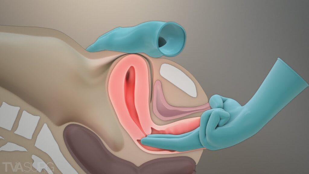
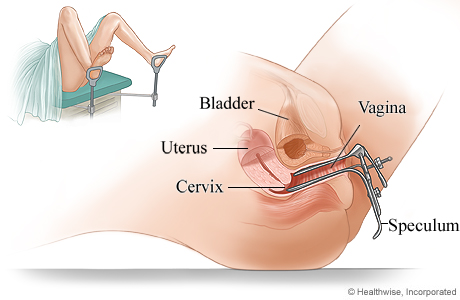
Explanation: Episiotomy is a surgical incision made on the perineum to enlarge the vaginal orifice prior to delivery of the baby. It is performed to expedite the delivery process in specific situations, such as maternal distress, fetal distress, cord prolapse, rigid perineum, forceps delivery, face-to-pubis delivery, and certain other conditions.

Question 2:
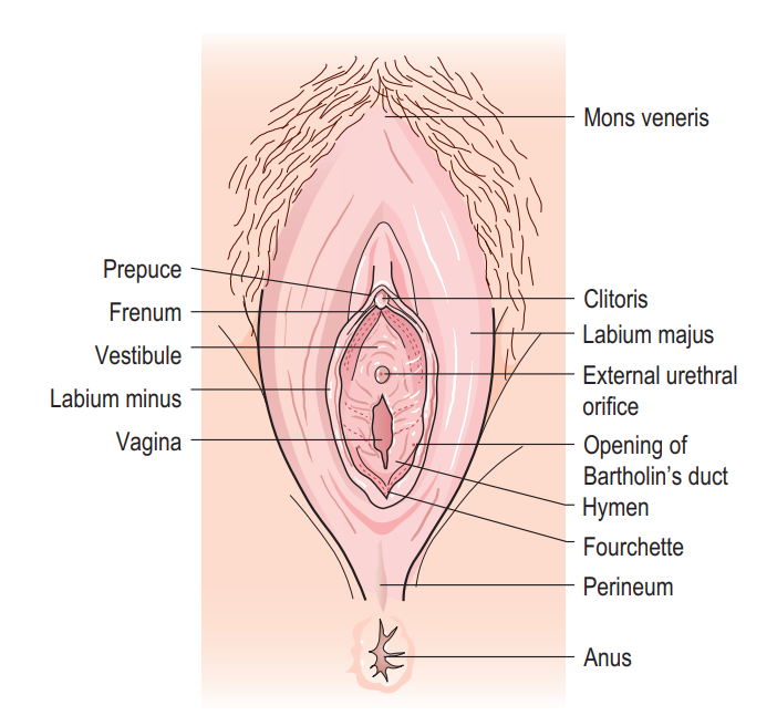
Which type of episiotomy incision is discouraged due to the risk of damage to the Bartholin’s glands?
A) Medial lateral
B) J-shaped
C) Lateral incision
D) Bilateral

Answer: D) Bilateral

Explanation: The “bilateral” episiotomy incision is done on either side and is similar to the lateral incision. However, it is discouraged because it can cause injury to the Bartholin’s glands, which are located on either side of the vaginal opening.

Question 3:
What is the primary purpose of controlled cord traction during the third stage of labor?
A) To expedite placental delivery
B) To prevent perineal tears
C) To encourage maternal pushing
D) To reduce postpartum hemorrhage

Answer: A) To expedite placental delivery

Explanation: Controlled cord traction is a method used during the third stage of labor to help deliver the placenta more efficiently by applying controlled downward traction on the umbilical cord. It aims to shorten the duration of the third stage and reduce the risk of postpartum hemorrhage.

Question 4:
What is the average volume of blood flow through the placental site before the baby is born?
A) 100-200ml/min
B) 300-500ml/min
C) 500-800ml/min
D) 1000-1200ml/min

Answer: C) 500-800ml/min

Explanation: The normal volume of blood flow through the placental site before the baby is born is approximately 500-800ml/min. This blood flow decreases significantly once the baby is delivered and the placental separation begins.

Question 5:
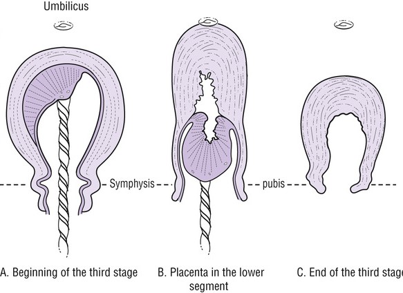
Which stage of labor involves the separation, descent, and expulsion of the placenta and membranes?
A) First stage
B) Second stage
C) Third stage
D) Fourth stage

Answer: C) Third stage

Explanation: The third stage of labor involves the separation, descent, and expulsion of the placenta and membranes after the delivery of the baby.

Question 6:
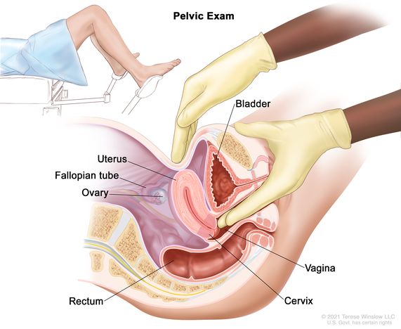
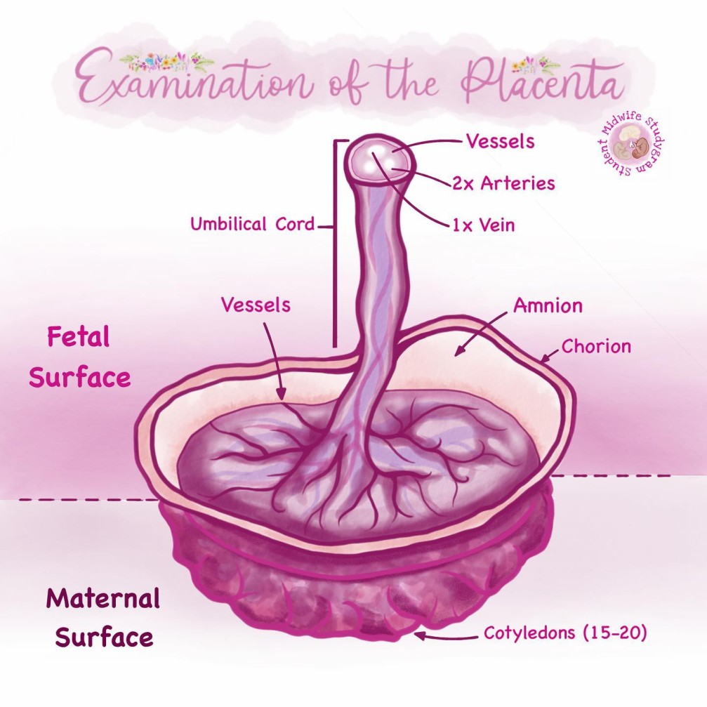
What is the main purpose of the placental examination after delivery?
A) To check the baby’s health status
B) To detect any abnormalities in the placenta
C) To ensure proper positioning of the baby
D) To assess maternal blood loss

Answer: B) To detect any abnormalities in the placenta

Explanation: The placental examination after delivery aims to assess the completeness of the placenta and membranes and to detect any abnormalities that may have occurred during pregnancy or delivery.

Question 7:
What is the recommended method of placental separation during the third stage of labor?
A) Mathew Dankan method
B) Active management with controlled cord traction
C) Passive management with maternal efforts
D) Schultze method

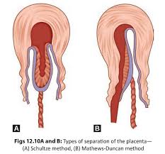
Answer: D) Schultze method

Explanation: The Schultze method is the recommended method of placental separation during the third stage of labor. It involves separation starting centrally, resulting in the formation of a retroplacental clot that aids in the separation process.

Question 8:
Which type of episiotomy incision is done by doctors and involves curving away from the anal sphincter?
A) J-shaped
B) Medial lateral
C) Bilateral
D) Lateral incision

Answer: A) J-shaped

Explanation: The J-shaped episiotomy incision is done by doctors and starts from the center of the fourchette, curving away from the anal sphincter.

Question 9:
What is the primary aim of uterine contractions during the third stage of labor?
A) To facilitate placental separation
B) To promote fetal descent
C) To prevent maternal discomfort
D) To dilate the cervix

Answer: A) To facilitate placental separation

Explanation: Uterine contractions during the third stage of labor help to facilitate placental separation and expulsion by reducing the area of the placental site and exerting pressure on the torn blood vessels.

Question 10:
Which stage of labor involves the arrest of hemorrhage?
A) First stage
B) Second stage
C) Third stage
D) Fourth stage

Answer: C) Third stage

Explanation: The third stage of labor involves the arrest of hemorrhage as the uterus contracts and closes the spiral arterioles after placental expulsion, reducing bleeding.

Question 11:
What is the main indication for performing an episiotomy in a forceps delivery?
A) To reduce fetal distress
B) To prevent cerebral damage in premature babies
C) To quicken delivery
D) To prevent excessive bleeding

Answer: C) To quicken delivery

Explanation: In a forceps delivery, an episiotomy may be performed to quicken delivery, allowing for easier extraction of the baby using forceps and reducing the risk of prolonged labor.

Question 12:
Which type of episiotomy incision is the commonest, safest, and recommended for use by midwives?
A) J-shaped
B) Medial lateral
C) Bilateral
D) Lateral incision

Answer: B) Medial lateral

Explanation: The medial lateral episiotomy is the commonest, safest, and recommended incision for use by midwives. It starts from the fourchette to the medial lateral direction of the perineum.

Question 13:
What is the typical healing time for an episiotomy?
A) 1-2 weeks
B) 2-4 weeks
C) 4-6 weeks
D) 6-8 weeks

Answer: C) 4-6 weeks

Explanation: The typical healing time for an episiotomy is around 4-6 weeks, depending on the size of the incision and the type of suture material used.

Question 14:
What is the average volume of blood flow through the placental site after the baby is born?
A) 100-200ml/min
B) 300-500ml/min
C) 500-800ml/min
D) 1000-1200ml/min

Answer: A) 100-200ml/min

Explanation: After the baby is born, the average volume of blood flow through the placental site reduces to approximately 100-200ml/min.

Question 15:
What is the primary aim of rubbing the fundus during the third stage of labor?
A) To assess the size of the uterus
B) To promote uterine contractions
C) To monitor the baby’s heart rate
D) To assess cervical dilation

Answer: B) To promote uterine contractions

Explanation: Rubbing the fundus during the third stage of labor helps to promote uterine contractions and assist in the expulsion of the placenta and membranes. It aids in preventing postpartum hemorrhage and achieving hemostasis.

Examination of THE placenta Read More »

Normal third stage of labour

Normal Third Stage of Labour

The third stage of labor is a critical phase that involves the separation, descent, and expulsion of the placenta and membranes, as well as the prevention of hemorrhage.

Physiology of the Third Stage of Labour:

  1. Contraction and Retraction:
    • The placental separation is initiated by the contraction and retraction of the uterine muscles. These contractions thicken the uterine wall, reducing the capacity of the upper uterine segment and decreasing the area of the placental site.
    • Separation starts from the center of the placenta. As the blood sinuses tear, a retroplacental clot forms, aiding in further placental separation.
  2. Descent of the Placenta:
    • The placenta descends due to the force of gravity acting like a piston on the clot. This propels the placenta from the upper uterine segment into the lower uterine segment.
  3. Separation of Membranes:
    • The membranes become separated as the weight of the placenta peels them off the decidua. However, the membranes may remain adherent around the cervix until the placenta is expelled from the vagina.
  4. Haemostasis (Preventing Hemorrhage):
  • At placental separation, swift control of blood flow is crucial to prevent serious hemorrhage. Several physiological processes play a role in achieving haemostasis:


a
. Retraction of oblique uterine muscle fibers leads to the thickening of the uterine muscles, acting as a clamp and securing a ligature action on the torn vessels.

b
. Vigorous uterine action after separation brings the uterine walls into opposition, exerting further pressure on the placental site.

c
. A fibrin mesh rapidly covers the placental site after separation, utilizing 5-10% of the circulating fibrinogen to aid in clot formation and control bleeding.

d
. Breastfeeding stimulates the release of oxytocin, which enhances uterine contractions, contributing to haemostasis.

Separation of the Placenta normal

Separation of the Placenta:

The separation of the placenta during the third stage of labor can occur in two ways, known as the Schultze method and the Mathew Dancan method.

  1. Schultze Method:
  • Separation usually starts centrally, resulting in the formation of a retroplacental clot. This clot exerts pressure at the midpoint of the placental attachment, aiding in the separation process and helping to strip the placenta’s adherent lateral borders.
  • The increased weight of the placenta also assists in peeling the membranes off the uterine wall, creating a membranous bag enclosing the clot. As the placenta descends, the fetal surface comes out first.
  • This method is associated with more complete shearing of both the placenta and membranes, leading to less fluid blood loss. It is a quick and clean method.
  1. Mathew Dancan Method:
  • Placental separation begins from the sides, and blood escapes from the sides during the process, without the assistance of a retroplacental clot.
  • The placenta descends slipping sideways, with the maternal surface coming out first.
  • This method takes longer and is associated with ragged and incomplete expulsion of membranes, leading to a higher fluid blood loss.

After separation, the uterus contracts strongly, forcing the placenta and membranes to fall into the lower uterine segment and eventually into the vagina.

Signs of Placenta Separation:

  1. The uterus becomes hard, round, and mobile.
  2. The fundus rises to or above the umbilicus.
  3. The cord lengthens or elongates.
  4. There may be a gush of blood.
  5. The placenta can be felt on vaginal examination (VE).
  6. Presence of the placenta at the vulva.
  7. If suprapubic pressure is applied, the cord does not recede into the vagina.

Mechanism of Placental Separation:

  • Placental separation is facilitated by a combination of uterine contractions and involution. 
  • After the delivery of the fetus, the uterus continues to contract approximately every 3-4 minutes. These contractions, along with the process of involution (the shrinkage of the uterus), lead to the site of implantation of the placenta undergoing shrinkage as well.
  • Within 10-15 minutes after the baby’s delivery, most of the placenta detaches from the uterine wall. This results in an increase in vaginal bleeding from the exposed implantation site, which signals the impending delivery of the placenta.
  • As the placenta is delivered, the uterus continues to contract, closing the spiral arterioles and reducing bleeding. The ongoing contraction of the uterus helps in preventing excessive blood loss.

N.B: It is important to note that the average blood loss from a vaginal delivery is approximately 250-300mls.

Management of the Third Stage of Labour:

Methods of Delivering the Placenta:

  • Controlled cord traction. (active)
  • Maternal efforts(passive management).
  1. Active Management (Controlled Cord Traction):
    • Palpate the abdomen to exclude undiagnosed twin pregnancies.
    • Administer oxytocin 10 IU intramuscularly to enhance uterine contractions.
    • Extend the cord clamp slightly to the vulva to get a good grip. Place the left hand over the fundus of the uterus.
    • During the first contraction, turn the palm of the left hand facing the fundus and apply counter traction above the pubic bone.
    • The right hand grasps the cord clamp and applies steady downward and outward traction until the placenta is visible at the vulva, then applies upward traction to receive the placenta in a cupped hand.
    • Take care to roll the membranes to prevent them from breaking.
    • Deliver the membranes in upward and downward movements.
    • Note the time of placenta and membranes delivery.
    • Rub the fundus to promote further uterine contractions.
    • Quickly examine the placenta for completeness and place it in a receiver.
    • Clean the vulva to remove any blood, and examine the cervix and vagina for lacerations or an extension of an episiotomy.
    • Repair any lacerations or tears.
    • Place a clean pad and ensure the mother is comfortable.
  2. Passive Management (Maternal Efforts):
    • This method is only used when the placenta has already separated from the uterine wall.
    • Wait for signs of placental separation.
    • Once the placenta has separated, ask the mother to push during contractions.
    • Place a flat palm over the mother’s abdomen to provide resistance for her to push against.
    • Receive the placenta in both hands and deliver it complete.
    • Administer oxytocin 10 IU intramuscularly.

Note: It is essential to keep the mother warm as she has undergone strenuous physical exercise during the first and second stages of labor, leading to significant heat loss from her body.

Normal third stage of labour Read More »

Get Pen and a Paper and write,

No copy and Paste Allowed!

Scroll to Top