Minor disorders of pregnancy are a series of commonly experienced symptoms related to the effects of pregnancy hormones and the consequences of enlargement of the uterus as the fetus grows during pregnancy.
These are referred to as minor because they are not life threatening. The causes can be;-
Hormonal changes
Accommodation changes
Metabolic changes and
Postural changes
I. Digestive System Disorders:
A. Nausea and Vomiting (Morning Sickness):
Causes: Primarily attributed to hormonal surges, specifically elevated levels of human chorionic gonadotropin (hCG), oestrogen, and progesterone during early pregnancy (weeks 4-16). These hormones affect the gastrointestinal tract’s sensitivity and motility. The exact mechanism remains unclear but likely involves alterations in brain neurotransmitters and gastrointestinal hormone levels. Decreases as the placenta takes over hormone production.
Symptoms: Range from mild to severe vomiting (hyperemesis gravidarum, a severe form requiring medical attention). Symptoms often peak in the morning but can occur throughout the day.
Management:
Dietary Modifications: Small, frequent meals; consuming bland foods like crackers, toast, or rice; avoiding strong smells or triggers; consuming carbohydrates.
Lifestyle Changes: Getting out of bed slowly, staying hydrated, eating before getting out of bed, regular, gentle exercise.
Pharmacological Interventions: In cases of severe nausea and vomiting, antiemetics (medications to control nausea and vomiting) may be prescribed by a healthcare provider. Vitamin B6 supplements are sometimes recommended.
Acupressure: Wristbands with pressure points can sometimes help alleviate nausea.
B. Heartburn (Pyrosis):
Causes: Relaxation of the lower esophageal sphincter (LES) due to progesterone, allowing stomach acid to reflux into the esophagus. Increased intra-abdominal pressure from the growing uterus further exacerbates this. Most troublesome between 30-40 weeks gestation.
Symptoms: Burning sensation in the chest, often radiating upwards. Can be worsened by lying down, bending over, or consuming certain foods.
Management:
Dietary Modifications: Small, frequent meals; avoiding fatty, spicy, or acidic foods; avoiding eating before bed.
Lifestyle Changes: Elevating the head of the bed with extra pillows, avoiding tight clothing, maintaining an upright posture after meals.
Pharmacological Interventions: Antacids (e.g., magnesium trisilicate, calcium carbonate) can neutralize stomach acid, providing temporary relief. H2 blockers or proton pump inhibitors (PPIs) may be prescribed for more severe cases.
C. Excessive Salivation (Ptyalism):
Causes: Likely hormonal influences, although the exact mechanism is unclear. Often associated with nausea and vomiting. It may also be caused by anxiety or psychological factors.
Symptoms: Excessive production of saliva.
Management: Rinsing the mouth frequently, sucking on ice chips or hard candies, avoiding trigger foods. Counseling may be helpful to address underlying anxiety.
D. Constipation:
Causes: Progesterone’s relaxing effect on the smooth muscles of the intestines, leading to slowed bowel movements (decreased peristalsis). Iron supplementation can also contribute. Decreased physical activity may play a role.
Symptoms: Infrequent bowel movements, hard stools, straining during bowel movements.
Management:
Dietary Modifications: Increased intake of fiber (fruits, vegetables, whole grains), fluids (water), and gentle exercise. Bulk-forming laxatives (psyllium) may be used under medical supervision.
Lifestyle Changes: Regular exercise, particularly walking, can stimulate bowel movements.
Pharmacological Interventions: Stool softeners or mild laxatives should be used cautiously and only when dietary changes and exercise are insufficient, under the guidance of a healthcare professional.
E. Pica:
Causes: Unknown. Possible links to nutritional deficiencies (iron, zinc), psychological factors, or hormonal imbalances.
Symptoms: Craving and consumption of non-nutritive substances (e.g., clay, ice, starch). This can lead to serious health consequences.
Management: Addressing any underlying nutritional deficiencies through dietary changes and supplementation under medical supervision. Psychological counseling may also be beneficial.
II. Musculoskeletal System Disorders:
A. Leg Cramps:
Causes: Exact cause is unknown but various factors have been suggested, including:
Changes in electrolyte balance: Decreased calcium or magnesium levels can make muscles prone to cramping.
Compression of nerves: The growing uterus may compress nerves, affecting muscle function.
Reduced blood circulation: Restricted blood flow can lead to cramping.
Increased weight: Added weight puts pressure on the muscles.
Symptoms: Sudden, sharp pain in the calf muscles, often at night.
Management:
Stretching exercises: Regular stretching of calf muscles.
Hydration: Adequate fluid intake.
Dietary changes: Addressing any potential electrolyte imbalances through diet or supplementation (calcium, magnesium, potassium). A balanced diet is key.
Foot elevation: Raising legs above heart level. Dorsiflexion (pulling toes towards shin) can also provide relief.
B. Backache:
Causes: Shifting center of gravity due to the growing uterus, relaxation of ligaments and joints due to relaxin hormone, and changes in posture.
Symptoms: Aching or pain in the lower back, often radiating to the buttocks or legs.
Management:
Postural adjustments: Maintaining good posture, avoiding high heels, using supportive footwear.
Exercise: Low-impact exercises such as walking, swimming, or prenatal yoga.
Rest: Regular periods of rest throughout the day.
Supportive measures: Using a maternity support belt, applying heat or ice packs, pelvic floor exercises.
Pelvic floor exercises: Strengthening pelvic floor muscles can help improve support and reduce back pain.
III. Circulatory System Disorders:
A. Fainting (Syncope):
Causes: In early pregnancy, vasodilation from progesterone can cause a temporary drop in blood pressure before the body compensates by increasing blood volume. Orthostatic hypotension (a sudden drop in blood pressure when standing up) can also occur. Dehydration can contribute.
Symptoms: Dizziness, lightheadedness, loss of consciousness.
Management: Avoiding prolonged standing, changing positions slowly, lying down immediately if feeling faint, staying well-hydrated. Avoiding lying on the back, except during necessary medical examinations.
B. Varicose Veins:
Causes: Progesterone relaxes the smooth muscles in the veins, leading to reduced blood flow and pooling of blood. Increased blood volume and pressure from the growing uterus also contribute. They may occur in legs, vulva, and anus.
Symptoms: Enlarged, twisted veins; aching, heavy, or swollen legs; pain or cramping in the legs.
Management:
Compression stockings: Wearing compression stockings to improve circulation.
Elevation: Elevating legs regularly.
Exercise: Regular exercise to promote circulation.
Avoiding prolonged standing or sitting: Frequent movement to improve blood flow.
Managing constipation: Preventing constipation helps reduce pressure on the veins.
Medical intervention: In severe cases, a doctor may recommend other treatments.
C. Hemorrhoids:
Causes: Increased pressure on the pelvic veins due to constipation and the growing uterus.
Symptoms: Painful, swollen, and inflamed veins in the rectum or anus.
Management: High-fiber diet to prevent constipation; topical treatments (e.g., creams, ointments); warm sitz baths; stool softeners (as advised by a healthcare provider).
D. Heart Palpitations:
Causes: Increased cardiac output to supply the growing fetus with blood and nutrients. Hormonal changes also affect heart rate and rhythm. Anxiety and stress can also exacerbate palpitations.
Symptoms: Feeling of a racing heart, fluttering, or pounding in the chest. Can be associated with shortness of breath or dizziness.
Management: Identifying and managing underlying anxiety or stress. Regular exercise, maintaining a healthy weight, and avoiding caffeine and nicotine can help regulate heart rate. In cases of persistent or concerning symptoms, medical evaluation is necessary to rule out other causes.
IV. Urinary System Disorders:
A. Urinary Tract Infections (UTIs):
Causes: The changing hormonal environment of pregnancy can make women more susceptible to UTIs. The expanding uterus can also compress the ureters, slowing urine flow and increasing the risk of bacterial growth.
Symptoms: Frequent urination, burning sensation during urination, urgency, pain in the lower abdomen or back. Fever and chills may indicate a more serious infection.
Management: Prompt medical attention is crucial for UTIs in pregnancy. Treatment usually involves antibiotics.
B. Frequency of Micturition:
Causes: In early pregnancy, hormonal changes increase blood flow to the kidneys, leading to increased urine production. In later pregnancy, the enlarging uterus compresses the bladder, reducing its capacity and leading to more frequent urination.
Symptoms: Increased urge to urinate, often with small amounts of urine being passed.
Management: Regular voiding to prevent bladder distension, drinking plenty of fluids throughout the day but avoiding excess fluid close to bedtime. Kegel exercises to strengthen pelvic floor muscles may help improve bladder control.
C. Stress Incontinence:
Causes: Weakening of pelvic floor muscles due to hormonal changes and the pressure exerted by the growing uterus.
Symptoms: Leakage of urine during coughing, sneezing, laughing, or physical exertion.
Management: Pelvic floor exercises (Kegel exercises) to strengthen the pelvic floor muscles. Avoiding activities that increase intra-abdominal pressure. In some cases, medical intervention may be necessary.
V. Integumentary System Disorders:
A. Itching of the Skin (Pruritis):
Causes: Stretching of the skin due to weight gain, hormonal changes, and cholestasis of pregnancy (a liver condition that can cause intense itching). Poor hygiene, heat rash, or minor skin rashes also contribute. Stretch marks (striae gravidarum) can also be itchy.
Symptoms: Itching, particularly on the abdomen, breasts, thighs, and buttocks. The degree of itchiness can range from mild to severe.
Management: Keeping the skin moisturized, cool baths or showers, wearing loose-fitting clothing made of breathable fabrics, topical creams or lotions (as advised by a physician). Medical attention is required if itching is severe or persistent or if it is accompanied by other symptoms (jaundice, dark urine, pale stools).
B. Stretch Marks (Striae Gravidarum):
Causes: Rapid stretching and thinning of the skin due to weight gain during pregnancy. Genetic predisposition plays a role.
Symptoms: Red or purple streaks on the abdomen, breasts, thighs, and buttocks. They eventually fade to silvery white.
Management: Keeping the skin well-hydrated with lotions or creams may help minimize the appearance of stretch marks. There is no known cure.
C. Melasma (Chloasma):
Causes: Hormonal changes during pregnancy stimulate increased melanin production, resulting in hyperpigmentation. Exposure to sunlight exacerbates the condition.
Symptoms: Dark brown patches, usually on the face. Often seen on cheeks, forehead, and upper lip.
Management: Sunscreen protection is crucial to prevent further darkening. Topical treatments may be recommended. The discoloration usually fades after delivery.
VI. Other Disorders:
A. Emotional Instability:
Causes: The physiological changes, lifestyle adjustments, anxieties and fears associated with pregnancy can significantly impact emotional well-being. Hormonal shifts play a crucial role.
Management: Support from family and friends, stress management techniques (yoga, meditation, etc.), prenatal yoga, counseling or therapy, if needed. Open communication with a healthcare provider is vital.
VII. Disorders Requiring Immediate Medical Attention:
The following symptoms warrant immediate medical attention as they could indicate serious complications:
Vaginal Bleeding: Could indicate placenta previa, placental abruption, or other serious complications.
Reduced Fetal Movements: May signify fetal distress.
Severe or Persistent Headache (especially frontal or recurrent): Can be a sign of preeclampsia or eclampsia.
Sudden Swelling or Edema (especially in face or hands): A possible symptom of preeclampsia.
Early Rupture of Membranes (PROM): Increased risk of infection and premature delivery.
Premature Onset of Contractions: Risk of preterm labor.
Maternal Exhaustion (to any extent): Can indicate underlying health issues.
Fits or Seizures: Potentially indicative of eclampsia.
Excessive Nausea and Vomiting (Hyperemesis Gravidarum): Severe dehydration and electrolyte imbalance.
Epigastric Pain: Can be a symptom of preeclampsia.
Quick Quiz
Minor Disorders Quiz
Midwifery - mobile-friendly and focused practice.
Privacy: Your details are used only for quiz tracking and certificates.
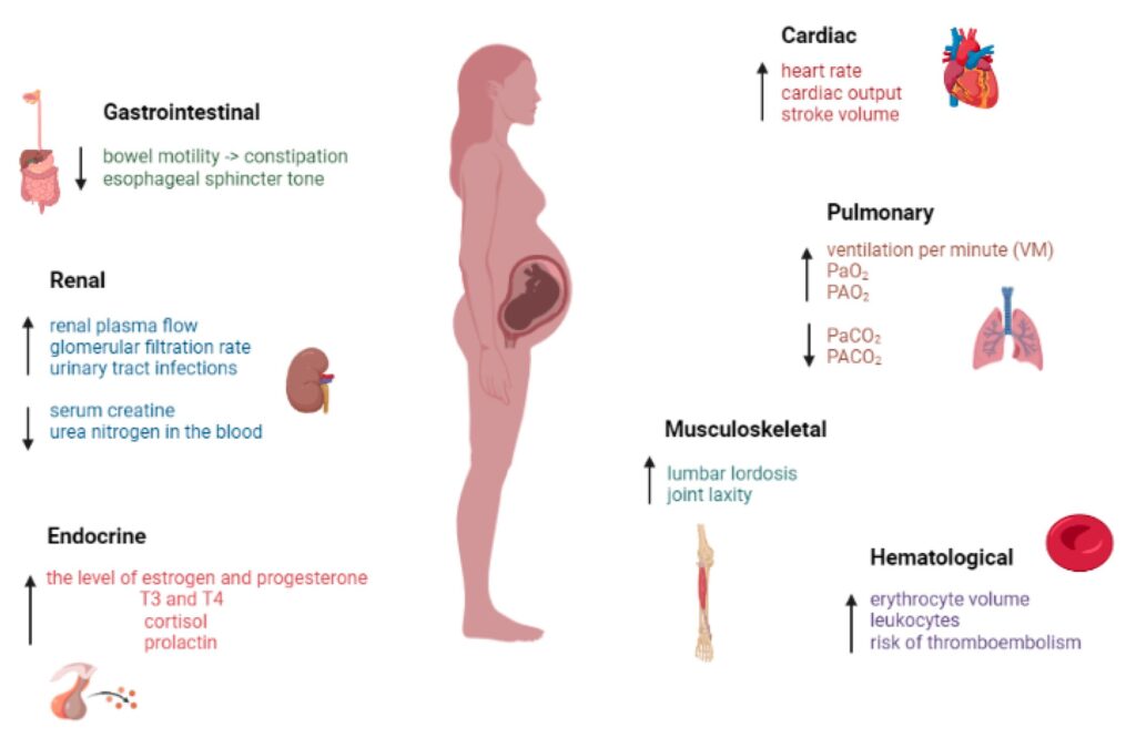
Overview: The Physiology of Pregnancy covers all the normal, natural changes that take place in a mother's body because she is pregnant. These changes happen mainly because of different hormone levels and changes in how the body uses energy (metabolism). Everything is designed to support, protect, and grow the baby.
1. Changes in the Endocrine System (Hormones)
The placenta produces several hormones that cause many physical changes. The successful adjustment of the mother's body is made possible by changes in hormone production by the mother's endocrine system and the trophoblast (the outer layer of cells of the early embryo).
Human Chorionic Gonadotropin (HCG): This hormone is produced by the trophoblast. HCG levels increase very fast in early pregnancy, reaching their highest levels at 8 to 10 weeks of gestation. The main job of HCG is to keep the corpus luteum working so it can keep producing Progesterone and Oestrogen. It does this until the placenta is ready to produce enough hormones on its own (around 10 to 12 weeks). After that, HCG levels slowly drop until it completely disappears 2 weeks after birth.
Progesterone Hormone: This is mainly produced in the corpus luteum in the beginning. Its job is to make the lining of the uterus (decidua) thick, soft, and ready to receive the fertilized egg. It also helps to increase the glandular tissue and milk ducts in the breasts, and the muscle fibers in the uterus.
Oestrogen: This hormone causes the uterus to grow and also develops the duct system in the breasts during pregnancy. It is passed out of the body in the urine, and measuring the amount of oestrogen in the urine can show how well the baby is doing (fetal wellbeing).
Relaxin Hormone: During the last few weeks of pregnancy, this hormone acts on the ligaments and joints, making the pelvis relax and "give" way for the baby to pass. It is also produced by the decidua and the trophoblast to help the uterine muscles (myometrium) relax. It also plays a role in softening the cervix and breaking the water membranes.
Human Placental Lactogen (HPL): This hormone stimulates breast growth and helps prepare the breasts for making milk (lactogenic properties). It also causes metabolic changes that make sure plenty of glucose (sugar) is available for the growing baby's brain and body, protecting the baby from poor nutrition.
Pituitary Hormones: The Follicle Stimulating Hormone (FSH) and Luteinizing Hormone (LH) are stopped from working because of the high levels of Oestrogen and Progesterone. The adrenal gland only grows slightly in size, but it increases its hormone production.
Thyroid Function: In a normal pregnancy, the thyroid gland gets bigger because its tissue grows and it gets more blood supply. The body takes up more iodine during pregnancy to make up for the iodine lost through the kidneys, which stops the blood iodine levels from dropping too low.
2. Changes in the Reproductive System
Changes in the Uterus
The uterus stretches and expands greatly to make room for and feed the growing baby. This growth happens in the middle muscle layer called the myometrium. The body of the uterus grows to create a safe, feeding environment for the baby to develop.
Uterine Muscle Layers
1. Endometrium: Menstruation completely stops. During pregnancy, this inner lining is called the decidua. It becomes very thick, soft, spongy, and gets a very rich blood supply.
2. Myometrium (Muscle Layer): The growth of the uterus body is caused by two main factors:
Hypertrophy: The actual muscle fibers increase in size. They become about 10 times longer and 3 times wider.
Hyperplasia: New muscle cells are created and grow next to the original muscle cells. This means an increase in the total number of cells.
Size, Weight, and Shape Changes:
Size and Weight: Before pregnancy, the normal uterus is 7.5cm long, 5cm wide, and 2.5cm thick, weighing just 60g. By the end of pregnancy, it becomes 30cm long, 23cm wide, and 20cm thick, weighing a massive 960g.
Shape: A healthy pregnancy needs enough space for the baby, the amniotic fluid (liquor), and the placenta. At the start of pregnancy, the uterus is shaped like a pear. At 12 weeks, it becomes round (globular). From 12 to 38 weeks, it becomes oval-shaped. After 38 weeks, when the baby drops into the pelvis (lightening), it turns back to a round globular shape.
The Three Muscle Layers of the Myometrium
Outer Longitudinal Layer: This layer starts in the front wall of the upper uterus, goes over the top (fundus), and down the back wall. When this muscle layer contracts and pulls back (retracts), it pushes the baby out during labor.
Middle Oblique Layer: Here, muscles are arranged in a criss-cross way, looking like a "figure of 8" around the blood vessels. After the baby is born and the placenta separates, these muscles squeeze the blood vessels tight to stop bleeding and prevent Postpartum Haemorrhage (PPH). Because of this action, they are called living ligatures.
Inner Circular Layer: This is the weakest of the three layers. The muscle fibers run straight across the uterus. They are well developed around the cervix, lower uterus, and fallopian tubes. They help the cervix to open (dilate) during labor.
3. The Perimetrium: This is the outer layer of the peritoneum that covers most, but not all, of the uterus. It folds over the bladder in the front to form the utero-vesical pouch, and folds in the back to form the pouch of Douglas. After 12 weeks, the uterus grows so big it rises out of the pelvis and becomes an organ in the abdomen. It loses its forward-bending position and stands straight up, leaning slightly to the right side.
Clinical Observations of the Growing Uterus
As the pregnancy progresses, midwives check the size of the uterus (fundal height) to ensure the baby is growing well:
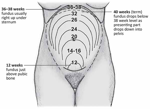
At 12 weeks: The uterus comes out of the pelvis and stands straight. It can be felt just above the symphysis pubis (pubic bone) and is about the size of a grapefruit.
At 16 weeks: The top of the uterus (fundus) becomes shaped like a dome. As it grows up, it twists slightly to the right side (dextrorotation) because the colon is on the left side of the pelvis. The uterus becomes more round.
At 20 weeks: The fundus can be felt right at the level of the mother's umbilicus (belly button).
At 30 weeks: The fundus is halfway between the umbilicus and the xiphisternum (the bottom tip of the breastbone). The large uterus pushes the mother's intestines to the sides and upwards. If the mother lies flat on her back, the heavy uterus falls back against her spine and major blood vessels.
At 36 weeks: The enlarged uterus fills the entire abdominal space. The fundus touches the very tip of the xiphoid cartilage at the ribcage.
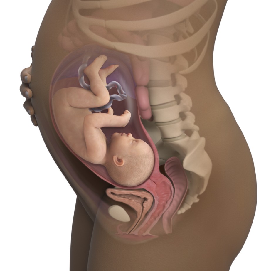
At 38 weeks: Between 38 and 40 weeks, the lower part of the uterus becomes smooth and soft. The uterus looks rounder, and the fundal height actually drops lower. This drop happens because the baby's head enters the pelvis, a process called lightening.
Diagram showing the clinical landmarks of fundal height at 12, 16, 20, 36, and 40 weeks.
Changes in Other Reproductive Organs
Blood Supply: The blood vessels in the uterus get much wider, and new blood vessels form because of Oestrogen. Blood flowing to the uterus and ovaries increases to an amazing 750ml per minute at the end of pregnancy to feed the hard-working placenta.
Fallopian Tubes: They are stretched out on both sides of the big uterus and get a rich blood supply. The end connected to the uterus closes, but the finger-like ends (fimbriae) stay open.
The Isthmus: It becomes very soft and stretches out from just 7mm to 23mm long. This stretched part becomes the lower uterine segment in late pregnancy.
The Ovaries: The Follicle-Stimulating Hormone (FSH) stops working because of high levels of estrogen and progesterone. This stops ovulation and menstrual periods. Both ovaries get bigger due to extra blood flow, and they are lifted out of the pelvis. The corpus luteum grows big in early pregnancy (sometimes forming a harmless cyst) to produce progesterone, which protects the early pregnancy until 10-12 weeks when the placenta takes over. After that, it shrinks.
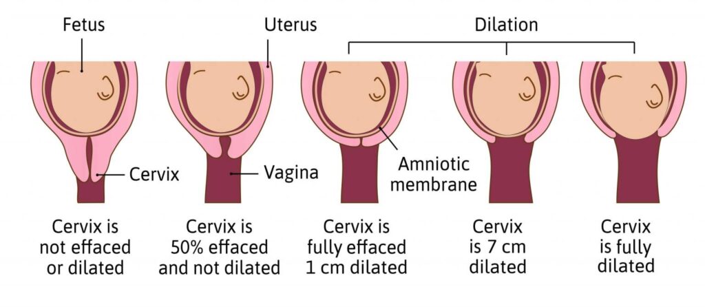
The Cervix: It stays tightly closed to protect the baby from infections and hold the baby inside when the mother stands up. It grows slightly and becomes very soft due to extra blood and relaxing hormones. The glands secrete a very thick mucus that blocks the cervical canal, called the operculum (mucus plug). Near labor, the cervix becomes part of the lower uterine segment (effacement) and opens slightly. A soft, slightly open cervix is called a "ripe cervix."
The Vagina: The muscles grow thicker, and the vagina becomes bigger and very elastic so it can stretch during birth. It produces more normal white discharge called leucorrhea. The cells have a lot of glycogen (sugar), which interacts with normal bacteria (Doderlein’s bacillus) to make the vagina very acidic. This acid kills bad bacteria but makes the mother more likely to get yeast infections like Candida albicans. The extra blood makes the vagina look purple or violet.
The Vulva: It also appears bluish in color because of the heavy blood flow and pelvic congestion.
Breast Changes
In early pregnancy, breasts feel full, heavy, or tingle, and they grow bigger as pregnancy continues.
The nipples stick out more easily (become more erectile).
The dark circle around the nipple (areola) becomes much darker and wider.
Small oil glands on the areola grow big and look like small bumps. These are called Montgomery’s tubercles. They produce oil (sebum) to keep the nipple soft and lubricated for breastfeeding.
Blue blood vessels become very easy to see on the skin of the breasts because of the increased blood flow.
A clear or yellowish sticky fluid called colostrum can sometimes be squeezed from the nipples after the first 3 months (1st trimester).
3. Changes in the Cardiovascular System (Heart and Blood)
The Heart and Blood Pressure
The Heart: Because it has to work so hard, the heart muscle grows slightly bigger (hypertrophies), especially the left side. The growing uterus pushes the heart upwards and to the left side of the chest. Heart sounds change, and harmless heart murmurs are very common. The amount of blood the heart pumps (cardiac output) increases by 40%. The heart beats faster by an average of 15 extra beats every minute.
Blood Pressure (BP): In the first 3 months, BP stays normal. In the second trimester, BP actually drops because the hormone progesterone causes blood vessels to relax and open wide (vasodilation). It is at its lowest between 16 and 20 weeks, which can cause the mother to feel dizzy or faint. Near the end of pregnancy, BP goes back to normal levels.
⚠️ Clinical Alert: Supine Hypotensive Syndrome
A pregnant woman should not lie flat on her back (supine position). The heavy baby and uterus will press down on the large vein in the back (inferior vena cava). This traps the blood, stopping it from returning to the heart. This causes a sudden, dangerous drop in blood pressure, making the mother faint. It also causes swelling (oedema) in the legs, swollen leg veins (varicose veins), and swollen veins in the anus (hemorrhoids). Always encourage the mother to sleep on her side.
Blood Volume and Composition
Blood Flow: Much more blood flows to the uterus, kidneys, breasts, and skin. Blood flow to the liver and brain stays the same. Blood flow to the kidneys goes up by 70-80%.
Total Blood Volume: The total amount of blood in the mother's body increases a lot, usually by 35% to 45% above normal. She needs this huge amount of extra blood to:
Give the placenta enough blood to feed the baby.
Meet the extra energy needs of the growing baby.
Ensure the kidneys and other organs get enough oxygen.
Protect the mother from danger when she loses blood during delivery.
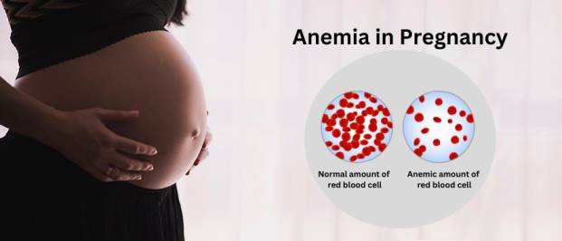
Physiological Anaemia: The liquid part of the blood (plasma) increases by 40%, but the red blood cells only increase by 20%. Because there is so much more water than red cells, the blood becomes diluted. This is perfectly normal and is called physiological haemodilution. The acceptable Hemoglobin (Hb) level during pregnancy is 11-12g/dl.
Iron Metabolism: The mother needs a massive 1000mg of iron. She needs 500mg to make extra red blood cells, 300mg to give to the baby, and 200mg to replace iron naturally lost every day. Because the body only absorbs 20% of the iron she eats, she must take iron tablets. The goal of iron tablets is to prevent true anemia, not just to raise her Hb levels artificially.
Clotting Factors: Proteins in the blood that cause clotting (Fibrinogen and factors 7, 8, 9, and 10) increase heavily. Because of this, her blood clots much faster (clotting time drops from 12 minutes to 8 minutes). This is the body's smart way of preventing heavy bleeding (PPH) when the placenta tears away after birth.
White Blood Cells: These cells fight infection and are slightly increased during pregnancy (from normal levels up to 10,500/mm) and can jump up to 16,000/mm during labor. This keeps her immune system active.
4. Systemic Changes (Breathing, Urine, and Digestion)
Respiratory System (Breathing)
The body burns more energy at rest (basal metabolism increases). The mother breathes in slightly more air with every breath because the baby, the mother's working heart, and her lungs need more oxygen.
In late pregnancy, the mother's ribcage opens up wider (flares out). The huge uterus pushes the breathing muscle (diaphragm) high into the chest, squeezing the bottom of the lungs. This can make the mother feel slightly short of breath.
Urinary System
Blood flow to the kidneys and the speed at which they filter waste (glomerular filtration rate) increase by 50%.
Because of the large uterus pressing on the bladder, the mother has to urinate very often (frequency) in early and late pregnancy.
The tubes connecting the kidneys to the bladder (ureters) become long and bent (kinked) because of the relaxing effects of progesterone. This causes urine to get trapped or move slowly, making pregnant women very easy targets for Urinary Tract Infections (UTIs).
Gastrointestinal System (Digestion)
Gums: They become swollen with fluid, soft, spongy, and can bleed easily when brushing teeth.
Ptyalism: This is a condition where the mother produces too much saliva.
Nausea and Vomiting: This is very common, affecting 70% of pregnant women, especially early in the morning.
Taste: Foods might taste different, sometimes leaving a metallic taste in the mouth.
Pica: A strange craving to eat things that are not food, like soil, clay, or wall plaster.
Appetite: Most women feel much hungrier.
Heartburn: As the uterus grows, it leaves less space for the stomach. Stomach acid gets pushed up into the chest, causing a burning pain.
Constipation: Progesterone relaxes the bowel muscles, so food moves very slowly through the intestines, causing hard stools.
5. Metabolism, Weight Gain & Muscles/Bones
Weight Gain in Pregnancy
The mother's metabolism speeds up to provide plenty of nutrients to the baby. Because of this, she steadily gains weight, which is a good sign that the baby is growing healthy. The average weight gain for a normal pregnancy is about 12.5 kg.
How the weight is gained:
In the first 20 weeks: Gains about 4 kg (0.2 kg per week).
In the last 20 weeks: Gains about 8.5 kg (0.4 kg per week).
Body Part
Maternal Weight Gain (kg)
Fetal Weight Gain (kg)
Total Weight Gain (kg)
Uterus
1.0
-
1.0
Breasts
0.4
-
0.4
Fat Stores
3.5
-
3.5
Blood Volume
1.5
-
1.5
Extracellular Fluid
1.5
-
1.5
Fetus (Baby)
-
3.4
3.4
Placenta
-
0.6
0.6
Amniotic Fluid
-
0.6
0.6
Total
7.9
4.6
12.5
Important Factors That Influence Weight Gain
Many details affect exactly how much weight a mother will put on during her pregnancy. A midwife must monitor these closely:
Maternal Oedema (Swelling): Edema, or swelling, can directly affect weight gain. It involves the gathering and trapping of extra body fluid in the tissues (especially in the legs and feet). This extra water weight adds pounds on the scale.
Maternal Metabolic Rate: The speed at which the mother's body burns energy (metabolic rate) impacts weight gain. A mother with a very fast, high metabolic rate uses up more energy quickly, which may result in her gaining less weight overall.
Dietary Intake: The amount (quantity) and the healthiness (quality) of the food the mother eats play a massive role. Eating a well-balanced, highly nutritious diet ensures that the weight she gains is healthy and beneficial for the baby.
Vomiting or Diarrhea: If a mother suffers from frequent vomiting (like in severe morning sickness) or diarrhea, it can cause weight loss or stop her from gaining the weight she needs. This happens because the body loses water and fails to absorb important calories and nutrients from food.
Amount of Amniotic Fluid: The volume of the water surrounding the baby (amniotic fluid) is heavy. If the mother has an unusually large amount of fluid (a condition called polyhydramnios), it will lead to a much higher number on the weight scale.
Size of the Fetus: Simply put, larger babies weigh more. A fast-growing or naturally large baby will cause the mother's total pregnancy weight gain to increase.
Maternal Physical Activity Level: How much a mother exercises or moves around during the day impacts her weight. A mother who maintains regular physical activity burns more calories, which helps her keep her weight gain in a healthy range.
Maternal Genetics: Family history and genes also play a part. Genetic factors can make a woman naturally more likely to gain extra weight quickly or hold onto weight longer during her pregnancy.
Musculo-Skeletal System (Muscles and Bones)
The hormones Progesterone and Relaxin cause the ligaments, joints, and muscles in the pelvis to soften and relax. This relaxation allows the pelvic bones (like the symphysis pubis in the front and the sacroiliac joints in the back) to spread out slightly. This increases the capacity of the pelvis, giving the baby enough room to pass through during labor (this process is known as a "give"). Because the joints are loose and the heavy uterus pulls the body forward, the mother's balance changes, leading to the typical "waddling" pregnant walk (gait).
6. Skin Changes
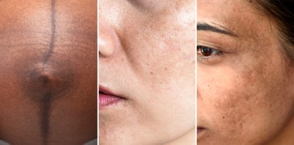
The pituitary gland produces more melanin-stimulating hormone, which causes different parts of the pregnant woman's skin to become dark (pigmentation). This usually starts in the 2nd month and lasts until birth. The areas most affected are the nipples, the middle line of the stomach, the perineum, and the armpits. This happens because the pigment cells become highly sensitive.
Linea Nigra: This is a dark, visible line that runs straight down the middle of the belly, from the umbilicus to the pubic bone (and sometimes up to the chest). It is caused by hormones and will fade away after delivery, though it may never completely vanish.
Chloasma (Mask of Pregnancy): This is the brownish darkening of the skin on the mother's face, usually over the forehead, nose, and cheeks. It gives the skin a bronze look, especially in women with dark complexions.
Striae Gravidarum: These are the common stretch marks that appear on the skin of the growing belly, breasts, and thighs as the skin stretches rapidly.
Sweat Glands: The sweat glands all over the body become much more active. This causes the pregnant woman to sweat more heavily. Also, because she has extra blood flowing and her base body temperature rises by 0.5 degrees, the mother will frequently feel hot.
❓ Quick Review
Why do pregnant women often complain of dizziness when lying flat on their back?
Answer: When lying flat, the heavy uterus presses against the large inferior vena cava vein in the back. This blocks the blood from returning safely to the heart, dropping her blood pressure and causing dizziness (Supine Hypotensive Syndrome).
Quick Quiz
Physiology of Pregnancy Quiz
Midwifery - mobile-friendly and focused practice.
Privacy: Your details are used only for quiz tracking and certificates.
Normal Pregnancy refers to the growth and development of a fertilized ovum. It begins from the exact moment the ovum (egg) is fertilized by a sperm, and it lasts until the fetus is safely expelled from the mother's uterus.
Term Delivery: Normally, the fetus is expelled at term, which means 9 months, 40 weeks, or 280 days.
Abortion: If the fetus is expelled before 28 weeks of pregnancy.
Premature Labour: If the fetus is expelled after 28 weeks but before 37 weeks.
Post-mature: If the baby is born after 42 weeks, we use the term post-mature.
Conditions for a Normal Pregnancy
A pregnancy is clinically considered to be normal only when the following simple conditions are met:
The fertilized ovum is growing safely inside the cavity of the uterus (not in the tubes).
One fetus is forming, along with exactly one placenta and two membranes.
There is a normal amount of amniotic fluid, which is about 1000 to 1500ml of liquor amnii.
The baby is in a vertex presentation (meaning the head is pointing downwards, ready for birth).
There is absolutely no bleeding from the vagina until the "show" appears during the very first stage of labour.
The mother must remain healthy and strong, with no serious diseases or disorders related to the pregnancy.
Signs and Symptoms of Pregnancy
When a woman misses one or two of her regular menstrual periods, she may start to suspect that she is pregnant. In most cases, her guess is correct with a very high accuracy of about 98%, especially if she has always had a regular menstrual cycle.
To make it easy to understand, midwives classify the signs of pregnancy into three main groups:
Presumptive signs (Things the mother feels)
Probable signs (Things the health worker can see or feel)
Positive signs (100% proof that there is a baby)
1. Presumptive Signs
These are the very first signs a woman will notice. They make us "presume" or guess she might be pregnant, but they are not 100% sure because other sicknesses can cause them.
Amenorrhea: This simple word refers to the absence of menstruation (missing a period). A woman may report missing one or two periods, which is a very strong indicator of pregnancy. However, we must be careful because amenorrhea can also be caused by using family planning methods, changing environments, long illnesses, or even high emotional stress.
Breast changes: Many women feel a tingling and prickling sensation in their breasts. The breasts will also start to enlarge and feel very tender or painful to touch. These changes are very common early signs of pregnancy as the body prepares for breastfeeding.
Morning sickness (nausea and vomiting): About 30% to 50% of pregnant women will feel sick to their stomachs and vomit. This usually happens between the 4th and 14th weeks of pregnancy. While bad food or other sicknesses can cause vomiting, if a woman has both nausea and a missed period, it strongly suggests she is pregnant. This sickness usually stops completely by the end of the first three months (first trimester).
Increased frequency of urination: Because the uterus is growing, it sits directly on top of the bladder and presses down on it. This pressure causes the mother to make many trips to the bathroom to pass small amounts of urine. This is usually felt before 12 weeks of pregnancy. It gets better and decreases once the uterus grows big enough to rise out of the pelvic bone area at around 12 weeks.
Skin changes: As the pregnancy grows, the mother's skin goes through many visible changes. The hormone responsible for all these dark skin changes is called the melanin hormone, and it is produced by the anterior pituitary gland in the brain.
Striae gravidarum: These are the common stretch marks. They begin to appear around the 16th week of pregnancy and are mostly found on the mother's abdomen, thighs, and breasts.
Chloasma (mask of pregnancy): Some women will develop dark, brownish patches of skin directly on their face. This looks like a mask.
Linea nigra: A very dark line will appear running straight down the middle of the mother's stomach, passing both above and below the umbilicus (belly button).
Darkening of areolas: The primary areolas (the dark circle around the nipple) will become even darker, and a second, lighter ring called the secondary areola may form around it.
Classic pregnancy skin changes: Chloasma on the face, Linea Nigra on the abdomen, and Striae Gravidarum.
Quickening: This refers to the very first time the mother feels the baby moving or kicking inside her womb. This usually happens around 18 to 20 weeks of pregnancy for a "primigravida" (a woman who is pregnant for the very first time). For a "multigravida" (a woman who has been pregnant before), she might feel it earlier, around 16 to 18 weeks. Quickening is very helpful for a midwife to estimate the age of the pregnancy if the mother does not remember the dates of her last period.
Fatigue: Pregnant women often feel extremely tired and sleepy. This is caused by the body working hard to produce extra blood, having lower blood sugar levels, and experiencing a drop in blood pressure caused by the hormone progesterone. Not sleeping well and morning sickness can also add to this feeling of severe tiredness.
Mood changes: Because of the physical stress, changes in the body's metabolism, tiredness, and big changes in hormones (especially progesterone and estrogen), pregnant women will often experience mood swings, crying easily or feeling sudden anger or sadness.
2. Probable Signs
Probable signs are strong physical signs that the midwife or doctor can feel or see during a medical examination. They make it highly likely that the woman is pregnant.
Hagar’s sign: This sign can be found between the 6th and 12th week of pregnancy. To detect it, the midwife performs a vaginal examination by inserting two fingers into the anterior fornix of the vagina, while her other hand gently presses the uterus from the outside of the abdomen. When the fingers from both hands meet, the midwife can feel a very distinct softening of the lower part of the uterus (the isthmus). This strongly indicates pregnancy.
Jacquemier’s sign: This refers to the bluish or purple discoloration of the walls of the vagina. It becomes easy to see from the 8th week onwards. This color change happens because there is a lot of extra blood flowing to the pelvic area (pelvic congestion).
Osiander’s sign: This is an increased, heavy heartbeat or pulsation that the midwife can feel on the side walls (lateral fornices) of the vagina. This sign can be felt from the 8th week onwards and happens because there are many more blood vessels bringing blood to the area to support the pregnancy.
Softening of the cervix (Goodell’s sign): Starting from the 8th week of pregnancy, the hard cervix becomes very soft. To understand this easily: a non-pregnant cervix feels firm and hard, exactly like the tip of your nose. But the cervix of a pregnant woman feels very soft, just like your lower lip.
Uterine soufflé: When a midwife uses a stethoscope to listen to the mother's abdomen, she might hear a soft, blowing sound. This sound starts around the 16th week of pregnancy and is simply the sound of extra blood rushing through the large blood vessels supplying the uterus.
Abdominal enlargement: The uterus grows quickly and steadily from the 16th week onwards. This growth can be clearly seen with the eyes and felt with the hands when examining the stomach. This helps prove it is a pregnancy and not just a stomach full of gas, a full bladder, or fibroids.
Braxton Hicks contractions: These are very mild, painless tightenings of the uterus that begin from the 16th week of pregnancy. They are perfectly normal practice contractions. The midwife can feel the stomach get hard approximately every 15 minutes during an examination.
Internal ballottement: This is a technique where the midwife places fingers inside the vagina and gives the cervix a sharp, gentle upward tap. This causes the fetus inside to float high up into the amniotic fluid. When the fetus slowly sinks back down, it bumps against the midwife's waiting fingers. This bouncing movement can be felt between the 16th and 28th weeks.
Presence of hCG (Human chorionic gonadotropin): This is the pregnancy hormone. It can be found in a woman's blood as early as 9 days after she gets pregnant, and it shows up in her urine about 14 days after conception. A positive test is a very reliable sign of pregnancy, though sometimes it can also be positive if a woman has a disease called a hydatidiform mole.
🧠 Easy Way to Remember Probable Pelvic Signs
Goodell's Sign = Gooey or Soft cervix (like a soft lip). Jacquemier's (also known as Chadwick's) = Wearing a Blue Jacket (the vagina turns blue). Osiander's Sign = Oscillating pulse (feeling a strong heartbeat in the vagina).
3. Positive Signs
Positive signs give us 100% undeniable proof that the woman is pregnant. These signs come directly from the baby itself.
Fetal heart sounds: The baby's heart actually starts beating around the 4th week, but we can hear it clearly by the 24th week using a fetoscope (Pinard horn). If we use a modern Doppler machine, we can hear it very early, at just 10 weeks. A normal baby’s heart beats very fast, ranging between 120 and 160 beats per minute. The midwife must carefully listen to make sure she is hearing the fast baby's heart, and not the slower whooshing sound of the mother's own blood (uterine soufflé).
Ultrasound scanning of the fetus: Using a scan machine, we can take real pictures of the baby. As early as the 4th week, the tiny embryo can be seen. By the 10th week, the baby’s head, arms, and legs begin to show clearly on the screen.
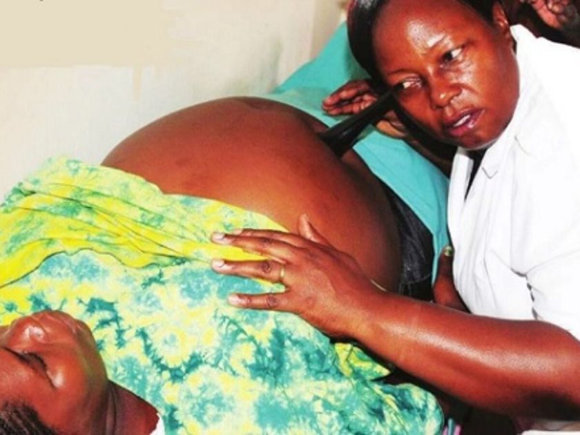
Palpation of the entire fetus: A trained midwife can use her hands to feel the mother's stomach and clearly identify the baby's actual body parts. She will be able to feel the hard, round head, the long smooth back, and the small kicking arms and legs. This confirms the baby’s position and size.
Palpation of fetal movement: After the 24th week of pregnancy, a skilled health worker can place their hands on the mother's stomach and clearly feel the baby kicking, rolling, and moving around.
X-ray: An X-ray machine can show the baby's full bone skeleton as early as the 12th week. However, this is not a recommended method. We never use X-rays just to confirm pregnancy because the dangerous radiation can pass into the womb and harm the growing baby, causing birth defects (genetic alterations) or damaging the baby's reproductive organs. Ultrasound is much safer.
Actual delivery of the baby: The final and most absolute confirmation of pregnancy is when the mother goes into labour and actually delivers a live newborn baby into the hands of the midwife!
⚠️ Attention: Dangers of X-Rays in Pregnancy
Even though an X-ray can easily show the baby's skeleton, midwives and doctors must avoid it. The radiation from X-rays is very dangerous to a developing fetus and can cause permanent genetic damage. Always use ultrasound scans instead.
Differential Diagnosis (Things That Look Like Pregnancy)
Sometimes, a woman's stomach gets big and her periods stop, but there is no baby inside. The midwife must think about other sicknesses that can mimic pregnancy. Some of these false alarms include:
Ovarian cysts: A woman can have large bags of fluid (cysts) growing on her ovaries, causing her stomach to swell. When the midwife feels the stomach, this swelling feels different from a pregnant uterus. Most importantly, if we test her urine, the pregnancy test will be negative.
Fibroids: These are hard, non-cancerous lumps of muscle that grow inside the walls of the uterus. They can make the stomach look very large, just like a pregnant mother. However, fibroids feel like hard, bumpy masses, and the pregnancy test will always remain negative.
Distended urinary bladder: Sometimes a person fails to pass urine, and the bladder fills up with so much fluid that the stomach swells out like a pregnancy. The solution is simple: the health worker inserts a catheter tube to drain the trapped urine, and the stomach immediately goes back to being flat. There will be no other signs of pregnancy.
Pseudocyesis (False or Phantom Pregnancy): This is a psychological condition. It happens when a woman wants a baby so badly, or is so stressed and afraid of being pregnant, that her mind tricks her body. She will stop seeing her periods, her stomach might swell, and she will feel like she has morning sickness. However, when the doctor does a scan or checks for the baby's heartbeat, nothing is there, and the urine pregnancy test is completely negative.
💡 Practice Question for Midwives
Question: A mother comes to the clinic and happily reports that she has felt her baby moving for the very first time. You know this is called "Quickening." Under which classification of pregnancy signs does Quickening fall?
Answer:Presumptive Sign. Even though the mother is sure she feels it, it is a presumptive sign because it is felt only by the mother. It strongly suggests pregnancy, but the midwife cannot prove it just by the mother's word.
Quick Quiz
Normal Pregnancy Quiz
Midwifery - mobile-friendly and focused practice.
Privacy: Your details are used only for quiz tracking and certificates.
Overview: Mastering these foundational Midwifery Terminologies is essential for accurate documentation, effective communication among healthcare teams, and delivering safe maternal and neonatal care. This glossary has been grouped by category for easier studying.
1. General & Professional Concepts
Core definitions shaping the practice of maternal and child healthcare.
Midwifery: The profession of providing assistance and medical care to women undergoing labor and childbirth during the antenatal, prenatal, and postnatal periods.
Obstetrics: A branch of medicine dealing with pregnancy, labor, and the postpartum period.
Antenatal care: Medical care and monitoring provided to pregnant women before childbirth.
Midwifery-led care: A model of care in which midwives are the primary providers for pregnant women, providing continuity of care throughout pregnancy, labor, and postpartum.
Birth plan: A written document created by the pregnant woman outlining her preferences and expectations for labor, delivery, and postpartum care.
Neonatal intensive care unit (NICU): A specialized medical unit providing care for newborns with serious health conditions or premature babies.
2. Maternal History: Gravidity & Parity
Understanding a woman's obstetric history is a critical daily skill on the maternity ward.
Term
Clinical Definition
Gestation / Pregnancy
The period from conception to the delivery of the baby, or the maternal condition of having a developing fetus.
Gravida
A woman who is or has been pregnant, regardless of pregnancy outcome.
Primigravida
A woman pregnant for the very first time.
Multigravida
A woman who has been pregnant more than once.
Nullipara
A woman who is not currently pregnant and has never been pregnant.
Parity (PARA)
The number of pregnancies resulting in a viable birth (≥28 weeks gestation), regardless of whether the baby was born alive or stillborn.
Primipara
A woman who has given birth to one child.
Multipara
A woman who has given birth to two or more children.
Grand Multipara
A woman who has given birth to five or more children.
🧠 Memory Trick: Gravida vs. Para
To avoid confusing the two, remember:
Gravida = Going to have a baby (Total Conceptions).
Para = Parted with the baby (Deliveries past 28 weeks).
3. Fetal & Neonatal Terminology
Fetus: Refers to the human conceptus from the 9th week of gestation up to delivery.
Viability: The capability of the fetus to live outside the womb (usually accepted between 24 and 28 weeks, although survival is rare at the lower limit).
Neonate: A newborn infant up to 28 days old.
Infant: A child from birth to one year of age.
Toddler: A child between one and two years of age.
Preterm birth: Delivery of a baby before completing 37 weeks of gestation.
Intrauterine growth restriction (IUGR): A condition in which the fetus fails to grow at the expected rate inside the uterus.
Vernix caseosa: A greasy, cheese-like white substance that covers and protects the baby’s skin at birth.
Meconium: The very first stool of the neonate. It is present in the lower bowel at 16 weeks of gestation, passed within 3 days following birth, and is greenish-black in color.
Amniotic fluid: The fluid surrounding the fetus within the amniotic sac, providing protection, temperature regulation, and cushioning.
Placenta: A temporary organ that develops during pregnancy, providing oxygen and nutrients to the fetus and removing waste products.
Umbilical cord: The flexible cord connecting the fetus to the placenta, through which nutrients and oxygen are transferred.
4. Labor, Delivery, & Fetal Mechanics
Terms defining the physical and physiological processes of childbirth.
The Process of Labor
Antepartum: The period before birth.
Intrapartum: The period during labor and delivery.
Parturition: The process of giving birth.
Labour: The physiological process of expelling the products of conception from the uterus after 28 weeks of gestation.
Lightening: The descent of the baby into the pelvis, resulting in a noticeable drop in maternal fundal height.
Show: The blood-stained mucoid discharge seen at the onset of labor (loss of the cervical mucus plug).
Crowning: When the largest transverse diameter of the fetal skull emerges under the subpubic arch and does not recede back between contractions.
Fetal Mechanics in the Pelvis
Lie: The relationship between the long axis of the fetus and the long axis of the uterus. (Can be longitudinal, transverse, or oblique).
Attitude: The relationship of the fetal head and limbs to its trunk. (Can be complete flexion, flexion, partial extension, or extension).
Presentation: The fetal part that enters the maternal pelvis first. (Common presentations include cephalic/head, breech/buttocks, face, brow, and shoulder).
Denominator: The specific part of the fetal presenting part used to describe fetal position (e.g., occiput in a cephalic presentation, sacrum in a breech presentation).
Position: The relationship of the denominator to the maternal pelvis (e.g., ROA – right occiput anterior).
Presenting Part: The portion of the fetal presentation that lies over the internal os of the cervix.
Cephalic: A term directly referring to the head.
Diagram illustrating Fetal Lie, Attitude, and Presentation within the maternal pelvis. (Insert image via Elementor)
5. Obstetrical Complications & Interventions
Abortion: Termination of pregnancy before 28 weeks of gestation (Note: Week limits vary by country, 28 weeks is standard in the UNMEB curriculum).
Ectopic pregnancy: A pregnancy that occurs outside the uterus, most commonly in the fallopian tube.
Preeclampsia: A pregnancy complication characterized by high blood pressure and organ damage, usually occurring after 20 weeks of gestation.
Fetal distress: A condition in which the fetus is not receiving adequate oxygen, typically detected through abnormal heart rate patterns.
Episiotomy: A surgical incision made in the perineum during childbirth to enlarge the vaginal opening and facilitate delivery.
Caesarian section: An incision made on the uterus through the anterior abdominal wall to remove products of gestation after 28 weeks of gestation.
Haemorrhage Classifications
Antepartum Haemorrhage (APH): Bleeding from the genital tract between 28 weeks of gestation and the end of the second stage of labor.
Intrapartum Haemorrhage: Bleeding occurring during labor and delivery (e.g., bleeding that occurs after the delivery of a first twin).
Postpartum Haemorrhage (PPH): Significant blood loss from the genital tract after delivery of the baby and placenta.
⚠️ Clinical Alert: Postpartum Haemorrhage (PPH)
PPH is generally defined as a blood loss of ≥500mL, or any amount of bleeding that leads to maternal hemodynamic instability. It can occur up to 8 weeks postpartum and requires immediate emergency management to prevent shock.
6. The Postpartum Period (Puerperium)
Postpartum: After birth.
Puerperium: The period after childbirth or abortion, lasting approximately 6-8 weeks, during which the mother’s body undergoes physical and hormonal changes.
Lying-In Period: The period immediately following delivery, typically 14 days, during which the mother receives close postpartum care from a midwife.
Involution: The natural physiological process by which the uterus returns to its pre-pregnancy size and state.
Lochia: The vaginal discharge that occurs after childbirth or abortion, consisting of blood, mucus, and uterine tissue.
Colostrum: The first fluid found in the breasts, present from the 16th week of pregnancy up to the 2nd and 3rd day after delivery. Rich in antibodies.
Lactation: The production and secretion of breast milk.
Postpartum depression: A mood disorder characterized by feelings of sadness, anxiety, and exhaustion experienced by some women after giving birth.
7. Anatomical Terms & Vital Statistics
Anatomy Highlights
Cervix: The neck of the uterus.
Perineum: The area between the vagina and anus in females, which may stretch or tear during childbirth.
Maternal and Infant Mortality Rates
Statistical Term
Definition
Perinatal Period
Relating to the period around birth (typically from 28 weeks gestation to 7 days postpartum).
Mortality Rate
The number of deaths per 1,000 individuals in a specified population.
Maternal Mortality Rate
The number of maternal deaths attributed to pregnancy, childbirth, or the puerperium per 1,000 women of childbearing age.
Perinatal Mortality Rate
The number of stillbirths and neonatal deaths (within the first week of life) per 1,000 total births.
Neonatal Mortality Rate
The number of deaths of neonates within the first 28 days of life per 1,000 live births.
Infant Mortality Rate
The number of infant deaths within the first year of life per 1,000 live births.
❓ Quick Review Question
Scenario: A woman arrives at the antenatal clinic. She is currently pregnant. Her first pregnancy resulted in a healthy baby born at 39 weeks. Her second pregnancy ended in a spontaneous abortion at 12 weeks. What is her Gravida and Para status?
Answer: She is Gravida 3, Para 1. (She has had 3 conceptions/pregnancies in total, but only 1 viable birth past 28 weeks).
Guide to the Question Approach for Midwifery Exams
It is important for midwives preparing for the exam to be able to answer questions effectively.
This approach allows you to tackle questions in a systematic manner, ensuring that you cover all important points and provide concise and accurate answers. By following this structured approach, you will be able to effectively demonstrate your knowledge, critical thinking and analytical skills, leading to higher scores and overall success on your midwifery exams.
Whether you are facing questions that require you to EXPLAIN, OUTLINE, DESCRIBE, MENTION, IDENTIFY, STATE, LIST, WHAT and GIVE, this article is all you need!
Explaining Questions: Breaking Down Complex Concepts
When questions require explanations, it is essential to break down complex concepts into understandable parts. Start by introducing the topic you’re discussing and providing a concise definition if necessary. Then, proceed to explain on the key components or factors related to the topic. Use clear and simple language to ensure your explanation is easily understandable.
Example Question: Explain how you would admit a mother who has reported in active phase of first stage?
In response to this question, you can follow the question approach by giving the key points step by step:
Reception: the mother and the relatives are welcomed; mother is taken to the admission room while the relatives are offered seats. Rapport between the mother, attendants and the midwives is created.
History taking: if the mother has been attending ANC, her ANC record is obtained; to get the history and any risk factors like multiple pregnancies. If she has not been attending ANC, a full antenatal history is taken, which involves the mother‘s name, address, tribe, religion, husband‘s name and address, her gravida and parity, obstetrical, surgical, social, and medical history, the time and date of admission are entered in the admission forms of the mother.
Then, history of labour under the following headings is recorded:
Show: the mother is asked if she has seen any blood and mucus, her undergarments examined for any stain, vulva examined for the drainage of show which may appear a few hours before or after beginning of labour.
Uterine contractions: mother is asked when the regular pains began, how often and if she has backache. Her statement about the length, severity, or expulsive character of the contraction should be confirmed by observation and evaluation then
Membranes: She is asked whether her water (amniotic fluid) have ruptured or not; if she has noticed a gush or tickling of water → the amount and time are recorded. If in doubt of whether its liquor or urine, litmus paper is dipped into the draining fluid obtained from the vulva to confirm alkalinity or
Vaginal discharge or bleeding: the mother is also asked if she had any vaginal bleeding/ discharge which should be excluded
General examination of the mother: her general appearance is noted, that is healthy or ill, colour, any deformities like lame, presence of oedema, infections, varicose veins or enlarged neck veins. Breast examination is carried out to identify their sustainability for breast feeding
Observations; Vital signs are monitored like temperature, pulse, respiration and blood pressure to rule out eclampsia in She is also asked for bowel action, sleep and rest
Abdominal Examination: first, the bladder should be empty and this is carried out with the mother lying on the couch on supine position with a pillow under her head and This examination is carried out as follows:
On inspection: the shape of the abdomen is noted whether round or oval, the size should be correspond with the weeks of gestation, the foetal movements, skin changes like stria gravidarum and linea nigra and any scar are
On palpation: this can be superficial, fundal, lateral, pelvic, height of the fundus and hypochondriac. They are carried out to note the lie whether its longitudinal, transverse, or oblique; the position of the fetus which can be ROA, LOA, ROP, LOP; and engagement plus enlargement of the spleen and the liver.
On auscultation: the fetal heart is listened, if its heard and regular
Vulva shave, toilet and examination: the shave is done on women whose cultures allow keeping the vulval area with pubic hair. Any abnormal discharges, oedema, or paleness are noted if present
Vaginal examination: this is done to mothers who have no history of APH with the current pregnancy under strict asepsis. It‘s done to confirm the onset of labour, the presentation, position, engagement, station of the presenting part, to confirm whether membranes ruptured or intact, exclude cord prolapsed, assess the pelvis if adequate or inadequate and also progress of labour and it‘s the one that determines the admission of a mother on the partograph
Investigations: routine samples are obtained for example:
Blood: for routine counseling and testing (RCT), rapid polymerase reaction (RPR), HBsAg, haemoglobin estimation, Grouping and cross matching → rational to confirm presence of any disease so as to prevent mother to child transmission (MTCT) and for blood transfusion (BT) in case of anaemia
Urine: for analysis to test for albumin, sugar, acetone that might complicate labour
Personal hygiene: a shower is both hygienic and pleasant. If the mother‘s membrane have ruptured or in advanced first stage of labour, she is sponged down on a couch and given a clean
Admission: the mother is then admitted on a partograph, all the necessary information recorded and the continuous observation of the mother takes place accordingly
Outlining Questions: Structuring Information
When faced with outlining questions, it is important to structure your response in a logical and organized manner. Begin by providing an the main points or components related to the topic. Use subheadings to break down the information further, making it easier for the examiner to follow your answer.
Example Question: Outline the changes that take place in the uterus during the first stage of labour?
Before answering the proposed question above, it‘s vital to first define the following terms:
Labour: is the process by which the foetus, placenta and the membranes are expelled out of the birth canal after 28th weeks of gestation
First stage of labour: is the period of dilatation of the cervix lasting from the onset of true labor till full dilatation.
During the first stage of labour the following occurs:
Effacement or take up of the cervix: this is made possible by the work of muscle fibres surrounding the internal OS which are drawn upward by the retracted upper segment.
Pacemaker / Fundal dominance: each contraction begins from the pace maker situated at the cornua of the uterus. From the fundal region it spreads downwards being stronger and persisting longer in the upper region on reaching the lower region, the wave of contraction weakens and allows the cervix to dilate.
Dilatation of the cervix: this is the opening of the external OS to allow the passage of the fetal head; it occurs as a result of uterine action and the pressure from fore bag of waters and the well fitting presenting part
Contraction and Retraction: is the special ability of the uterine muscle where the contraction does not pass off completely and the muscle fibres, retaining some of the contractions, do not become completely relaxed, instead they become gradually shorter and thicker.
Polarity: is the term used to define the coordination between two poles of uterus throughout labour.
Development of upper and lower segment: the upper uterine segment is the thicker muscular contractile part. The lower part segment is the firm distensible are of 7.5cm – 10cm in length developed from isthmus to the uterus.
Development of retraction ring: is the ridge formed between the upper and the lower uterine segment. It‘s present in labour and normal as long as it‘s not marked enough to be visible above the symphysis pubis. NB: It is called bandl‘s ring in obstructed labour
Show: is the blood stained mucoid discharge seen a few hours within or a few hours after when labour has started.
General fluid pressure: while the membranes remain intact, the pressure of the uterine contraction is exerted on the fluid as the fluid is not compressible the pressure is equalized throughout the uterus.
Rupture of membranes: rupture of membranes may be early or later during the second stage of labour due to the malpresentation and pressure from the presenting part.
Fetal axis pressure: this is when the force of contractions from the uterus is transmitted via the long axis of the canal; this becomes more significant after the rupture of membranes and during second stage.
Describing Questions: Providing Detailed Information
Describing questions require you to provide detailed information about a specific topic or concept. When answering these questions, you have to offer a comprehensive and thorough response, including relevant facts, characteristics, and examples. Use clear language and provide specific details to enhance the depth of your description.
Example Question: Describe the vagina.
Definition: Vagina is a muscular fibrous canal which forms the part of the internal female reproductive organs.
Situation: It is a canal which extends from the vestibule below to the cervix above running in an upward and backward direction between the planes of the pelvic brim.
Shape: It is a potential tube which runs upwards and backwards with its walls in close contact but can be separated during coitus, menstruation, vaginal examination and child birth.
Size: The anterior wall measures 7.5cm. The posterior wall is longer and it measures 10cm.This is because the uterus enters the vagina at an angle of 90 degrees and bends forwards towards the anterior wall hence it encroaches on it
Structure
Gross structure
Superiorly; the upper end of the vagina is known as the vault, where the cervix protrudes into the vault it forms circular recess known as fournices.
The vagina is made up of four fournices that is to say;
The anterior fornix which is smaller and fairly deep The 2 lateral fournices which are shallow
The posterior fornix which is the longest and deepest
The lower end of the vagina is narrow and inferiorly we find the vulva, hymen enclosing the vaginal opening only present in virgins. If hymen is ruptured it leaves tags of membranes referred to as carunculae mytiformes. Vaginal orifice is also called introitus.
Microscopic structure
It is made up of four layers;
Squamous epithelium arranged in folds known as rugae and makes the inner most layer of the vagina, the rugae increase the surface area and offer the vagina ability to stretch when need be for example during coitus and child bearing.
Vascular connective tissue layer which is rich in blood vessels, nerves and lymphatics and is found just beneath the epithelium.
Muscular layer. This is thin but a strong layer which is divided into two; the weak inner circular and strong outer longitudinal fibres.
The pelvic fascial which is made up of loose connective It forms the outer protective coat and is continuous with the pelvic fascia.
Blood supply (arterial): The vagina is supplied by the branches of internal iliac artery which include vaginal artery and uterine artery.
Venous drainage: By the corresponding veins i.e branches of internal iliac veins which include vaginal veins and uterine veins.
Lymphatic drainage: Into the inguinal, the iliac and the sacro glands
Nerve supply: By the sympathetic and parasympathetic nerves which are branches from the lee Franken lanser plexus
Contents of the vagina
It doesn‘t contain any glands but its kept moist by cervical mucus and a transudation from the underlying blood vessels through the epithelium.
Its media is acidic (PH 3.8 to 4.5) and this is made possible by presence of lactic acid after action of doderleins bacilli on glycogen.
Relationships of the vagina
Anteriorly: Below, the base of the bladder rests on the upper ½ of the vagina and the urethra is embedded in the lower ½.
Posteriorly: Pouch of Douglas above, the rectum medial and perineal body below.
Laterally: Pubococcygeous muscles below and pubic fascial containing the uterus above.
Inferiorly: The structure of the vulva.
Superiorly: The cervix and the fournices.
Functions of the vagina
Exit from menstrual flow.
Entrance for spermatozoa.
Exit for products of conception.
Supports the uterus.
Prevents ascending infection due to acidic PH.
For assessing the pelvis.
Drug administration.
Mentioning, Identifying, and Stating Questions: Being Clear and Concise
When faced with questions that require you to mention, identify, or state specific information, it is essential to be clear, concise, and accurate in your response. Avoid unnecessary elaboration and focus on providing the requested information directly.
Example Question: State the major components of a comprehensive birth plan?
To answer this question effectively, you can provide a concise statement listing the major components of a birth plan:
A comprehensive birth plan typically includes the following components:
Preferred birth environment (hospital, birthing center, home birth)
Pain management preferences (medication, natural methods, water birth)
Support people and their roles during labor and delivery(husband, mother)
Positioning preferences for labor and birth(lithotomy)
Preferences for fetal monitoring during labor
Neonatal interventions and care preferences immediately after birth
Feeding preferences (breastfeeding, formula feeding)
Cultural or religious considerations
Preferences for postpartum care and rooming-in with the baby
Contingency plans for unexpected situations or interventions
By providing a clear and concise statement of the major components, you address the question directly and effectively.
Listing and Giving Questions: Providing Comprehensive Information
When asked to list or give information, it is important to provide a response that covers all the relevant points. Ensure that you include all necessary information without leaving out any key details.
Example Question: List and give examples of common obstetric emergencies that midwives may encounter?
In response to this question, here is a comprehensive list of common obstetric emergencies along with examples:
Postpartum Hemorrhage (PPH): This is excessive bleeding following childbirth. Examples include uterine atony (lack of uterine contractions), retained placenta, or trauma to the birth canal.
Shoulder Dystocia: It occurs when the baby’s shoulders become stuck behind the mother’s pubic bone during delivery. This can lead to complications such as brachial plexus injury or fetal hypoxia.
Umbilical Cord Prolapse: The umbilical cord slips through the cervix ahead of the baby, potentially cutting off the baby’s oxygen supply. This requires immediate action to relieve pressure on the cord.
Amniotic Fluid Embolism: This is a rare but life-threatening condition where amniotic fluid enters the mother’s bloodstream, triggering an allergic reaction. It can result in cardiac arrest, respiratory failure, or disseminated intravascular coagulation (DIC).
Pre-eclampsia/Eclampsia: Pre-eclampsia is characterized by high blood pressure and organ damage during pregnancy, while eclampsia is the development of seizures in a woman with pre-eclampsia.
Placental Abruption: This occurs when the placenta separates from the uterine wall before delivery. It can cause severe bleeding and compromise fetal oxygen supply, necessitating emergency delivery.
Fetal Distress: This refers to a compromised fetal condition during labor, usually due to inadequate oxygen supply. It may require interventions such as changing maternal positions, administering oxygen, or performing an emergency cesarean section.
Cord Compression: The umbilical cord becomes compressed during labor, restricting blood flow to the baby. This can occur due to cord entanglement, excessive cord length, or abnormal positioning.
Maternal Infections: Infections such as chorioamnionitis (infection of the placental membranes), sepsis, or genital tract infections can pose risks to both the mother and the baby.
Maternal Hypertensive Disorders: These include gestational hypertension, chronic hypertension, and HELLP syndrome.
Write Short Notes: Concise and Informative Summaries
When encountering “Write Short Notes” questions, the aim is to provide concise yet informative summaries of the given topic. These questions require you to summarize the key points and present them in a clear and organized manner. Avoid excessive details and focus on providing a brief but comprehensive overview.
Example Question: Write short notes on the following
(a) Causes of pain in labour.
(b) Factors that affect pain perception during labour.
(a)CAUSES OF PAIN
There are two major causes of pain
Hormonal factors
Mechanical factors
Hormonal factors These include;
Oxytocin stimulation. This increases the strength, intensity, duration and frequency of the contractions leading to pain.
Progesterone withdrawal. The reduced levels of progesterone lead to an increase in estrogen levels which stimulates the muscles of the deciduae muscles of the uterus to produce prostaglandin which stimulates the smooth muscles of the uterus to contract.
Mechanical factors These include;
Strength and frequency of Braxton hick‘s contractions occurring in late pregnancy which leads to over stretching of the uterus which irritates the uterine muscles to contract leading to pain.
Pressure of the presenting part on the sacro-nerves and lumbar nerves which has pain receptor.
Pressure of the presenting part on the cervix. The presenting part exerts pressure on the cervix muscles hence leading to pain.
Displacement of the pelvic floor muscles. The advancing presenting part distends the vagina and displaces the pelvic floor muscles which are over stretched and the nerves are compressed leading to pain.
(b) PERCEPTION.
Is the process of becoming aware of the environment through the five senses.
Factors that affect pain perception during labour
These factors are emotional experience involving physical and psychological mechanism and can be contributed by the mother, fetus, health workers and structural environment.
Mother
Maternal medical; conditions like pre-clampsia and eclampsia, cardiac conditions which can affect pain perception.
Compromised immunity due to chronic conditions like HIV and cancer which can affect pain perception.
Size of the pelvis. Any pelvic deviation from normal leading to cephalous pelvic disproportion which can affect pain perception.
Age. Young prime gravidae below 18years their pelvic bones are not fully developed and the mother above 35years their pelvic bones are contracted and this hinders normal progress of labour and affects pain.
Parity. Prime gravidae‘s their muscles are still intact and sensitive to pain which leads to strong contractions. The multipara mothers their muscles are laced which leads to uterine itial.
Past obstetrical history like caesarian section which increases the risks of uterine rapture and affects normal labour
Social economic factors for example lack of support which can affect pain perception.
Cultural factors like use of native drugs can affect pain perception.
Past experience can also affect pain perception
Level of education, occupation, religion can also affect pain perception.
Fetus
Fetal abnormalities like hydrocephalous, macrosomic babies can lead to cephalopelvic disproportion hence affecting pain.
Lie, position and presenting pain can affect pain perception during labour
Size of the fetus that is to say big babies which cannot pass through the pelvis hence affecting pain perception
Gestation age. In past maturity the sutures and the fontanels are closing and molding can hot take place hence affecting pain perception.
Health workers
Poor screening of mothers during antenatal Poor management during labour
Poor attitude towards the mother
Structural environment
Hospital setting where there is no privacy for mothers, all stages of labour in one room which can affect pain perception.
Lack of recreation in labour rooms like newspapers, television to occupy the mothers.
What Questions: Providing Clear Definitions and Explanations
“What” questions typically require you to provide clear definitions, explanations, or descriptions of a specific concept, procedure, or pathophysiology These questions aim to test your understanding and knowledge of the subject matter. When answering “What” questions, it is important to be precise and concise in your response, while still providing sufficient information to address the question accurately.
Example Question: What is the role of the midwife in the immediate postpartum period?
To answer this question effectively, you can provide a concise definition and description of the midwife’s role during the immediate postpartum period:
Monitoring maternal and neonatal vital signs: Midwives closely monitor the mother’s blood pressure, heart rate, and bleeding. They also assess the baby’s breathing, heart rate, and overall well-being.
Assisting with breastfeeding initiation: Midwives provide support and guidance to initiate breastfeeding, ensuring proper positioning. They offer education on breastfeeding techniques, addressing any concerns or difficulties that may arise.
Providing emotional support: Midwives offer emotional support to new mothers, addressing any anxieties, fears, or questions they may have. They create a nurturing and supportive environment for the mother and her newborn.
Assessing postpartum recovery: Midwives conduct physical examinations to assess the mother’s postpartum recovery, including uterine involution, healing of perineal tissues, and overall well-being. They provide guidance on self-care practices and postpartum contraception options.
Identifying and managing postpartum complications: Midwives are identify and manage any postpartum complications that may arise, such as postpartum hemorrhage, infection, or breastfeeding difficulties. They collaborate with healthcare providers if further interventions are required.
By explaining the midwife’s role in the immediate postpartum period, you address the “What” question while providing a clear understanding of the topic.
What information must you note on vaginal examination?
On inspection
State of the vulva, note any abnormal discharges like pus, blood, abnormal growths like warts, oedema and scars.
On examination
Note condition of the vagina. Normally the vaginal walls feel warm and moist and dilatable. If dry may be a sign of infection or obstruction.
State of the cervix. If thin, thick, whether soft or rigid and whether its well applied to the presenting part. Note dilatation and cervical effacement.
State of the membranes. Whether intact or ruptured. If ruptured check colour and smell of liquor
Presentation and presenting part. Note level of presenting part in the pelvis. Confirm position by finding or palpating sutures and fontanelles and relate them to the maternal pelvis. Note moulding.
Do internal pelvic assessment and note
-sacro promontary if protruding
-hollow of the sacrum if well curved
-sciatic notches if well rounded
-ischial spines if prominent
-sub pubic arch-if it accommodates 2 ½ to 3 fingers
-inter tuberous diameter if it accommodates 4 knuckles
Tips for Success:
a. Understand the instructions: Carefully read and follow the instructions.
b. Plan your response: Take a moment to brainstorm and outline your ideas before starting to write. This will help you organize your thoughts and ensure a great structured response.
c. Provide relevant examples: Whenever possible, support your answers with real-life examples, or evidence-based practices to demonstrate your understanding and application of midwifery knowledge.
d. Use clear and simple language: Write in a clear and simple manner, avoiding unnecessary elaboration. Focus on delivering information effectively while maintaining clarity.
e. Practice time management: Allocate time for each question based on its difficulty. This will help you ensure that you have enough time to answer all questions within the given time.
f. Review your answers: Before submitting your answer sheet, review your answers to check for any errors, omissions, or areas that need further clarification or elaboration. Don’t forget to write your NSIN Number!
Anaemia means a reduction in oxygen carrying capacity or in quantity of red blood cells.
This may be due to: > A reduction in number of the red blood cells > A low concentration haemoglobin > A combination of both Degrees
Mild – 80% = 11 g/dl – 12.5 g/dl
Moderate – 70% = 8-10.3
Severe – 60% = 7 g/dl and below
OR
Mild – 9.0g/dl
Moderate – 7.8g/dl
Severe – 6g/dl
Causes
Social and economic factors;
Ignorance about utilization of food
Poverty unable to buy the high protein foods
Native medicine
Unstable country / Insecurity
Beliefs – cultural superstition which forbid women from taking certain foods for example chicken and eggs
Obstetrical causes
Frequent child bearing
Repeated haemodilution
Multiple pregnancy due to high fetal demand
Hyperemesis gravidarum leading to poor absorption of Vitamin B12
Abortions, ruptured ectopic, PPH, APH, and heavy periods
Medical causes
Frequent attacks of malaria
Hookworm infestation
Infections such as septicaemia, TB
Sickle cell anaemia
Drugs like Chloramphenical
Types of Anaemia
Physiological anaemia.
Nutritional anaemia.
Aplastic anaemia.
Haemorrhagic anaemia.
Haemolytic anaemia.
Pernicious anaemia.
Physiological anaemia: This is the type of anaemia that occurs during pregnancy due to haemo-dilution that occurs during pregnancy where the blood plasma is increased by 25-30%.
Nutritional anaemia: This is the type brought about about by poor diet whereby there may be deficiency in: > Folic acid which is responsible for the RBC development bringing about megoblastic anaemia, (Immature Red blood cells) > Iron deficiency anaemia which is due to the increased fetal demand from the 28th week of pregnancy or due to excessive morning sickness, vitamin B12 and vitamin c deficiency with lack of protein leading to pernicious anaemia.
Aplastic anaemia: This is due to the damage of the red bone marrow caused by prolonged use of chloramphenical depressing the bone marrow > Radiation where insufficient protection from the x-rays was not provided > Diseases such as cancer and leukaemia > Poison from insecticides
Haemorrhagic anaemia: This when there is increased blood loss as may occur in the following cases > Frequent child bearing that the mother does not get time to regain her haemoglobin levels > Worm infestation such as hookworms > Abortions, PPH and APH > Ruptured ectopic pregnancy > Trauma and accidents > Haematemesis and haemoptysis
Haemolytic anaemia: This is when there is increased blood destruction due to: > Infections such as septicaemia, Pyelonephritis and Bacterial Streptococcus > Diseases for example malaria > Mis-crossmatched transfusion > Sickle cell disease – big spleen disease > Drugs for example primaquine
Pernicious anaemia This is when the intrinsic factor is missing leading to mal absorption of vitamin B12. It may occur in the following conditions: > Diseases of the stomach for example cancer > Hyperemesis gravidarum > Surgical operations for example gastrectomy. However, this condition rarely occurs during child bearing age
Signs and symptoms
On history taking
Patient gives history of general body weakness
Dizziness
Faintness
Palpitations
Loss of appetite (Anorexia)
Headaches
Breathlessness
History of heavy bleeding may be there
On Examination
Pale mucus membranes and conjunctiva for example gums, kips, tongue, soles of the feet and palms of the hands
Distention of the jugular veins
In severe cases oedema of the ankles, feet or it may be generalized
On abdominal palpation, there may be enlarged spleen and liver
Jaundice
Laboratory Tests Haemoglobin level will be low below 12.5 g/dl
Diagnosis
Based on three factors:
History taken from the patient about her home including the surrounding diet, parity and hygiene.
Examination of the patient to detect pale mucus membranes and venous return for example oedema of the upper limbs, wedding rings and puffiness of the face.
Investigations: Investigations carried out
(a) . Haemoglobin estimation that any haemoglobin of 10 g/dl or below is regarded as anaemia (b) . Packed cell volume (Normal is 40%) (c) . Blood film – thick to identify the shape, maturity and consistency, thin red blood cells. (d) . BS for malarial parasites (e) . Sickling test to exclude abnormal cells (f) . Blood film for the thin:
Microcytosis and hypochromia for iron deficiency
Megaloblastic cells – Normochromic for vitamin B12 and folic deficiency
Sickle cell disease
Target cells
Reticulocytes for haemolysis whether its going on mainly in the spleen, whether it is a mixture of no. 1 and 2 which will be indicating nutritional anaemia
(g) . Coombs test for haemolytic anaemia to see whether the mother s developing antibodies against the red blood cells. Usually gets recurrent anaemia. (h) . Bone marrow – to confirm the shape of the cells (i) . Urinalysis for protein indicating damage to the kidney. Microscopic – for put cells in case of severe pyenehiritis
(j) . Stool for intestinal parasites, the commonest is hookworm anaemia (k) . Haemoglobin: Electrophoresis to confirm SCD
Effects of anaemia on pregnancy and labour
General body fatigue with tiredness, breathlessness, palpitations and headache.
Placental insufficiency due to lack of oxygen may lead to: > Intra-Uterine Fetal Death – intra uterine fetal death > Small for dates > Neonatal death > Abortion and premature labour
Post partum haemorrhage
Stress of labour may not be tolerated by a very anaemic and even minor blood loss may be fatal
Fetal and maternal distress leading to instrumental delivery
May go into heart failure
More likely to have venous thrombosis
Less resistance to infection
Poor lactation
Management
Will depend on the severity of the grade of anaemia, stage of gestation and investigation or cause.
Early pregnancy with mild or moderate anaemia in a maternity centre and hospital A mother with mild or moderate anaemia treated as an out patient.
Put the mother in bed
Take history from the mother concerning the type of diet way of living and her surrounding to know the cause of anaemia
Carry out general examination for degree of anaemia by use of a tallquist book
The midwife is only allowed to treat mild and moderate anaemia in early pregnancy
The condition is managed according to the cause
If no clinical examination the haemoglobin is found to be below 60% is sent to the hospital for investigations
Active Treatment For a mother with haemoglobin of 60% and above may be treated with the following:-
Where malaria is common, the mother is given three doses of Fansidar 960 mgs tablets.
Mebendazole 200 mgs bd x 3 days for hookworm
Iron therapy of ferrous Sulphate and folic acid then review after 2 months, ferrous sulphate 200 mgs bd, forric acid 5 mgs od NB. In the maternity centre moderate anaemia in late pregnancy, refer to hospital.
In the hospital
Admit in the hospital in Antenatal ward
Take history about the diet, environment and hygiene
Observations – temperature, Pulse, Respirations and blood pressure taken
Any underlying cause will be treated accordingly
The mother is given routine nursing care
Proper hygiene
Given a high protein diet
Severe anaemia in early pregnancy and late pregnancy In maternity Centre – refer to hospital please
In Hospital
The mother is admitted and history taken
Observations and investigations carried out
Patient is resuscitated immediately with: (a) Blood transfusion or parenteral iron dextran (Inferon) infusion if blood is not available. (N.B Total dose of inferno is given slowly and only in severe anaemia nearly to the time of delivery, and after delivery, should be transfused with packed cells under Lasix.)
Dueretics are given for example lasix 120 mg IV
The patient with severe anaemia should be nursed propped up in bed and given all the care of a very ill patient.
Mouth should be given special attention as stomatitis and glossitis are common in anaemia patients diet – high protein diet with green vegetables plus fresh fruit
Strict fluid balance chart, observe for signs of impending cardiac failure which are raising pulse and respirations. Should report breathlessness if the patient has tuberculosis N.B. IV inferon – 5 ampoules of 250 mgs each in 100 mls of dextrose 5% or normal saline 500mls.
Prevention of anaemia
Good antenatal care – by detecting anaemia and malaria early and treat them.
Health education about diet, personal hygiene, environmental, sanitation by proper use of latrines
Protection against malaria
Reduce blood loss in 3rd stage even after by good management of all the stages of labour
Replace proteins at least during lactation by giving extra protein
Administration of extra Iron and folic acid
To carry out the routine examination of blood for haemoglobin
Advice to the mother
Explain to the mother the reason why she has become anaemic, dangers of anaemia and how to prevent it.
Rest to avoid overworking
Diet and types of food
Advise the mother to take any treatment ordered regularly
The need to prevent mosquitoes to avoid malaria
To avoid frequent child bearing (family planning)
To deliver in the hospital
In labour
Good management of 3rd stage of labour to prevent much blood loss
Administration of iron during puerperium
On discharge
Tell the mother to report immediately when they become pregnant in order to receive appropriate prophylactic treatment of iron therapy
Overview: The First Stage of Labour is the period when the uterus starts regular, painful contractions that cause the cervix to open completely (fully dilate). This module explains exactly how the uterus works to push the baby down, how a midwife manages a mother when she arrives at the hospital, and the step-by-step rules for checking the mother safely.
1. Physiology of the First Stage of Labour
A. Uterine Action (How the Womb Works)
Fundal Dominance: Each contraction of the uterus starts from the very top (the fundus) near the corners (cornua) and spreads across and downwards. The contraction lasts longer at the top where it is strongest, but the peak of the pain is felt all over the uterus at the same time. This strong top action allows the cervix to open and pushes the baby down.
Polarity: This is a natural teamwork (neuromuscular harmony) between the top and bottom parts of the uterus during labor. During a contraction, the upper part contracts strongly and pulls back to expel the baby, while the lower part relaxes and opens up to let the baby pass. If this teamwork is disorganized, labor will stop progressing.
Contraction and Retraction: During labor, when a contraction ends, the muscle fibers do not relax completely. They keep some of their shortness. This is called retraction. Because of this special ability, the upper uterus becomes shorter and thicker, making the space inside smaller, which helps push the baby out.
Formation of Upper and Lower Uterine Segments: By the end of pregnancy, the uterus divides into two parts:
Upper Segment: Mainly for strong contractions; it is thick and muscular.
Lower Segment: Mainly for stretching and opening; it is thin and develops from the lower neck of the womb (the isthmus), becoming about 8-10cm long.
Retraction Ring: This is a small ridge formed between the thick upper segment and the thin lower segment. As labor goes on, this normal ridge rises higher. You cannot normally see it on the outside. However, in dangerous obstructed labor, it rises very high and becomes clearly visible above the pubic bone. This dangerous sign is called a Bandl’s Ring.
B. Cervical Action (How the Cervix Opens)
Cervical Effacement: This means the cervix gets pulled up and thins out. The muscle fibers around the inner opening (internal os) are pulled upwards by the upper uterus until the cervix merges completely into the lower uterine segment.
Cervical Dilatation: This is the process where the tight, closed opening of the cervix enlarges into a hole big enough for the baby's head to pass. It is measured in centimeters (cm). A fully open cervix at term equals 10cm. The pressure from the bag of waters and the baby's well-bent head pushing down helps the cervix open smoothly.
Show: This is the blood-stained thick mucus seen just before or at the start of labor. It is the mucus plug (operculum) that blocked the cervix during pregnancy. The blood comes from tiny broken blood vessels when the bag of waters separates from the opening cervix.
C. Mechanical Factors
Formation of Fore Waters: As the lower uterus stretches, the bag of waters (chorion) detaches from it. The baby's head drops down and blocks the fluid. The small amount of water trapped in front of the baby's head is called the fore waters, and the water behind the baby's body is called the hind waters.
General Fluid Pressure: When the water bag is still intact, the squeezing pressure of the contractions is spread evenly through the fluid over the baby's whole body. Since water cannot be compressed, this protects the baby. If the waters break early, the uterus squeezes directly on the baby and the umbilical cord, which can dangerously reduce the baby's oxygen supply.
Rupture of Membranes (Breaking the Waters): The normal time for the waters to break is at the end of the first stage of labor when the cervix is fully open (10cm). Sometimes they break days before labor, or early in labor if the baby is sitting in a bad position. Rarely, the bag never breaks, and the baby is born completely covered in the water sac. This is called being born in a Caul.
Fetal Axis Pressure: During a contraction, the force from the top of the uterus travels straight down the baby's spine, pushing the baby's presenting part hard against the cervix.
Descent of the Presenting Part: This is the downward movement of the baby through the mother's pelvis. The head twists, turns, bends, and stretches to fit through three main obstacles:
Pelvic Inlet / Brim: When the widest part of the head passes the brim, it is called engaged. The baby is now at Station 0.
Pelvic Cavity: When the head reaches deep down near the perineum, it is at Station +2.
Pelvic Outlet: The head finally passes under the pubic arch and out of the body.
2. Management of the First Stage of Labour
🎯 Aims of Management
To strictly monitor the progress of labor.
To prevent the mother from becoming completely exhausted.
To prevent infections.
To give comfort to the mother and keep her morale and spirits high.
To relieve her pain.
To prevent and catch any complications early.
Admission of a Mother in Labour
When a mother arrives at the health facility, the midwife must systematically admit her to ensure total safety.
Welcome: Greet the mother and her relatives warmly to reduce their fear and anxiety. Build a good relationship (rapport).
History Taking: While she sits or lies down comfortably, review her antenatal card and ask:
Demographic data (Name, age, etc.)
Exact date and time of admission.
Exactly when the contractions started.
How often the contractions come (frequency) and how strong they are.
Whether her waters (membranes) have broken.
Consent: Explain what you will do and get her permission (consent). Ensure she has enough clear information before deciding.
Vital Observations:
Pulse: Check every 30 minutes. If it is over 100 beats/min, it indicates severe pain, anxiety, infection, lack of energy (ketosis), or bleeding.
Blood Pressure (BP): Check every 2 hours.
Temperature & Respiration: Check every 4 hours (normal breathing is 16-20 breaths/min).
Investigations: Test her urine for acetones (dehydration), glucose, and proteins. Draw blood for Hemoglobin (Hb) levels, grouping, and cross-matching just in case she needs a blood transfusion.
Examinations on Admission
General Examination: Check the mother from head to toe. Notice if she looks healthy or sick. Look for body deformities, pale eyes (anemia), yellow eyes (jaundice), swelling (oedema), dehydration, infections, swollen leg veins (varicose veins), and swollen neck veins. Examine her breasts to see if they are ready and suitable for breastfeeding.
Abdominal Examination: Ensure her bladder is totally empty first.
Inspection: Look at the size, shape, scars, and signs of pregnancy.
Palpation: Feel for pain (tenderness), measure the height of the fundus, find the presentation, lie, position, and how deep the baby is (descent). Feel the contractions (frequency, length, and strength).
Auscultation: Listen to the fetal heart rate, checking if it is regular and strong.
3. Vaginal Examination (VE)
This is a strictly clean (sterile) procedure carried out by a midwife by inserting fingers into the mother's vagina to check the progress of labor and rule out any abnormalities.
Indications (Reasons to do a VE)
During Pregnancy: To confirm pregnancy, rule out tumors like fibroids, assess the size of the pelvis, check if the cervix is closed, confirm if a miscarriage is happening, and check abnormal discharges.
During the First Stage of Labour:
To find out how many centimeters the cervix has opened (dilatation).
To make sure the umbilical cord has not dropped down (cord prolapse) when the waters break.
To confirm the cervix is fully open (10cm) when the mother feels the urge to push.
To check if the cervix is soft before forcing labor to start (induction).
To rule out a blocked delivery in cases of very long, prolonged labor.
To positively identify what part of the baby is coming first (presentation) and if it is engaged.
During the Second Stage: To confirm full dilatation, check why the baby is not descending, and check the position of a second twin after the first is born.
During the Third Stage: If the placenta is delayed, to feel if it is stuck in the birth canal. To check for torn tissues (lacerations) or remove large blood clots. For manual removal of a trapped placenta.
During the Puerperium (After Birth): To check if stitches have healed after 6 weeks, ensure organs have returned to normal, take swabs for lab testing, or find the cause of a bad discharge.
Contraindications (When NEVER to do a VE)
Active Vaginal Bleeding (APH): If the mother is bleeding heavily, inserting fingers can tear the placenta and cause fatal bleeding.
Recent Pelvic Surgery or Trauma: The tissues are delicate and easily damaged.
Threatened Miscarriage or Ectopic Pregnancy: It may cause the water to break early or rupture an ectopic tube.
Cervical Incompetence: If the cervix is very weak, touching it can force it open and cause a premature birth.
Active Pelvic Infection (PID): It can push bad bacteria deeper into the womb.
Severe Pain: If the mother is in extreme pain, stop immediately.
Patient Refusal: The mother has the absolute right to refuse the procedure.
Elective Caesarean Section: If she is already planned for surgery, there is no need.
Complications of Vaginal Examination
Discomfort or Pain: Especially if the mother is frightened or anxious.
Vaginal Bleeding: A small amount of spotting is normal, but rough checking can cause cuts (lacerations).
Infection: It can accidentally push outside bacteria into the clean vaginal canal.
Premature Rupture of Membranes (PROM): Pushing too hard can pop the water bag by mistake, causing premature labor.
Requirements for a VE
On the Tray: A gallipot for swabs with antiseptic, 2 receivers, sterile gloves, vaginal speculum, sterile bowl for lotion, clean perineal pads, a sheet and a mackintosh (waterproof sheet), clean gloves, and lubricating gel.
At the Bedside: A privacy screen, hand washing water and soap, and a bedpan.
Procedure for Vaginal Examination
Welcome the mother and explain what you will do. Ask her to empty her bladder.
Screen the bed for privacy and assemble your tray. Encourage the mother to relax her body. Her arms should rest by her side so her stomach muscles stay soft.
Help her lie on her back with knees bent and wide apart (dorsal position). Cover her legs with a sheet (drape).
Place the waterproof mackintosh and draw sheet under her buttocks.
Put on clean gloves. Observe the outside (vulva) before cleaning. Check hygiene, swollen veins, warts, sores, old scars, or female circumcision. Check any fluids coming out. If the fluid smells bad, it means infection. If it is green (meconium), the baby is suffocating (fetal distress).
Clean (swab) the vulva using your left hand, wiping purely from front to back (towards the anus) to prevent transferring feces into the vagina.
Remove clean gloves, wash hands thoroughly, and put on highly sterile gloves.
Dip the two fingers of your right hand into the sterile lubricant. Use your left hand to hold the vaginal lips open. Gently insert the two fingers downwards and backwards. Point them along the front wall of the vagina.
Do not remove your fingers until you have felt everything you need to know. Note: Never touch the clitoris as it causes severe discomfort to the mother.
Findings During VE (What the Midwife Feels For)
Condition of the Vagina: It should feel warm and moist. A completely hot and dry vagina means the labor has been stuck for hours (obstructed labor) and the mother is in deep danger.
Condition of the Cervix: A normal cervix in labor is very thin and stretches like a rubber band tightly around the baby's head. If the cervix feels thick and spongy, it might be the placenta blocking the way (Placenta Previa). If the cervix is not tight against the baby's head, the baby is in a bad position. You must assess how much it has opened (dilatation) and how thin it is (effacement).
The Membranes (Water Bag): If the bag is not broken, it feels tight like a balloon during a contraction.
Level / Station: You feel exactly how deep the baby's head has dropped by comparing it to the mother's side bones (ischial spines).
Fetal Position: Feel the baby's skull bones. You will usually feel a straight line (the sagittal suture) and trace it to a soft spot (fontanel). If you feel the skull bones overlapping strongly, it tells you how the baby's head is squeezing to fit.
Pelvic Capacity: Feel the mother's inside bones again to be absolutely sure the birth canal is wide enough.
After finishing, gently remove your fingers, check them for fresh blood or colored fluid, clean the mother, remove your gloves, and record every single finding on the Partograph.
4. Nursing Care Plan & Sample Questions
📝 Practice Scenario & Nursing Diagnoses
Question 1: Formulate 3 actual and 2 potential nursing diagnoses for a mother in the first stage of labour.
Actual Nursing Diagnoses:
Pain related to strong uterine contractions and cervical dilation, as evidenced by the patient verbally complaining of pain and crying out.
Anxiety related to fear, uncertainty, and anticipation of labor, as evidenced by the mother asking many restless questions about whether she will deliver safely.
Fluid volume deficit related to increased fluid loss (sweating) and frequent urination during labor.
Potential Nursing Diagnoses:
Risk for fetal distress related to the strong physical changes and pressure on the baby during contractions.
Risk for ineffective coping related to severe labor pain and emotional stress.
Question 2: Outline 10 nursing interventions for this mother, giving the rationale (scientific reason) for each.
Assess the pain level (scale 0-10):Rationale: This helps the midwife know exactly how much pain the mother is feeling so they can give the right pain relief.
Teach deep breathing and relaxation:Rationale: Proper breathing keeps the mother calm, stops her muscles from going tight, and reduces her anxiety.
Educate her about the labor process:Rationale: Knowing what is happening removes the fear of the unknown and makes her feel in control.
Provide constant emotional support:Rationale: Having a caring midwife by her side provides a deep sense of security and safety.
Encourage frequent walking and changing positions:Rationale: Moving around helps blood flow, reduces pain, and uses gravity to pull the baby into a good position.
Give frequent sips of clear fluids/water:Rationale: Labor is heavy exercise. Drinking prevents dehydration and replaces water lost through sweating.
Monitor her vital signs (BP, Pulse, Temp) regularly:Rationale: Catching any sudden changes early stops small problems from becoming emergencies.
Listen to the Fetal Heart Rate regularly:Rationale: This is the only way to know if the baby is getting enough oxygen or is suffocating inside.
Provide pain relief (warm massages or safe drugs):Rationale: Reducing extreme pain prevents shock and keeps the mother comfortable.
Collaborate with doctors if labor stops:Rationale: Good teamwork ensures that if the mother needs surgery (C-Section), it is done quickly before the baby gets tired.
Quick Quiz
First Stage Quiz
Midwifery - mobile-friendly and focused practice.
Privacy: Your details are used only for quiz tracking and certificates.
Labour is the natural process by which the fetus, placenta, and membranes (the products of conception) are expelled from the mother's womb through the birth canal after 28 weeks of pregnancy.
In medical terms, labour is defined as the rhythmic contraction and relaxation of the uterine muscles. This causes the cervix to progressively thin out (effacement) and open up (dilatation), eventually pushing the baby out into the world.
What is Normal Labour?
For a midwife to classify a mother's labour as Normal Labour, it must carefully meet all the following conditions:
Occurs at term: The pregnancy must be between 37 and 42 weeks.
Spontaneous in onset: Labour starts on its own naturally, without the use of drugs or artificial methods to induce it.
Fetus presenting by vertex: The baby is coming out head-first (the head is pointing downwards).
Completed within normal time: The whole process should be completed within 12 to 18 hours.
No complications arise: Everything goes smoothly without emergencies.
No injury: Both the mother and the fetus suffer no harm or injury during the process.
No artificial assistance is given: The baby is born naturally without the need for vacuums, forceps, or surgery.
The Three P’s of Normal Labour
For normal labour to be successful, three important factors must work perfectly together. We call these the "Three P's":
Powers: This refers to the strength of the uterine contractions. Strong, regular contractions are needed to push the baby down.
Passage: This refers to the mother's pelvis, including its size and shape, as well as the soft tissues of the birth canal. It must be wide enough for the baby to pass through.
Passenger: This refers to the fetus itself (its size, position, and presentation) as well as the bag of fore-waters (the amniotic sac).
🧠 Easy Memory Aid for the 3 P's
Imagine a train journey: The Powers are the engine pushing, the Passage is the railway track, and the Passenger is the baby taking the ride!
Types of Labour (True vs. False)
It is very common for mothers to feel pains and think they are in labour when they are actually not. Midwives must know how to tell the difference between True Labour and False Labour.
Feature
True Labour
False Labour
Contractions
Regular rhythm. Start mild but increase in severity (strength) and frequency.
Irregular. No clear rhythm. Pain is continuous without rhythm.
Cervical Dilatation
Causes the cervix to progressively open (dilate).
No cervical dilatation happens at all.
Pain Location
Starts in the back and radiates to the front of the abdomen. Associated with stomach hardening.
Pain remains stationary in the lower abdomen. No associated hardening of the abdomen.
Effect of Movement
Pain gets stronger when walking or moving around.
Pain appears stronger when in bed and weaker when the mother is up and walking.
Show (Mucus plug)
Present (Bloody show is seen).
No show.
Effect of Enema/Rest
Pain continues and gets stronger.
Pain often reduces or stops after an enema, resting, or drinking water.
Signs of Impending Labour (Pre-Labour)
These are changes that happen in the mother's body during the last few weeks of pregnancy to prepare for birth. This period is termed as pre-labour.
Lightening: About 2 to 3 weeks before labour starts, the lower part of the uterus expands. This allows the baby's head to sink lower into the pelvis. The pelvic bone (symphysis pubis) widens, and the pelvic floor relaxes to let the uterus descend further. Because the baby drops, the mother feels lighter and can breathe easier.
Cervical changes: As labour gets closer, the cervix becomes "ripe." It changes from being hard like a nose to being soft like a lower lip. It also starts to thin out (effacement) and open very slightly.
False labour: As explained above, the mother may experience painful uterine contractions that do not open the cervix. These are exaggerated Braxton Hicks contractions (practice contractions) which have been occurring painlessly since 6 weeks. False labour can happen on and off for days, or even 3 to 4 weeks before true labour begins.
Premature Rupture of Membranes (PROM): Normally, the water breaks (membranes rupture) at the end of the 1st stage of labour. But if the water breaks before labour starts, it is called PROM. This happens in about 12% of women. For 90% of these women, true labour will start on its own within 24 hours.
Bloody show: Throughout pregnancy, a thick mucus plug sits in the cervix to block infections. As the cervix softens and slightly opens, this mucus plug falls out, mixed with a little blood. This is called the bloody show.
Energy spurt: Many women experience a sudden burst of energy about 24 to 48 hours before labour. After feeling tired for weeks, they suddenly wake up full of energy and vigor (often cleaning the house, known as the "nesting instinct").
G.I.T upset: Some mothers experience unexplained diarrhea, nausea, vomiting, and indigestion just before labour begins. If there is no illness causing this, it is considered a sign that the body is preparing for labour.
Signs of Labour
The signs that labour has actually begun are divided into two main categories: Premonitory signs and Actual signs.
1. Premonitory Signs (Warning Signs)
Lightening: As discussed, it occurs 2-3 weeks before labour. The head sinks to engage in the pelvis, taking pressure off the mother's lungs, giving her relief.
Frequency of micturition (Urination): As the baby drops deep into the pelvis, it limits the space for the bladder. The mother will need to pass urine very often.
Effacement of the cervix: This is the "taking up" of the cervix. The cervix is pulled up and smoothly merges into the lower part of the uterus, becoming very thin.
Braxton Hick’s contractions: These practice contractions become stronger and make the mother anxious. She will feel backache and pain while walking because her pelvic joints are relaxing.
2. Actual Signs (True Proof of Labour)
Regular uterine contractions: The mother feels painful, rhythmic contractions that start out mild but quickly increase in severity (pain) and frequency (how often they come).
Dilatation of the cervix: The external opening of the cervix (the OS) enlarges from a tiny circle into an opening wide enough to allow the baby to pass through (up to 10cm).
Show: The bloody, mucus-like discharge comes out. As the cervix opens, tiny blood vessels (capillaries) tear, mixing blood with the mucus plug.
Rupture of membranes: The breaking of the water. However, midwives do not rely on this as a sure sign of early labour, because the water can break very late in the 1st stage or even spontaneously right as the baby is born.
Causes of the Onset of Labour
The exact trigger that starts labour remains a medical mystery, but it is highly believed to be a combination of hormonal factors and mechanical factors.
Hormonal Factors (Theories)
Oxytocin stimulation theory: As pregnancy reaches its end, the uterus becomes highly sensitive to the hormone oxytocin. Oxytocin causes strong muscle contractions.
Progesterone withdrawal theory: Progesterone is the hormone that relaxes the uterus and keeps the pregnancy safe. Near the end of pregnancy, progesterone levels drop. This removes the relaxing effect, allowing the uterus to start contracting.
Oestrogen stimulation theory: Oestrogen makes the uterine muscles irritable and ready to contract. A rise in oestrogen forces the lining of the womb (decidua) to release prostaglandins. Both prostaglandins and oxytocin work together to make the uterus contract.
Fetal cortisol theory: The baby's own body produces cortisol (a stress hormone) when it is ready to be born. This fetal cortisol affects the mother's oestrogen levels, triggering labour.
Prostaglandin stimulation theory: Prostaglandins directly stimulate the smooth muscles of the uterus to contract violently, pushing the baby down.
Mechanical Factors
Over-stretching of the uterus: By 9 months, the uterus is stretched to its absolute maximum limit by the baby and the fluid, causing it to naturally want to contract and empty itself.
Pressure on the cervix: The heavy baby's head presses down on the nerve endings in the cervix (the cervical ganglion). This nerve pressure sends a message to the brain to start labour.
Braxton Hicks strength: The constant increase in the strength and frequency of practice contractions eventually blends into true labour.
The progression of the cervix during the first stage of labour, thinning (effacement) and opening (dilatation).
The Stages of Labour
Normal labour is divided into four distinct stages to help midwives monitor the mother safely:
1. First Stage (Stage of Cervical Dilatation)
This stage begins with the onset of regular, rhythmic uterine contractions and is complete when the cervix is fully open (dilated to 10cm). It is broken down into three phases:
Latent phase: A slow period where the cervix opens from 0 to 3cm. In mothers having their first baby, this slow phase can take 6 to 8 hours.
Active phase: The cervix begins to open much faster. This phase starts when the cervix is 4cm dilated and ends when it reaches 8cm.
Transitional phase: The hardest and final part of the first stage. It starts at 8cm and ends when the cervix is fully dilated (10cm).
2. Second Stage (Stage of Expulsion)
This is the stage where the baby is actually pushed out. It begins when the cervix is fully dilated and ends when the baby is completely born. It has two phases:
Propulsive phase: The baby's head descends from the fully opened cervix down onto the pelvic floor muscles.
Expulsive phase: The mother feels a strong, uncontrollable urge to push (bear down). This phase ends with the actual delivery of the baby.
3. Third Stage (Placental Stage)
This stage begins immediately after the birth of the baby and ends with the complete separation and expulsion of the placenta and membranes. It involves controlling the mother's bleeding. Naturally, it takes 5 to 30 minutes, but with active management by a midwife, it is usually completed within 5 to 15 minutes.
4. Fourth Stage (Recovery Stage)
This is the critical observation period. It is defined as the first one hour immediately after the placenta is delivered. The midwife closely monitors the mother for bleeding and checks her vital signs to ensure she is recovering well.
💡 Quick Practice Check
Question: A pregnant mother comes to the ward complaining of abdominal pain. Upon assessment, you notice she is having painful contractions, but her cervix is completely closed (0cm dilated), and the pain stops when you advise her to walk around. What is she experiencing?
Answer:False Labour. The key signs are no cervical dilatation, and the pain reduces with movement.
Overview:Antenatal Care (ANC) is the planned, methodical care and supervision given to a pregnant woman by a midwife or obstetrician from her very first clinic visit until the beginning of labor. It is followed by Pelvic Assessment near the end of pregnancy to ensure the mother's bones are wide enough for a safe delivery.
1. Aims of Antenatal Care
The primary goals of providing antenatal care are to ensure a safe pregnancy and a healthy outcome for both mother and baby. These aims include:
To monitor the progress of the pregnancy in order to support maternal health and ensure normal fetal development.
To prepare the mother for labor, lactation (producing breast milk), and the subsequent care of her newborn baby.
To detect early and treat appropriately any high-risk conditions—whether medical or obstetrical—that could endanger the life of the mother and the baby.
How These Aims Are Achieved
A midwife achieves these goals through the following practical steps:
Developing a strong partnership with the pregnant woman.
Providing a holistic (complete) approach to the woman’s care that meets her individual and personal needs.
Promoting awareness of public health issues for the woman and her family.
Exchanging information with the woman and her family, enabling them to make informed and smart choices.
Being an advocate for the woman and her family, supporting her right to choose care that is appropriate for her own needs and those of the family.
Recognizing complications of pregnancy early and appropriately referring women to specialists within the multi-disciplinary healthcare team.
Facilitating the woman to make an informed choice about methods of infant feeding, and giving appropriate and sensitive advice to support her decision.
Facilitating the woman and her family in their preparations to meet the physical and financial demands of birth by making a clear birth plan.
Offering continuous health education to prepare the couple for parenthood.
Activities Done in the Antenatal Clinic (ANC)
Every ANC clinic must offer a specific set of services to every mother:
Registration: Officially recording the mother into the clinic system.
Booking: Taking a comprehensive medical and social history.
Special Tests and Investigations: Conducting blood and urine tests.
Health Education: Teaching about healthy pregnancy habits.
Immunization: Giving vaccines such as the Tetanus Toxoid.
Treatment of Minor Disorders: Managing common pregnancy complaints like nausea or heartburn.
Provision of Supplements: Giving essential vitamins like Iron and Folic Acid.
Examination: Performing full physical and abdominal checks.
Orientation of Mothers: Showing them around the maternity unit.
Formulating a Birth Plan: Preparing for the day of delivery.
Counseling: Offering emotional and psychological support.
Referral of Cases: Sending complicated or high-risk cases to better-equipped hospitals.
2. Initial Assessment (The Booking Visit)
The first day a mother comes to the clinic is called the "Booking Day." It is the most detailed visit.
Objectives for the Initial Assessment
To assess the mother's level of health by taking a detailed history and to offer appropriate screening tests.
To ascertain baseline data (starting measurements) of her blood pressure, urinalysis, uterine growth, and fetal development. This baseline is used as a standard for comparison as the pregnancy progresses.
To identify risk factors by taking accurate details of her past and present obstetric, medical, family, and personal history.
To provide an opportunity to discuss any fears or concerns the woman has.
To give advice pertaining to pregnancy in order to maintain the health of the mother and the developing fetus.
To build the foundation for a trusting relationship in which the woman and the midwife are partners in care.
To make appropriate referrals when additional health care or support needs have been identified.
Comprehensive History Taking
The midwife must ask detailed questions across multiple categories to ensure nothing is missed.
Demographic Data
Name, Age, Address, and Occupation.
Next of Kin (NOK): Their relationship to the mother, occupation, and contact details.
Level of Education (LOE), Tribe, and Religion.
Nearest health facility and the exact distance from her home.
Social History and Habits
Smoking: Cigarettes contain nicotine which constricts (narrows) blood vessels leading to placental insufficiency. This can result in fetal hypoxia (lack of oxygen for the baby), small for dates babies, and abortions. The woman should be advised to reduce the number of sticks gradually to avoid withdrawal syndrome.
Alcohol: Drinking alcohol carries a risk of trauma (accidents) which can result in abortion or placenta abruption. It also causes a loss of appetite leading to maternal malnutrition and small for dates babies.
Marital Status: Is she married or single? Find out the number of years spent in the marriage and try to find out if she is happy and safe at home.
Home Environment
House: Is it rented or her own? Note the number of rooms and the total number of occupants to check for overcrowding.
Environmental Hygiene: How clean is the home surrounding?
Resources: What is her primary source of water and food?
Family History
The health status of the woman’s parents and her siblings. (If they are deceased, note the exact cause of death).
Familial diseases (diseases that run in the family) for example, a history of cancer, diabetes, cardiac (heart) diseases, allergies, etc.
Other serious illnesses in the family like mental illnesses or severe complications with pregnancy.
History of multiple pregnancies (twins, triplets) in the family line.
Past Surgical and Medical History
Past Surgical History: History of accidents involving the spine, pelvis, and lower limbs which would reduce the pelvic diameters. History of major operations like Caesarean Section (C/S) and pelvic operations. History of blood transfusion (which carries a risk of exposure to HIV/AIDS and iso-immunization).
Past Medical History: Medical conditions that may complicate or be complicated by pregnancy, labor, and puerperium (e.g., sickle cell disease, Diabetes Mellitus, Hypertension). Childhood illnesses like rickets or poliomyelitis which can reduce pelvic diameters, hence leading to a contracted pelvis. Infectious diseases like TB and Hepatitis B. Sexually Transmitted Infections like syphilis and gonorrhea.
Gynecological and Menstrual History
Gynecological Conditions: History of abortions, ectopic pregnancy, fibroids, etc.
Gynecological Operations: Previous surgeries like myomectomy (removing fibroids), D and C (Dilation and Curettage), or evacuation of the uterus.
Menstrual History: Menarche (when periods first started), length of periods, interval between periods, and amount of blood flow. Any history of Dysfunctional Uterine Bleeding (DUB) or pre-menstrual spotting.
Family Planning: Any method of family planning ever used, any complaint she had about it, and the reason for stopping it.
Past Obstetrical History
Previous Pregnancies: Ask about any abnormalities such as abortions, stillbirths, living children, their general health status, and their immunization status. Ask for the interval between pregnancies, the length of gestation, birth weight, fetal outcome, length of labor, presentation (how the baby came out), and type of delivery. Ask about any prenatal and postnatal complications, and if the previous babies were breastfed and for how long.
Labor History: Ask about any operations, induction of labor, assisted delivery (vacuum/forceps), and Postpartum Hemorrhage (PPH).
Puerperium (Postnatal) History: Check if her recovery was normal, or if she had any history of sepsis (severe infection) or PPH.
Present Obstetric History
Gravidity: Total number of pregnancies.
Parity: Number of deliveries past 28 weeks.
LMNP: Last Normal Menstrual Period.
WOA (Weeks of Amenorrhea): How many weeks she has gone without her period (gestational age).
📅 Calculating the Expected Date of Delivery (EDD)
The EDD is calculated by adding 9 calendar months and 7 days to the date of the first day of the woman’s last menstrual period. This is known as Naegele’s Rule.
This method assumes that:
The woman takes regular note of the regularity and length of time between her periods.
Conception occurred exactly 14 days after the first day of the last period (true if the woman has a regular 28-day cycle).
The last period of bleeding was true menstruation (because sometimes early embryo implantation can cause slight bleeding).
She has not been taking contraceptive pills, as break-through bleeding and lack of ovulation (anovulation) caused by pills can impact the accuracy of the LNMP.
Present Health Status
Finally, the midwife must check how the mother's body is functioning right now:
Appetite: It is important to know because a poor appetite leads directly to malnutrition and anemia.
Sleep: Find out if the mother sleeps well. If not, find out the cause, which could be due to worries, insects in bed, physical pain, or signs of illness.
Micturition (Urination): It’s good to know whether the woman passes urine well because Urinary Tract Infections (UTI) are common in pregnancy due to the stagnation of urine in dilated and kinked ureters. In case of increased frequency without pain, the mother is counseled in relation to the normal physiology of pregnancy.
Bowel Action: As constipation is very common in pregnancy, the mother is reassured and advised to take plenty of fluids and roughages (fiber-rich foods).
NB: Always conclude the history by asking the mother if she has anything else she would like to tell you.
3. Investigations and Clinical Tests
On the first day, every single pregnant woman should receive the following laboratory investigations to ensure her safety.
Test / Investigation
Reason / Clinical Interpretation
Urinalysis
Tested for Albumen (indicative of Pre-eclampsia/PET), Acetone (indicative of dehydration), and Sugars (indicative of diabetes).
RPR / VDRL
Done routinely to test for and exclude Syphilis.
HIV Screening
Done to ensure the Elimination of Mother-to-Child Transmission (EMTCT) of HIV.
Blood Grouping
Done to know her blood type in case of an emergency requiring blood transfusion.
Hb Level (Hemoglobin)
It should be done on the booking day, then at 32-34 weeks, and lastly at 36 weeks to rule out anemia.
Coombs Test
It is done to detect harmful antibodies circulating in the mother's blood.
Clinical Tests Done in the Clinic
Weight: This is taken on every visit to ANC. The mother is expected to gain about 12.5kg during the whole pregnancy (4kg in the first 20 weeks and 8.5kg in the last 20 weeks). Excessive weight gain could be due to twins, a very big baby, or excess amniotic fluid (polyhydramnios). Failure to gain weight could be due to poor fetal growth.
Height: It’s done on the booking visit, or in labor if the mother has not been attending ANC. The normal height should range between 152-170cm. A height below 150cm indicates a small pelvis, and a height above 170cm indicates a narrow pelvis.
Shoe Size: Normal shoe size ranges between 5 and 8. If the shoe size is below 5, it strongly indicates a small pelvis.
Blood Pressure (BP): This is done on every visit to ANC. The BP of a pregnant mother normal ranges from 90/60 to 140/90 mmHg. A raised BP is a danger sign and may be due to PET and eclampsia. Any rise of 30 mmHg (systolic) and 15-20 mmHg (diastolic) from what has been considered normal is dangerous, and the mother’s urine should be tested for proteins immediately. The mother is asked how she feels generally, especially her sight (checking for blurred vision), and then referred to the doctor.
4. Full Physical Examination
This includes a full head-to-toe review of the physical systems to ascertain the woman’s general health. The breasts, pelvis, and abdomen receive particular attention. The examination is carried out systematically beginning with the head and ending with the pelvis and abdomen.
General Appearance: Observe her body type, weight, energy level, grooming (cleanliness), and posture. This is noted when the mother is entering the room or when she is sitting.
Head: Check the scalp, check the hair (whether treated), and check the hair pattern distribution.
Eyes: Look at the conjunctiva (inner eyelids) to check for anemia. Look at the sclera (white of the eyes) to check for jaundice. Ask about her vision and look for abnormal eye discharge.
Nose: Check her sense of smell. Look for bleeding, obstruction, abnormal growths, and discharge.
Oral Cavity: Check for toothache, check dentures, and observe the state of her lips. Ask about chewing or swallowing problems. Check the tongue and gums for paleness (anemia), and ask about her sense of taste.
Ears: Check for any discharges or any hearing loss.
Neck: Check neck movement. Palpate for swellings or enlarged salivary glands (parotid, sub-mandibular, sublingual), the thyroid gland, and lymph nodes (superficial cervical, deep cervical glands, and sub-clavicles). Palpate and observe the jugular veins and pulsation of the thyroid gland. Swelling of the thyroid gland may be due to iodine insufficiency, though during pregnancy there is a slight enlargement of the glands which may be due to a chronic cough. Extended jugular veins may be due to cardiac problems or anemia.
Upper Limbs (Arms): There should be two arms with the same size and length. Check skin texture and look for muscle wasting. Examine the palms for their color. Check the fingernails to see if capillary refill is good, and look for oedema (swelling).
Chest: Observe exactly how the mother is breathing to detect if she has problems with her respiratory system, like pneumonia.
Breast Examination
This is done carefully to promote proper breast feeding and exclude hidden abnormalities.
Inspection (Looking): Observe for size, equality, shape, and pulling of the breasts. Look for signs of pregnancy and signs of abnormalities like changes in the skin (e.g., redness, orange-like discoloration). Look at the nipple for prominence, dimpling, retraction (pulled inwards), size, and whether it is flat or well protracted. Check for presence of scars, cracks, sores, and axillary extension (breast tissue under the arm).
Palpation (Feeling): The breasts are examined by touch to find breast abnormalities and deep-seated masses.
Back: Check for any fungal infections, surgical scars, and sacral oedema (swelling at the bottom of the spine, which may indicate dangerous PET or Eclampsia).
Lower Limbs (Legs): Check size, muscle wasting, pain or stiffness of joints, pain in the calf muscles, oedema, varicose veins, extra digits (toes), any infections, and tibia/ankle oedema.
Feet: Check hygiene, any fungal infections, and nails (check for venous return and color). Check the sole of the feet for cleanliness and color.
Perform a Homan’s Sign: Homans’s sign is often used in the diagnosis of deep venous thrombosis (DVT) of the leg. A positive Homans’s sign (which is calf pain when dorsiflexing/bending the foot upwards) is thought to be associated with the presence of dangerous blood clots (thrombosis).
Assess for maternal efforts.
Vulva: Check for sores, warts, varicose veins, and abnormal discharges. Request the mother to cough while observing to check for stress incontinence or discharges.
Midwife performing an abdominal examination to establish fetal growth.
5. Abdominal Examination
It is carried out from 24 weeks gestation to establish and affirm that the fetal growth is consistent with the gestational age during pregnancy.
Aims of Abdominal Examination
To observe signs of pregnancy.
To assess fetal size and growth.
To assess fetal health by auscultating (listening to) the fetal heart.
To detect any deviations from normal.
To diagnose the location of fetal growth.
To locate fetal parts.
Preparation and Procedure
Ensure the mother has emptied her bladder within the last 30 minutes before the abdominal examination.
Ensure privacy.
The mother should be lying comfortably on a couch.
Wash your hands, expose only the area of the abdomen that needs to be palpated, and cover the remainder of the woman to provide her privacy and protect her dignity.
The 3 Steps of Abdominal Examination
1. Inspection
Stand at the foot of the bed while the mother is on her back with her abdomen exposed from the xiphisternum up to the symphysis pubis. Look at the size, shape, operational scars, signs of pregnancy like darkening of the linea nigra below and above the umbilicus, fetal movements, and striae gravidarum (stretch marks).
2. Palpation (Leopold’s Maneuvers)
Stand at the right side of the mother. Ensure the pads of your fingers, and not the sharp tips, are used. Palpate as follows:
Superficial palpation: For localized tenderness.
Hypochondriac palpation: For enlarged organs under the ribs.
Height of fundus estimation: To check growth.
Pelvic palpation: To check for presentation (which part of the baby is entering the pelvis).
Fundal palpation: To check for the lie (how the baby's spine aligns with the mother's).
Lateral palpation: To check for position (where the baby's back is facing).
🤲 Pawlik’s Grip (Second Pelvic Grip)
During a deep pelvic palpation, a midwife grips the fetal head between the thumb and fingers of one hand to check for engagement (if the head has dropped into the pelvis). This specific maneuver is termed as Pawlik’s grip or the second pelvic grip.
3. Auscultation
This is the way of listening to the fetal heart to determine fetal wellbeing by use of a feto-stethoscope.
Abdominal Summary
Conclude your abdominal exam by noting: Height of fundus, Presentation, Lie, Position, and Fetal heart.
Case Summary
Comment on all histories, general and abdominal examination.
Give feedback to the mother.
Give necessary advice.
Provide the return date for the next clinic visit.
6. Ongoing Antenatal Care
Purpose of Ongoing Visits
To continue to observe for maternal health and freedom from infections.
To assess fetal wellbeing continuously.
To ascertain that the fetus has adopted a lie and presentation that will allow a normal vaginal delivery.
To offer an opportunity for the mother to express any fear or worries about pregnancy and labor.
To ensure that the mother and family are confident enough to decide when true labor starts.
To discuss any views about the conduct of labor and formulate a birth plan if required.
Risk Factors Arising During Pregnancy
Be highly alert if any of the following occur:
Change in fetal movement pattern - increased or reduced movements.
Hb (Hemoglobin) less than 10g/dl (severe anemia).
Poor weight gain or sudden weight loss.
Proteinuria (protein found in urine).
BP (Blood pressure) above 140/90 mmHg.
Uterus is either too large or too small for the calculated dates.
Excess or decreased liquor (amniotic fluid).
Malpresentation (baby is in the wrong position for birth).
Any form of vaginal bleeding.
Premature contractions.
Vaginal infection.
Head not engaged by 38 weeks in PGs (Primigravidas - first-time mothers).
On Each Visit, Do The Following:
Review the clinic card and assess any past complaints.
Take Blood Pressure, weight, and test urine.
Carry out a general and abdominal examination.
Give drugs (like iron, folic acid, anti-malarials) accordingly.
Indicators of Fetal Wellbeing
Increased maternal weight in association with increasing uterine size.
Fetal movements which follow a regular pattern throughout the pregnancy.
Fetal heart rate is steady between 120-160 beats per minute.
The Ministry of Health (MOH) Goal-Oriented Antenatal Care Protocol.
The MOH Goal Oriented ANC Protocol
Goals are different depending on the timing of the visit. A minimum of 8 Contacts are aimed for in an uncomplicated pregnancy. If a woman books later than in her first trimester, the preceding missed goals should be combined and attended to in the current visit. At all visits, the midwife must address any identified problems, check the BP, and measure the Symphysio-Fundal Height (SFH). Women must receive Hb, HIV testing, and Syphilis testing (RPR) routinely.
7. The Individual Birth Plan & Health Education
The Individual Birth Plan
A structured birth plan must be made to prepare the mother and her family. The plan includes:
A birth place where there is a skilled birth attendant.
Identifying someone to take care of the family in her absence.
Knowing her EDD (Expected Date of Delivery).
Her choice of a birth companion.
Identifying a blood donor in case of emergency bleeding.
Her choice of clothes for labor.
Strategies for labor pain relief.
Position for labor and childbirth.
Place of delivery.
Transportation to use and how it will be available.
How to raise funds for transport and the cost of delivery.
Family security and feeding provisions while she is away.
Family planning goals for after the baby is born.
Where to go after delivery.
When her next appointment is.
NB: Involve the partner in the birth planning process. Teach the mother how to recognize the onset of true labor.
Health Education Topics
During the ANC visits, the midwife must continuously teach the mother about:
Nutrition (eating a balanced diet).
Sleep and resting.
Sexual counseling.
Hygiene.
Daily activities.
Healthy weight gain.
Postnatal follow-up.
6. Immunization:
TT (Tetanus Toxoid): Ensure the mother receives her Tetanus Toxoid injections to protect her and the newborn against tetanus.
Record and Interpret Findings
After taking proper history, doing a thorough physical examination, and relevant investigations, record all findings clearly in the antenatal card.
Interpret the findings accurately so as to identify any risk factors.
Give care and management accordingly based on your findings.
Give an appointment for the next visit accordingly.
📝 Assignment
Discuss the Goal Oriented Antenatal Protocol. Review the minimum 8 contacts, what is achieved at each contact, and how to combine goals for late bookings.
Assessing the maternal pelvis to ensure it is adequate for childbirth.
8. Pelvic Assessment
This is the estimation of the pelvic cavity so as to see whether it is adequate for that particular baby to pass through safely. OR, it is an examination done by a doctor or midwife on a pregnant woman at or after 36 weeks to see that both the mother and baby are out of danger at the time of delivery.
It is always done at 36 weeks because of the natural relaxation of the pelvic joints due to the Relaxin hormone.
Aims of Pelvic Assessment
To rule out a poor obstetric history.
To ensure normal delivery of the mother without any assistance (like vacuum or forceps).
To rule out bone abnormalities like prominent ischial spines or a narrow sub-pubic arch.
To reduce the infant and maternal mortality rate.
To reduce injuries to both the mother and the fetus.
Pelvic assessment is done in 2 distinct ways: External Pelvic Assessment and Internal Pelvic Assessment.
A. External Pelvic Assessment (External Pelvimetry)
This is done on the 1st visit. It includes:
1. History Taking
Age: A woman of the age of 18 years is expected to have a mature pelvis, but below 18 years, the bones are not fully ossified (hardened). A primigravida (PG) who is 35 years and above is expected to have a difficult delivery because the ligaments of the pelvis are already fused; therefore, the necessary "give" of the pelvis is impossible.
Tribe: It’s important to know the tribe because different tribes have different types of pelvis structures. For example, the Bakiga and Banyankole have a large normal pelvis, but the Basoga and Baganda are at risk of having a contracted pelvis.
Marital Status: It’s important to know the size of the husband because small women marrying giant men may carry big babies which can lead to CPD (Cephalopelvic Disproportion).
Medical History: It’s important to know because some diseases like poliomyelitis may permanently affect the growth of the pelvic bones and muscles.
Surgical History: Ask the mother if she has ever had any accident involving her spine, pelvis, and lower limbs.
Past Obstetrical History: If the previous labor and delivery were completely normal, and if the baby weighed at least 3kg and over, she is expected to have an adequate pelvis. A history of instrumental delivery or C/S may give a strong suspicion of an inadequate pelvis.
2. Observations
Gait: Always be alert on a woman who walks with a limp or who has muscle wasting of the legs. A poor gait means a deformed pelvis hence a reduced diameter. It often indicates congenital hip deformity.
Height: The normal average height in women is between 152-170 cm. Height below 152cm may indicate a contracted pelvis, and if above 170cm, it indicates a narrow birth canal.
Palms: Those with short palms usually indicate a small pelvis.
Shoe Size: The normal shoe size is 4-8. A shoe size below 4 indicates a small pelvis.
Stature: A woman of very small stature and a tiny waist is expected to have an inadequate pelvis.
3. Abdominal Examination: Engagement of the Fetal Head (Head Fitting)
NB: It’s no longer heavily practiced for fear of HIV transmission through aggressive handling.
Procedure for Head Fitting:
Explain the procedure to the woman.
The bladder should be completely emptied.
The mother is relaxed flat on the bed with support on a pillow.
The midwife with the right hand locates the symphysis pubis while the other hand is under the mother’s head.
The mother takes a deep breath in and out.
The baby's head is pushed downwards and inwards.
The fingers of the right hand should feel if the largest transverse diameter of the fetal head is passing easily through the pelvic brim as the mother is supported to sit upright without relaxing her elbows.
If the transverse diameter can be pushed through the pelvic brim, the top of the pelvis is adequate. This test is called head fitting.
NB: It’s incredibly important that from 36 weeks onwards, the abdomen is palpated to see if the head is engaged or can be made to engage.
B. Internal Pelvic Assessment (Digital Pelvimetry)
It’s done under strict aseptic (sterile) technique. The midwife must first clearly know the exact measurement of her own fingers.
Procedure:
Explain the procedure thoroughly and ask the mother to empty her bladder and rectum.
Prepare a Vaginal Examination (VE) tray and put it on the side of the bed.
Screen the bed for absolute privacy.
Ask the mother to lie on her back and carry out a routine abdominal examination first.
The midwife measures the length of her working fingers.
Position the mother in a dorsal position (knees bent, legs apart) and drape her to protect dignity.
The right hand is gloved, and two fingers of the gloved hand are heavily lubricated, introduced, and passed high into the vagina. The following key pelvic areas are assessed:
Areas Assessed Internally
Pelvic Structure
Assessment Detail
Sacral Promontory
An attempt is made to reach the sacro-promontory bone at the back by assessing the diagonal conjugate, which is normally 12-13cm deep. If the midwife's short fingers (less than 12-13cm) easily reach it, that directly shows it’s too prominent and the pelvis is small.
Hollow of the Sacrum
This is the curve of the lower back bone. It should be well-curved and smooth. It should not be too long; if it’s flat, the internal cavity is severely reduced and internal rotation of the fetal head will be very difficult.
Pelvic Walls
These side walls are felt, and they should be smooth and straight down (flat). If they converge (slant) downwards like a funnel, the mid-cavity is reduced.
Greater Sciatic Notches
These gaps should feel wide. If they are reduced or tight, internal rotation of the baby's head will be difficult.
Ischial Spines
These two side bones are palpated to see whether they are sharp or prominent. The distance between them is estimated to ensure the baby won't get stuck.
Sub-Pubic Arch
The front arch is measured and should not be less than 90 degrees. It should comfortably accommodate 2-3 fingers. A narrow sub-pubic arch reduces the anteroposterior (AP) diameter of the pelvic outlet, trapping the baby.
Inter-Tuberous Diameter
The distance between the 2 ischial tuberosities (sitting bones) can be assessed by inserting a closed fist between them externally; it should freely admit 4 knuckles.
NB: After the assessment, record all your findings clearly and give prompt, honest feedback to the mother.
Quick Quiz
Antenatal Care Quiz
Midwifery - mobile-friendly and focused practice.
Privacy: Your details are used only for quiz tracking and certificates.