
Gametogenesis: Oogenesis & Spermatogenesis
1. Introduction to Gametogenesis
In the human body, normal body cells (somatic cells) contain 46 chromosomes. These are called diploid cells. However, reproductive cells (gametes) must only contain 23 chromosomes so that they can combine during fertilization. These are called haploid cells. Gametogenesis is the journey of turning diploid germ cells into haploid gametes through meiosis.
- In females, the process is called Oogenesis (production of the ovum/egg).
- In males, the process is called Spermatogenesis (production of spermatozoa/sperm).
2. Oogenesis (Production of a Mature Ovum)
Oogenesis is accomplished through meiosis, but unlike in males, this process begins very early during the female's embryonic development (while she is still a fetus in her own mother's womb).
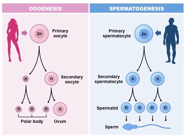
The Stages of Oogenesis
- 1. Fetal Stage (Before Birth): Inside the female fetus, primitive egg cells called Oogonia multiply rapidly by mitosis. They then grow into Primary Oocytes. These primary oocytes begin the first meiotic division (Meiosis I) but suddenly stop and go to sleep (arrest) in the prophase stage. A baby girl is born with all the primary oocytes she will ever have (about 1 to 2 million).
- 2. Puberty Stage: The primary oocytes remain dormant throughout childhood. When the girl reaches puberty, the hormones FSH and LH wake them up. Each month, a few primary oocytes resume meiosis.
- 3. First Meiotic Division: A primary oocyte successfully completes Meiosis I just before ovulation. However, the division of the cell's fluid (cytoplasm) is very unequal. It produces one large healthy cell called a Secondary Oocyte, and one tiny, useless cell called the First Polar Body.
Why unequal? The ovum needs to keep all the nutrients and cytoplasm to feed the early embryo if fertilization occurs. The tiny polar body eventually degenerates and dies. - 4. Second Meiotic Division (Fertilization): The secondary oocyte is released from the ovary during ovulation. It begins Meiosis II but stops again in the middle. It only finishes Meiosis II if a sperm successfully penetrates it. If a sperm enters, it completes the division, creating a fully Mature Ovum and a second polar body (which also degenerates).
🌟 Clinical Fact: The Size of the Ovum
The mature ovum is the largest cell in the entire human body. It is so large that it can almost be seen with the naked eye. It contains a massive amount of cytoplasm to provide energy for the fertilized egg as it travels down the fallopian tube to the uterus.
3. Spermatogenesis (Production of Sperm)
Spermatogenesis is the continuous formation of male sex cells (spermatozoa). Unlike oogenesis, this process does not start until the boy reaches puberty (around 14-16 years of age) and it continues uninterrupted throughout his entire adult life.
Location and Nourishment
- Sperms are produced deep within the coiled tubes of the testes, called the Seminiferous Tubules, specifically in the basal layer of the germinal epithelium.
- Inside these tubes are special "nurse" cells called Sertoli Cells. They protect, feed, and nourish the developing sperm cells. They also create a blood-testis barrier to prevent the man's immune system from attacking his own sperm.
The Stages of Spermatogenesis
This occurs through a continuous process of meiotic cell division:
- 1. Spermatogonia (Primitive Cells): These are the primitive, unspecialized stem cells at the edge of the tubule. They possess a diploid number of chromosomes (46). They divide to form Primary Spermatocytes.
- 2. First Meiotic Division: The primary spermatocytes undergo Meiosis I to form two equal-sized daughter cells known as Secondary Spermatocytes. These cells are now haploid (they have 23 chromosomes).
- 3. Second Meiotic Division: The secondary spermatocytes immediately undergo Meiosis II to form four round, immature cells called Spermatids.
- 4. Spermiogenesis: The round spermatids are not yet capable of swimming. They must undergo a massive physical transformation. They shed excess fluid, grow a long tail, and develop a sharp head. This specific final transformation from a round spermatid into a swimming Spermatozoon (sperm) is called Spermiogenesis.
Structure of a Mature Spermatozoon
A mature sperm is perfectly designed for its single mission: to swim to the egg and deliver DNA. It has three main parts:
- Head: This is the most critical part.
- It contains the Nucleus, which holds the 23 chromosomes (the father's genetic material).
- The tip of the head is covered by a cap called the Acrosome. The acrosome contains a powerful enzyme called Hyaluronidase. This enzyme is used to aggressively dissolve and break down the tough outer protective layer of the ovum (the zona pellucida), allowing the sperm to enter.
- Body (Midpiece): This is the "engine room" of the sperm. It is tightly packed with mitochondria, which produce massive amounts of ATP (energy/nutrients) required to power the tail for the long swim.
- Tail (Flagellum): A long, whip-like structure that propels and pushes the sperm forward after ejaculation into the female reproductive tract.
4. Male Hormones and Secondary Sexual Characteristics
The entire process of spermatogenesis is strictly controlled by a chain of command in the brain known as the Hypothalamic-Pituitary-Gonadal (HPG) axis.
The Hormonal Chain of Command
- Hypothalamus: The brain's master controller produces Gonadotropin-Releasing Hormone (GnRH).
- Anterior Pituitary Gland: GnRH travels down to the anterior pituitary gland, stimulating it to release two crucial hormones: FSH and LH.
- Follicle-Stimulating Hormone (FSH): FSH travels through the blood to the testes. It acts directly on the seminiferous tubules and the Sertoli cells to stimulate and maintain the heavy production of spermatozoa.
- Luteinizing Hormone (LH): LH travels to the testes and acts on special cells sitting between the tubules called Interstitial cells of Leydig. LH stimulates these cells to produce large amounts of Testosterone.
🧠 Memory Trick: Male Hormones
To remember which hormone does what in a male, use the letters:
- FSH = Stimulates Formation of Sperm (Spermatogenesis).
- LH = Stimulates Leydig cells to make Testosterone.
Effects of Testosterone (Secondary Sexual Characteristics)
Testosterone is the primary male sex hormone. When its levels surge in a boy at puberty, it transforms his body from a child to an adult man, preparing him for reproduction. It is responsible for developing the following secondary sexual characteristics:
- Musculoskeletal Changes: A massive increase in muscle mass, bone density, and a rapid gain in overall height and weight (the puberty growth spurt). The shoulders become broader.
- Vocal Changes: Enlargement and thickening of the larynx (the voice box or "Adam's apple"), causing the voice to break and become permanently deeper.
- Hair Growth: Sprouting of thick, terminal hair on the face (beard), armpits (axillae), chest, abdomen, and the pubic area.
- Genital Development: Significant enlargement and maturation of the penis, scrotum, testes, and the prostate gland.
- Skin Changes: The skin becomes thicker, tougher, and more oily (which often leads to teenage acne).
- Psychological Changes: An increase in libido (sexual drive) and general aggressiveness or energy levels.
5. Comparison: Oogenesis vs. Spermatogenesis
Understanding the differences between these two processes is highly examinable for midwifery students.
| Feature | Oogenesis (Females) | Spermatogenesis (Males) |
|---|---|---|
| Location | Inside the ovaries. | Inside the seminiferous tubules of the testes. |
| When it Starts | During embryonic/fetal development (before birth). | At the onset of puberty (around 14-16 years). |
| When it Ends | At menopause (around 45-50 years). | It is continuous and lasts throughout the man's entire life. |
| Cell Division Output | Unequal division: Produces one large functional ovum and 2-3 useless polar bodies. | Equal division: Produces four equal, fully functional swimming spermatozoa. |
| Process Speed | Very slow. Can take decades to finish (arrests in prophase and metaphase). | Fast and continuous. Takes roughly 64 to 72 days to complete entirely. |
| Size of Gamete | The ovum is massive, packed with nutrients and cytoplasm. | The sperm is microscopic, stripped of cytoplasm to swim fast. |
❓ Quick Midwifery Review
Question: Why is the acrosome of the sperm so clinically important for fertilization to happen?
- Answer: The acrosome is a cap on the sperm's head that acts like a chemical drill. It contains the enzyme hyaluronidase. Without this enzyme, the sperm would physically bounce off the egg. The enzyme digests and melts the tough outer shell of the ovum, allowing the sperm's nucleus to enter and fuse with the mother's DNA.
6. References
- Myles Textbook for Midwives (African Edition) - Anatomy and Physiology of Reproduction.
- UNMEB Curriculum for Diploma in Midwifery - Obstetric Anatomy & Physiology.
- World Health Organization (WHO) Training Manuals on Reproductive Health.
