midwivesrevision@gmail.com

delays in Safe Motherhood

 Safe Motherhood

Safe motherhood is defined as a series of initiative, practices and protocols and service  delivery guideline designed to ensure that women receive high quality gynecological,  family planning, prenatal, delivery and postpartum care in order to achieve optimal  health for the mother, fetus and infants during pregnancy, childbirth and postpartum

Safe motherhood means that no woman and child should die or be harmed by pregnancy  or birth. Safe motherhood begins with the assurance of basic safety living as a girl and a  woman in society.

Safe motherhood is founded on freedom to choose when and whether to have children  and family planning for all couples. 

Safe motherhood encourages active participation during health care. It is founded on the  freedom from discrimination of any form. 

Safe motherhood values the girl child. 

Safe motherhood implies the availability, acceptability and easy access to health care for  a woman’s prenatal, birth, postpartum, family planning and gynecological needs. 

Safe motherhood requires involvement and commitment from each community and the  nation to fairly allocate resources that promote the health of all women and infants.

Safe motherhood means: social equity for women, maternal health care within PHC and  access to emergency obstetrics and newborn care for management of complications when  they arise. 

Note: Safe motherhood is the concept that no woman or fetus or baby should die or be harmed by  pregnancy or childbirth. 

This is made possible by providing timely appropriate and comprehensive quality obstetric care  during: 

  • Preconception 
  • Pregnancy 
  • Childbirth 
  • Puerperium

The Road Map to Safe Motherhood 

This is the way the health of a woman is maintained throughout their child bearing age and  during pregnancy, labor and puerperium so that the mother remains in good physical and  mental conditions to avoid complications which may put her life at risk. 

In order to achieve a safe motherhood, the health of the mother has to be monitored during  pregnancy so that she remains in a good physical condition and delivers a normal healthy well  breastfed baby without any abnormality. 

During childhood, female children should have good nutrition so that they remain healthy as a good diet promotes good growth and adequate pelvis with fewer complications of future  deliveries. 

Children should be fully immunized against the killer diseases which may interfere with normal  development and growth of the children. 

Adequate and early hospitalization of children to avoid serious complications which may occur  due to diseases 

During adolescence, girls should be educated about safe sexuality and thus should be done  before the sexual period experiment and, to risk early and unplanned pregnancy with all its  risks of sexually transmitted diseases. 

Information and education to young girls about maternal and child health and family planning so  that mothers may avoid many children will make her work hard without having adequate rest. 

Community and family support. A woman needs to be valued and protected both in an emotional and  physical way. She should not be allowed to work too much hard especially when she is pregnant  in order to avoid complications which may put her life in danger.

Mothers during pregnancy should be encouraged to attend antenatal clinics early and regularly  so that the pregnancy and her condition is monitored , disorder detected and investigated, mother  is given adequate treatment and the more serious ones sent for advanced management. 

Education of traditional birth attendants (TBAs) and healers about safe motherhood,  management of mothers during pregnancy, labour and puerperium; and to identify at risk cases  in time and to refer them to hospital. 

Community should participate in organizing referral system in case of emergency Adequate management of delivery to avoid complications to the mother and baby 

Proper management during puerperium to detect early any complications so that proper  management is given in Post natal clinic.  

The history of global safe motherhood programs began in 1987, the global strategy for safe  motherhood was launched in Nairobi, Kenya in 1987 at the international conference on safe  motherhood. This conference was co-sponsored by the WHO in partnership with the World  Bank, the United nation Funds for Development Activity (UNFPA) and United Nation  

Development Program (UNDP). 

During the program of Action of the international conference on population and Development  (ICPD) in 1994, a consensus was reached that meeting the reproductive health needs of women  and men is a critical requirement for human and social development. The conference affirmed  that reproductive health care is an integral component of primary health care and should be  provided in that context. The elements (components) of reproductive health have a profound impact on the course and outcome of pregnancy and health service requirements for addressing  them are closely related. 

It was during this conference that consensus was built to adopt a strategy that addresses all  aspects of reproductive health and provides an opportunity to develop an integrated approach to  safe delivery and hence the WHO Mother Baby Package. 

After about 5 years of introduction of the Mother Baby Package, WHO and partners introduced  the need to improve maternal health and reduce maternal mortality through the making  pregnancy safer strategy highlighted below:- 

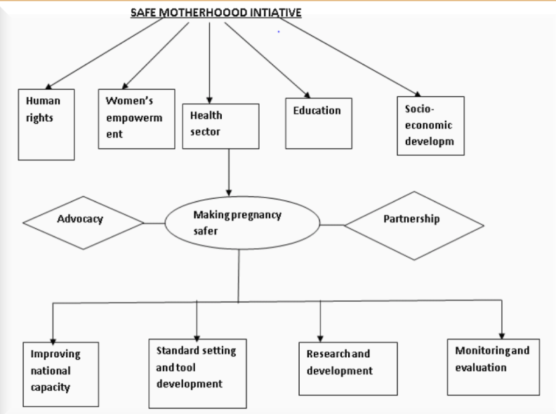
The making pregnancy safer strategy emphasizes the importance of the health sector  interventions highlighted: 

  • – Advocacy 
  • – Partnerships 
  • – Improving national capacity 
  • – Standard setting and tool development 
  • – Research and development 
  • – Monitoring and evaluation 

If these are well implemented they have the capacity to significantly reduce maternal mortality in  countries.

What is known worldwide about adverse maternal health is that a country’s overall economic wealth  is not the only important determinant. 

According to national and internal human right treaties, safe motherhood is considered a human  right issue. Therefore it is considered that maternal death is the reflection of ―social  disadvantage not merely a― health disadvantage.

Aims of Safe Motherhood

  1. To ensure that all deliveries are conducted hygienically and according to accepted medical  practices, thereby preventing complications that are caused or exacerbated by poor care. 
  2.  Identify complications promptly and manage them appropriately either by treating or  referring them to a higher level of care. 
  3. Provision of high quality, culturally appropriate care, ensuring necessary follow up and  linkages with other services including antenatal and post-partum care as well as family  planning, post abortion care and treatment of STIs. 
  4. To enhance the quality and safety of girls‘ and women‘ lives through adaptation of a  combination of health and non-health related strategies. 

Note: Maternal and child health promotion is one of the key commitments in the WHO  constitutions

Safe motherhood initiative is a global effort and it is designed to operate through its partner i.e. 

  • – Government agencies 
  • – NGOs 
  • – Other groups and individuals 

It aims to improve women‘s health through social, community and economic interventions.

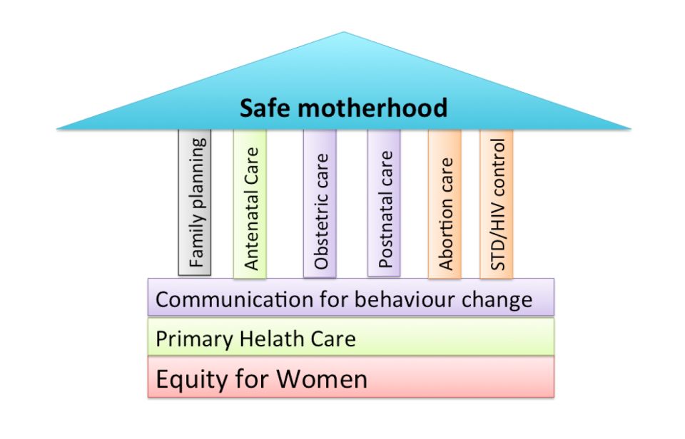
Pillars of Safe Motherhood 

  1. Family planning; to ensure that individuals and couples have the information and  services to plan the timing, number and spacing of pregnancies and thus the number of  unsafe abortion. 
  2. Antenatal care; to prevent complications where possible and ensure that complications  of pregnancy are treated appropriately and very serious conditions referred within the  shortest possible time. 
  3. Clean/safe delivery and postnatal care; to ensure that all birth attendants have the  knowledge, skills and equipment to perform a clean and safe delivery and provide  postpartum care to the mother and baby, all women should have access to basic maternity  care during delivery. 
  4. Emergency obstetric care; to ensure that essential care for high risk pregnancies and  complications is made available to all women and girls who need it. It is estimated that  about 15 % of all normal pregnancies end up with complications therefore the need to  always be prepared for emergency obstetric care. 
  1. Basic maternity care 
  2. Primary health care 
  3. Equity for women 

Components of safe motherhood 

  1. Per-conception care 
  2. Antenatal care 
  3. Postpartum care 
  4. Post abortion care 
  5. Emergency obstetric care  
  6. Care of the newborn
Requirements for safe motherhood 

Achieving safe motherhood and reducing maternal mortality requires a 3 way strong strategy:

  1.   All women have access to contraception to avoid unintended pregnancies. 
  2.  All pregnant women have access to skilled attendance at the time of birth. 
  3.  All women with complications have timely access to quality emergency obstetric care. 

The roles of community in safe motherhood 

The community can give support in several ways to make motherhood safer: 

  1.  Share the workload so that mother can avoid heavy physical work 
  2. Encourage pregnant mother to eat a balanced diet and rest than usual especially during  the last three months 
  3. Encourage mothers to take their non-pills or other medication as provided 
  4.  Help with looking after children so that mother can go for antenatal care and delivery in  the hospital 
  5. Establish transport readiness for emergency referral and obstetrical complications 
  6.  Encourage risk mothers to use maternity waiting areas, if advised to do so during  antenatal care 
  7. Creates inform and motivated community based safe motherhood groups

 Safe Motherhood Read More »

Integration of Reproductive Health Services

Integration of Reproductive Health Services

Integration; this is defined as an approach in which health care providers use opportunities  to engage clients in addressing broader health & social needs than those promoting health  encounters. 
  1. It‘s a phenomenon or a process where several services are made available to clients or  groups of people so that people who need specific RHS can access them within their  vicinity, for instance family planning with safe motherhood, Cancer of the cervix  screening. 
  2. This is the process of providing a variety of Reproductive Health services by either one or  more competent service providers within one facility on a daily basis. 

In order to maximize use of resources, Sexual Reproductive Health Services (SRHS) should be provided  as an integrated health care package that is convenient to clients and service providers. Clients  should be able to receive/access various SRHS during one visit at a given static or outreach  health units.  

Integration provides anticipatory assessment, plan and evaluates services relevant to the clients‘  desires, needs and/ or risks. 

When an integration approach is applied in RHS the goal is to provide more than one service other  than unique needs of the clients. Integrated services may be provided by one facility where the client  gets all of his/her health needs met during one encounter. Depending on the service capacity,  integrated services will be offered at the same facility or location during the same operating  hours. Services may be by the same provider in one visit or the provider of one services may  actively encourage the client to consider using another recommended services during that same  visit available within the same facility or if the needed services are beyond the capacity of the  facility or the skills of the attending provider then appropriate referral should be effected. 


However, for integration to be effective in the latter future an effective referral system must be in  place to provide accessible, timely and affordable coordinated care. 

Factors that can promote Integration of Sexual Reproductive Health Services 

Several factors can help in the smooth running of Reproductive Health Services in an integral  manner. It includes the following; 

  1. Capacity building (training).This involves improving the ability of the already existing  staffs and recruiting more skilled staffs to counter balance work load. 
  2. Improving infrastructures. The government and her partners in development should  improve on transport and communication networks as well as upgrading and improving  on her health centers, referral systems in a view to improve on clients‘ turn up and accessibility. 
  3. Increasing the range of commodities and sustaining availability. This can be achieved  by making constant and timely supply of Reproductive Health Services items to the  overwhelming number of clients. 
  4. Constant and timely integrated supervision, monitoring and evaluation to ascertain RHS  successes. 
  5. Facilitating effective referral across services. This will help to address and help clients  who need specialized care to be treated within the shortest time possible. 
  6. Community sensitization about the existence of integrated services in a bid to improve on  the health care seeking behaviors and make them aware of the available services. 

Reasons for Integration of Reproductive Health Services 

  1. To make various services available at the same time 
  2. To help reach the community who may not be able to access distant RHS 
  3.  To make people with specific reproductive related problems assisted and those with  complicated cases referred for specialized attention. 
  4. To create awareness and improve on community contact. 

 Benefits of integration 

  1. For clients: 
  • Convenient and time saving. 
  • Confidentiality is respected because information is shared with one provider.
  • The service is perceived to be complete because all clients‘ reproductive health  needs are addressed at the same time. 
  • Improve client provider relations. 
  • Increase client satisfaction.
  1. Providers: 
  • Better distribution and more effective sharing of duties. 
  • Resources accessible to every provider. 
  • Increased client confidence in the providers. 
  • Work load spread out over all service hours. 
  1. Service: 
  • Increase accessibility and availability of services. 
  • Complete-improves quality of care. 
  • Available and accessible ―ONE STOP SHOP
  • User- friendly. 
  • Efficient, effective and quick. 
  • Meet various clients‘ reproductive needs at the same time. 
  • Reduces missed opportunities. 
  • Maximizes utilization of the available resources, example; equipment, staff time.
  • Increases client satisfaction. 
  • Improves clients‘ provider relation. 

 Principles for integration of Reproductive Health Services 

  1. Build on existing opportunities for integration 
  • Assess the existing health services offered at the clinic particularly Reproductive  Health services. 
  • Type, age of clients being served and client load. 
  • Identify the strengths and limitations of the services offered and modes of offering  the services. 
  1. Involve other stakeholders. 
  2. Hold meetings with supervisors, colleagues as well as health unit management  committees to: 
  • Review personnel tasks and make a list for each cadre. 
  • Draw a work plan and re-allocate services according to providers‘ training and interest. 
  1. Reorganize services: 
  • Create space and ensure smooth client flow in order to: 

– Serve clients on first come first served basis 

– Prioritize the very ill clients who need immediate care

– Avoid clients to queue twice 

– Avoid unnecessary delays 

  • The waiting areas should include: 

Reading materials on RH issues 

Television and radio to help clients be educated as they are waiting and to  reduce on boredom 

Health talks by providers and peers 

  • Counseling /consultation rooms should: 

– Ensure privacy and confidentiality 

– Be well equipped with supplies 

– Minimize referrals 

  • Include recreation space/room to allow: 

Group discussion 

Peer education 

Indoor games especially for adolescent friendly services 

  1. Orient the community to create demand for services through: 
  • Client recruitment activities 
  • Identifying and offering services to young persons who come for other services
  • Putting up notices in public places about services offered at the health Centre 
  • Work with community leaders to reach the community 
  • Liaise with community health workers to spread the news and refer clients for  services 
  • Link up with peer educators and providers  

Note. Counselling and IEC (Information, education and communication (IEC)) form the backbone of all reproductive health services

Modes of Reproductive Health service delivery 

  1. Community outreaches: 
  • Health promotion and education 
  • Immunization 
  • Antenatal 
  • Family planning 
  • STI and HIV/AIDS screening and management
  • Malaria prevention and treatment 
  • Treatment of minor ailments 
  • Deworming 
  1. Static clinics

 All the above plus, 

  • Adolescent health 
  • Male friendly Reproductive Health services 
  • Infertility 
  • Screening of RH cancers example cancer of cervix, breast, prostate and testicles

      3. Community based services: 

  • Distribution of contraceptives and condoms 
  • Distribution of iron and folic acid distribution of anti-malarial 
  • Delivery services and referral 
  • Home-based care example. for HIV and postpartum mothers 
  1. Social marketing 
  • Health promotion and education 
  • Provision of family planning services 
  • Provision of medical supplies example. Mama kits, insecticide treated nets and  anti-malarial. 

Disadvantages of Integration 

  1. It increases workload especially where the number of staff is limited. 
  2. Tiresomeness since service providers have to spend great time serving clients. 
  3. It‘s costly especially where financial support is very poor. 
  4. It‘s very difficult to perform the outreach integration especially where geographical  barriers, impassable roads e.t.c.

Integration of Reproductive Health Services Read More »

reproductive system

Reproductive System

Reproductive system, also known as the genital system or the reproductive system, is a collection of organs and structures in the human body responsible for sexual reproduction.

Its primary function is to produce, store, and deliver gametes (reproductive cells) and facilitate the union of sperm and egg for the purpose of fertilization, leading to the creation of new life.

The male reproductive system

The Male Reproductive System

External genital organs 

External male reproductive organs are those outside and can be seen. They comprise of the;

  •  Scrotum. 
  • Testis. 
  • Penis.

The penis 

It is an organ that carries the semen with the sperm into the vagina. During sexual arousal, blood  is pumped into the muscles of the penis making it stiff/erect so it can easily enter the vagina. The penis additionally serves as the urethral duct. Although both semen and urine pass through  the urethra in the penis, at the time of ejaculation the opening from the bladder is closed so that  only semen comes out of the penis. After ejaculation, the blood quickly drains away into the  body and the penis returns to the normal state.  

The penis is enclosed by a foreskin (prepuce) that protects the glans penis. Usually the  penis produces a white creamy substance called smegma, which helps the foreskin to  slide back smoothly. When smegma accumulates under the foreskin, it causes a bad smell  or even infection. Therefore for men who are uncircumcised need to pull back the  foreskin and gently wash underneath it with clean water everyday 

The scrotum 

It is a sac of skin containing two egg-shaped organs called the testes, found in front of and  between the thighs. It protects the testes from physical damage and helps to regulate the  temperature of the sperm. 

The testes 

They are two sex glands that produce sperm and the male hormones, which are responsible for  the development of secondary sexual characteristics in men. 

The male internal reproductive organs 

  • Epididymis. 
  • Deferent ducts (vas deferens).
  •  Seminal vesicles.
  • Ejaculatory ducts. 
  •  Prostate gland
  • Urethra-bulbous glands.(bulbourethral  glands)

Vas deferens 

  • Prostate gland. 
  • Urethra-bulbous glands.(bulbourethral  glands) 

They are tubes through which the sperm passes from the testicles and penis.  

Epididymis 

  • They are cord-like structures coiled on top of the testes, it stores sperm.  
  • When sperm matures, it is allowed to pass into the vas deferens before being released  during ejaculation. 

Seminal vesicles  

  • They are glands where the white fluid, semen is produced.  
  • Semen is fluid that is released through the penis during ejaculation.  
  • It provides nourishment for the sperms and helps their movement. 
  • The seminal vesicles do not store sperm cells. 
  • They secrete a thick alkaline fluid that mixes with the sperm cells as they pass into the  ejaculatory ducts and then the urethra.  
  • These secretions provide most of the volume of the semen.  
Arterial supply, venous drainage and nervous supply

Arterial supply, venous drainage and nervous supply

  • The arteries are derived from the inferior vesical and middle rectal arteries.
  • The veins accompany the arteries.
  •  Nervous supply is by sympathetic and parasympathetic nerve fibers.


Prostate gland 

  • This is the largest accessory gland of the male reproductive system.  
  • It is situated below the bladder. 
  • The prostate is partly glandular and partly fibromuscular.  
  • The prostate produces fluid that makes up part of the semen; it helps create a good  environment for the sperm in the penile urethra and vagina 
  • Enables movement of sperm and provides nutrients for the sperm. 

Cowper’s gland 

  • It comprises two small glands situated below the prostate with ducts opening into the  urethra.  
  • Its function is to produce some fluids, which helps create a good environment for the  sperm in the penile.

The Female Reproductive System

The female external genital organs 

  • The Mons Pubis 
  • The Labia Majora 
  • The labia minora. 
  • The vestibule of the vagina. 
  • The External Urethral Orifice 
  • The Vaginal Orifice 
  • The Greater Vestibular Glands 
  • The Lesser Vestibular Glands 
  • The Clitoris 
  • The Bulbs of the Vestibule 

The mons pubis 

  • The mons pubis is a rounded fatty elevation located anterior to the pubic symphysis and  lower pubic region.  
  • It consists mainly of a pad of fatty connective tissue deep to the skin. 
  • The amount of fat increases during puberty and decreases after menopause.  
  • The mons pubis becomes covered with coarse pubic hairs during puberty, which also  decrease after menopause. 
  • The typical female distribution of pubic hair has a horizontal superior limit across the  pubic region.  

The labia majora 

  • The labia are two symmetrical folds of skin, which provide protection for the urethral  and vaginal orifices.  
  • These open into the vestibule of the vagina.  
  • Each labium majus, largely filled with subcutaneous fat, passes posteriorly from the mons pubis to about 2.5 cm from the anus.  
  • They are situated on each side of the pudendal cleft, which is the slit between the labia  majora into which the vestibule of the vagina opens.  
  • The labia majora meet anteriorly at the anterior labial commissure.  
  • They do not join posteriorly but a transverse bridge of skin called the posterior labial  commissure passes between them.  

The labia minora 

  • The labia minora are thin, delicate folds of fat-free hairless skin.  
  • They are located between the labia majora.  
  • The labia minora contains a core of spongy tissue with many small blood vessels but no  fat.  
  • The internal surface of each labium minus consists of thin skin and has the typical pink  color of a mucous membrane.  
  • It contains many sensory nerve endings.  
  • Sebaceous and sweat glands open on both of their surfaces.  
  • The labia minora enclose the vestibule of the vagina and lie on each side of the orifices  of the urethra and vagina.  
  • They meet just superior to the clitoris to form a fold of skin called the prepuce (clitoral  hood).  
  • In young females the labia minora are usually united posteriorly by a small fold of the  skin, the frenulum of the labia minora.  

The Vestibule of the Vagina 

  • The vestibule is the space between the labia minora.  
  • The urethra, vagina, and ducts of the greater vestibular glands open into the vestibule.  

The external urethral orifice 

  • This median aperture is located 2 to 3 cm posterior to the clitoris and immediately  anterior to the vaginal orifice.  
  • On each side of this orifice are the openings of the ducts of the paraurethral glands  (Skene’s glands).  
  • These glands are homologous to the prostate in the male. 

The Vaginal Orifice 

  • This large opening is located inferior and posterior to the much smaller external urethral  orifice.  
  • The size and appearance of the vaginal orifice varies with the condition of the hymen, a  thin fold of mucous membrane that surrounds the vaginal orifice.  

The greater vestibular glands 

  • These glands are about 0.5 cm in diameter.  
  • They are located on each side of the vestibule of the vagina, posterolateral to the vaginal  orifice.  
  • They are round or oval in shape and the bulbs of the vestibule partly overlap them  posteriorly.  
  • From the anterior parts of the glands, slender ducts pass deep to the bulbs of the  vestibule and open into the vestibule of the vagina on each side of the vaginal orifice. 
  • These glands secrete a small amount of lubricating mucus into the vestibule of the  vagina during sexual arousal.  
  • The greater vestibular glands (Bartholin’s glands) are homologous with the bulbourethral  glands in the male 

The clitoris 

  • The clitoris is 2 to 3 cm in length.  
  • It is homologous with the penis and is an erectile organ.  
  • Unlike the penis, the clitoris is not traversed by the urethra; therefore it has no corpus  spongiosum.  
  • The clitoris is located posterior to the anterior labial commissure, where the labia majora meet.  
  • It is usually hidden by the labia when it is flaccid.  
  • The clitoris consists of a root and a body that are composed of two crura, two corpora  cavernosa, and a glans.  
  • It is suspended by a suspensory ligament.  
  • The parts of the labia minora passing anterior to the clitoris form the prepuce of the  clitoris (homologous with the male prepuce).  
  • The parts of the labia passing posterior to the clitoris form the frenulum of the clitoris,  which is homologous with the frenulum of the penile prepuce.  
  • The clitoris, like the penis, will enlarge upon tactile stimulation, but it does not  lengthen significantly.  
  • It is highly sensitive and very important in the sexual arousal of a female.
Arterial supply of female external genitalia

Arterial supply of female external genitalia 

  • The rich arterial supply to the vulva is from two external pudendal arteries and one  internal pudendal artery on each side.  
  • The internal pudendal artery supplies the skin, sex organs, and the perineal muscles.  
  •  The labial arteries are branches of the internal pudendal artery, as are the dorsal and deep  arteries of the clitoris.  

Venous drainage  

  • The labial veins are tributaries of the internal pudendal veins and venae comitantes of the  internal pudendal artery.  

Lymph drainage of the female external genitalia 

  • The vulva contains a very rich network of lymphatic channels.  
  • Most lymph vessels pass to the superficial inguinal lymph nodes and deep inguinal nodes.

reproductive system Read More »

Introduction to Reproductive Health

Introduction to Reproductive Health

Reproductive health is defined as a state of complete physical, mental and social well being and not merely the absence of the disease or infirmity on all matters related to reproductive function  and its processes involved. 

This is a process concerned with peoples‘ ability to have a responsible satisfying and safe sex  life, their capability to reproduce and having the freedom to decide if, when and how often to do so. 

Reproductive health includes having: 

  1. Satisfying, safe sex life. 
  2. Ability to reproduce. 
  3. Successful maternal and infant survival outcome. 
  4. Freedom to control reproduction. 
  5. Information about and access to safer, effective and affordable methods of family planning. 6. Ability to minimize gynecological disease throughout life. 

The components of Reproductive Health 

  1. Safe motherhood: 
  • Preconception care 
  • Antenatal care 
  • Maternal nutrition 
  • Focused antenatal care 
  • Immunization for tetanus, hepatitis B etc. 
  • EMTCT of HIV/AIDS 
  • Clean safe delivery 
  • Emergency obstetric care 
  • Postnatal (newborn care) and postpartum care
  • Breastfeeding/ infant feeding 
  • IEC and community mobilization 
  • Post abortion care services 
  1. Family planning: 
  • Medical eligibility for family planning services 
  • Provision of contraceptives and natural family planning 
  • Emergency contraceptive 
  • Management and follow-up for side effects of contraceptives 
  • Infection prevention and quality care 
  1. STIs/HIV/AIDS: 
  • Behavioral change counseling 
  • Condom promotion and distribution 
  • Counseling and testing  
  • STI management and treatment 
  • Infection prevention and quality of care 
  • Partner notification and treatment 
  • Treatment compliance. 
  1. Sexual and adolescent health: 
  • Behavior change counseling 
  • Provision of adolescent friendly services 
  • Provision of contraceptive services 
  • Screening and management of STIs 
  1. Infertility: 
  • Investigation and treatment 
  • Partners‘ involvement 
  1. Reproductive organ cancers: 
  • Screening and referral 
  • Definitive management 
  • Palliative care 
  1. Gender related issues; such as gender based violence, sexual abuse, female genital  mutilation and fistulae. 
  • Advocacy 
  • Partner involvement 
  • Community involvement 
  • Specialized management
  • Multi-sectorial collaboration 
  • Legal support 
  1. Menopause and andropause: 
  • Symptomatic treatment 
  • Hormonal replacement 
  • Partner involvement 
  • Advise on exercise and nutrition

 Importance of reproductive health 

  1. Promotion of maternal and child health 
  2. Reduces maternal morbidity and mortality 
  3. Promotes free women‘s involvement in all matters related to reproductive health issues  e.g. family planning 
  4. Promotes prompt treatment and detection of life threatening cases throughout  reproductive life 
  5. It promotes safer sex practices and reduces the incidence of rampant sexual related abuses
  6. . Reduces on government expenditure on reproductive related health issues thus promotes  quality standard of living. 

Problems being faced during the implementation of Reproductive Health in Uganda 

The following are some of the problems being encountered during the implementation of  reproductive health services in Uganda; 

  1. Low socio-economic status (poverty): This is the major setback as many people in  Uganda live within poverty level which in turn makes them unable to access even the least  costly services. For instance, the Uganda Demographic Health Survey shows that  mortality rates are high in women from low socio-economic status as these women  are likely to be less privileged in the fields of nutrition, housing, quality education etc 
  2.  Improper/underutilization of the existing services 
  3. Delivery of substandard care i.e. when the care provided is below the generally  accepted level available at that particular coupled up shortages of resources and under-equipped facilities 
  4. Lack of communication and referral facilities: This could be due to poor coordination  between lower health facilities with the higher ones backed-up by geographical  barriers, transport means like ambulances etc. 
  5. Poor cultural perspectives on reproductive health; variety of cultural practices are the  basic obstacles to Reproductive Health Services for instance, female genital  mutilation, early marriages, denying women to eat certain foods etc. 
  6. Lack of awareness by the community on issues related to reproductive health.
  7. Inadequate supply of resources related to reproductive health. This therefore makes  the little existing services disproportionately consumed by the overwhelming  individuals who visit the health Centers. 
  8. Inadequate skilled staff specially trained on issues pertaining reproductive health.  The number of skilled staff to deliver various Reproductive Health Services in  Uganda is appalling as compared to the number of clients who desperately need the  scarce services. 
  9. Improper evaluation and supervision of reproductive health services to ascertain its  progress and successes 
  10. Lack of support from men, opinion leaders and development partners as they are  considered change agents in the community 
  11. Misappropriation and embezzlement of funds specially designed to facilitate  reproductive health services. 

Ways through which Reproductive Health Service can be improved in Uganda 

It is a coordinated long term effort within the families, opinion leaders, communities and health systems. 

It also involves the national legislation and policies where action may vary in respect of an  individual and the government ought to make Reproductive Health a priority of public  concern and to periodically evaluate the program to ascertain the successes. 

  1. A good quality of obstetric services and referral services are to be considered. 
  2.  Proper and timely evaluation of the Reproductive health related issues are to be taken  a priority. 
  3. Recruitment of skilled manpower at the functional referral points for proper  integration of reproductive health services to fill the pending gaps. 
  4. Decentralization of services to make them available in time. 
  5. Social inequalities and discrimination on grounds of gender, age and marital status  are to be removed. 
  6. Timely supply of essential reproductive health services to meet the overwhelming  number 
  7. Women should be empowered to gain access to education, economic and social status  in order to increase women’s decision making power in regards to their own health  and reproduction. 
  8. Sensitization to create awareness should be given to the community, society and decision makers about the values of reproductive health. 
  9. Improving on the standard delivery of care by organizing refresher courses (workshop  and for the health care personnel at various level others 
  10. Barriers to access of health facilities should be addressed 
  11. Proper utilization of services available
  12. Bad cultural practices and beliefs in the community should be discouraged. This can  be done by strict laws, for instance illegalization of female genital mutilation, sexual  gender based violence, early marriages etc. 
  13. Penalizing those who have been found culprits for misusing the funds. 
  14.  Encouraging men and community members to actively and wholeheartedly support  their partners through their active participation in RHS being provided 

Introduction to Reproductive Health Read More »

DOMICILIARY CARE

DOMICILIARY CARE

Domiciliary care is an obstetric care given to a mother in her home during pregnancy, labour and puerperium.

Types of Domiciliary Care

  1.  Type one domiciliary midwifery care “continuity:; In this type the woman is cared for in her home all through during antenatal period delivery and postnatal care. The woman will only visit a health unit or hospital only when there is a problem that requires specialized care or more gadgets to be used. This care is known as continuity of care or fragmented care. In this case one midwife provides all the care to the woman.
  2.  Type two, community, integrated or centralized care; In this care service is integrated (mixed) in a way that part of the care may be given at home and some in the health setting like a hospital. Usually antenatal or delivery may be offered in the hospital and puerperium period managed at home. This is the type of care that student midwives and nurses offer as part of their midwifery part two and is compulsory for them.
  3.  Employee or independent practitioner in domiciliary; This is a type of care in which a midwife practices as a private midwife in the community but not necessarily on one woman. The midwife may have a maternity Centre for all or part of the care or she may combine it with one to one community midwifery care. This is the commonest type of domiciliary care in Uganda.

Forms of Domiciliary Care
Characteristics of patterns of domiciliary care depend on a number of factors and these can be:

  • Decision of the midwife
  • Decision of the woman / family
  •  Location and nature of community
  •  Availability of basic requirements for domiciliary care

Objectives of Domiciliary Care.

  1.  Domiciliary midwifery care  to take midwifery near to the community thus increasing accessibility to services

  2.  To encourage full participation and involvement of male partners and family members in the birth process so as to get their full support

  3.  To reduce on maternal / infant morbidity and mortality as the midwife has less workload and concentrates on one woman.

  4.  To reduce on hospital/health facility over crowding

  5.  To promote midwife-mother relationship and mutual understanding between the woman and the midwife.

Domiciliary Care given by midwives
  1.  Care before conception
    >   Health education to young girls on good nutrition and hygiene
    >   Teaching young girls about life skills
    >    Immunization of young girls with tetanus toxoid
    >    Counselling adolescents on reproductive health and other social issues
  2.  Care during pregnancy
    >   Immunization
    >   Antenatal check ups
    >   Treatment of minor problems.    >   Health education on problems in pregnancy
  3. Care during labour
    >   Care of mother in Labour
    >   Use of partograph to monitor labour
    >   Delivering of the baby
    >   Infection prevention
  4. Care after delivery
    >   Immunization
    >   Care of mother and baby
    >   Postnatal exercises
    >   Family planning

Advantages of Domiciliary Services.

  • Domiciliary services promotes midwife – mother relationships and thus minimizing fears and phobias of childbirth
  • It promotes continuity of care and close supervision of the mother thus – contributing to the reduction of maternal / infant morbidity and mortality
  •  Increases access to health services as the woman is found in her home instead of herself looking for the services
  •  Domiciliary is cost effective to a certain level as only relevant care will be given to individual women and at the same time the woman will continue her responsibilities especially supervision of the home
  •  It gives peace of mind to the mother, husband children and other house members because the woman remains at home
  •  It promotes woman centered care including choice control over services rendered and also encourages continuity of care.
  •  It promotes privacy and security and respect the mother with less interference and exposure
  • Promotes good communication and openness. Only relevant information is given to the mother and her family. As the midwife knows the woman personally, she understands better their concerns, lives, and challenges and assists them accordingly.
  •  Promotes autonomy to the midwife and there is job satisfaction
  •  It promotes creativity, problem solving skills and maturity in service with good experience.
 

Brief History of Domiciliary Care

 Throughout the ages, women have depended upon a skilled person, usually another woman to be with them during child birth
 In United Kingdom, the midwives skills are increasingly valued and midwives are being urged to expand their role even further in the field of public health.

  • In Uganda in 1960’s(May 1968), this is when the midwife would look after the mother in the home environment.  Midwives would do antenatal care, deliver mothers in their own homes and continue to give post natal care in the mother’s home.
     >    This would also give opportunity for the midwife to give health education to the other family members.
    >     In the 1970s when the political system in Uganda changed, leading to a lot of insecurity, the midwives stopped delivering mothers at home  and instead delivered mothers in hospitals and maternity units. Then the midwives continued to nurse the mothers and their babies at the mother’s home.
     > These services have continued today and are being practiced by Private Midwives and the student midwives who are undertaking Registered Midwifery Course of Diploma in Midwifery Course.
Types/ Groups of mothers Needing Domiciliary care
  • Group 1: Women with less risk of getting complications
    Women who have ever delivered one baby but have not exceeded five – that is gravid two to four.
    This group of women if they did not experience any major complication in pregnancy labour and puerperium, can be care for in the community throughout, pregnancy labour and puerperium
  • Group 2: These are the women who are suspected of developing a complication, though they may not develop them at all. For examples: primigravida – pregnant for the first time,
    Grand multi para – has delivered more than four times, short women- less than 152cm high, women with previous complications that are likely to occur again e.g. cord prolapsed.
    This group of women may be cared for only for antenatal or delivery and puerperium depending on other factors as detected on history and assessment.
  • Group 3: These are the high Risk Mothers, women who come with obvious complications, or are highly suspected of developing various complications. Examples: Multiple pregnancy – those with medical conditions like cardiac diseases, diabetes mellitus, sickle cell disease.

Common Drugs used in Domiciliary 

  •  Ergometrine
  •  Ferrous sulphate
  •  Folic acid
  •  Panadol
  •  Chloroquine

How Domiciliary is carried out.

  •  Booking

A mother who has to be booked must be with the following
>  Must be normal with no risk factors like CPD,
>  Grande multi parity, multiple pregnancy

  •  Home delivery

The following must be put in consideration
(a).   Well ventilated home without without overcrowding
(b).   Clean house, good hygiene in and around the house
(c).   The house should have more than 4 bedrooms, toilets
and kitchen
(d).   The floor must be cemented
(e).   There must be tap water
(f).   There must be easy means of boiling water

  •  Enough equipment especially for the mother and baby(bathing)
  •  Husband and wife should be willing for the care
  •  The distance from the home to hospital should be less than 2 miles.
QUALITIES OF A MIDWIFE

In normal circumstances the midwife should be a qualified senior student midwife with enough knowledge
(a)  She must create a friendly relationship between her, the mother and family
(b)   She must remember that she does not belong to the family and is only a guest so she must adopt her behavior in relation to the family routine
(c)   No commands or orders should be given but advices, the midwife should be flexible
(d)   She should show interest in the family
(e)   Avoid embarrassing the mother in the family

(f)   She has to apply her professional code of conduct and stay in the home only as a midwife
(g)   Quick and correct judgment has to be applied in providing the best care expected


DOMICILIARY BAGS

The midwife must be equipped with the following

  •  Sphyginomanometer
  •  Stethoscope
  •  Urine testing strips
  •  Clinical thermometer
  •  Spirit for baby’s cord
  •  Swabs in the gallipot and cord ligatures
  •  Receivers, dissecting forceps, artery forceps, scissors
  •  Antiseptic lotion
  •  Plastic apron and tape measure
  •  Drugs like Panadol, and iron tablets


Care

Here in Uganda a mother is delivered in the hospital then cared for in her home for seven day including the 1st days in the hospital

ANTENATAL CARE
Normally a mother is booked on her 1st visit at 12wks.It should be during this time when the midwife inspect the home of the mother until the mother is delivered in the hospital and cared for the first 2 days and  then 5 days at home

PUEPERIUM
During puerperium the midwife continues to visit the mother daily at her home. If there is any indication of complication arising of the mother requires extra supervision and support additional visits will be made
The midwife observes the mother’s general condition both mentally and physically, ask her how she is feeling. Inquire about the baby particularly feeding, sleeping, passage of urine and stool.

If the mother appears stressed, depressed, or anxious about the baby or any other problem. The midwife should sit, listens and responds. The time spent listening and discussing problems with the mother invariably of great value to her wellbeing
The midwife inquires whether the mother is sleeping and eating well passing urine without difficult or discomfort and has had a bowel action.
She take the mothers vitals and carries out a full postnatal examination of the breast, abdomen to palpate the uterus, vulva to inspect lochia and perineum.

Any abnormality detected should be discussed with the mother and appropriate advice is given. Postnatal exercises are taught on the first day after delivery and the mother is encouraged to practice them dairy throughout puerperium
On the first postnatal visit the midwife usually assists the mother to bath there after the mother should have a bath on her own should be twice or more daily, mother should be advised to change her pads frequently.

Adequate rest and sleep are essential and though ambulating is good but the mother should rest and sleep at appropriate time each day.
The mid wife performs a daily examination on the baby and shows the mother how to bath and dress the baby and attend to the cord.


  • > She observes its general condition,  examine him from head to toe observing the skin, eyes, mouth and cord for any signs of infection or any abnormality.
  • > Stool should be observed and the passage of urine.
  • > Baby should be observed whether breastfeeding well
  • > At the last visit, the mid wife advises the mother when to go back to postnatal clinic and the baby to health clinic.
  • > Health educate and demonstrates to the mother the postnatal exercises. 

DOMICILIARY CARE Read More »

PARTOGRAPH

PARTOGRAPH

Partograph is a graph or tool used to monitor fetal condition, maternal condition and labour progress during the active 1st stage of labour so as to be able to detect any abnormalities and be able to take action.
It’s only used during 1st stage of labour. It is used for recording salient conditions of the mother and the fetus.

USES OF A PARTOGRAPH

  1. To detect labour that is not progressing normally.
  2. To indicate when augmentation of labour is appropriate.
  3. To recognize CPD when obstruction occurs.
  4. It increases the quality of all observations on the mother and fetus in labour.
  5. It serves as an “early warning system”
  6. It assists on early decision of transfer and augmentation.

Who should not use a partograph?

  • Women with problems which are identified before labour starts or during labour which needs special attention.
  • Women not anticipating vaginal delivery (elective C/S).

Parts of a Partograph

A partograph has 3 parts i.e. –

  • Fetal part
  • Maternal part
  • Labour progress part

Observations charted on a partograph:

  1. The progress of labour
    >  Cervical dilatation 4 hourly
    >  Descent 2 hourly
    >  Uterine contractions
  2. Fetal condition
    >  Fetal heart rate ½ hourly
    >  Membranes and liquor 4 hourly
    >  Moulding of the fetal skull 4 hourly.
  3.  Maternal condition
    >  Pulse ½ hourly
    >  Blood Pressure 2 hourly
    >Respiration and >  temperature 4 hourly
    Urine; – volume 2 hourly, acetone, proteins and sugars.
    >  Drugs
    >  I.V fluids 2 hourly and Oxytocin regimen.
Starting a partograph:
  • The partograph should be started only when a woman is in active phase of labour.
  • Contractions must be 1 or more in 10 minutes.
  • Cervical dilatation should be 4cm or more.
FETAL CONDITION
  1. Fetal heart;
    It is taken 1/2 hourly unless there is need to check frequently i.e. if abnormal every 15 minutes and if it remains abnormal over 3 observations, take action. The normal fetal heart rate is 120-160b/m. below 120b/m or above 160b/m indicates fetal distress.
  2. Molding;
    This is felt on VE. It is charted according to grades.
    State of moulding                                         Record
    Absence of moulding.                                     (-)
    Bones are separate and sutures felt   (0)
    Bones are just touching each other   (+)
    Bone are over lapping but can be Separated (++)
    Bones are over lapping but cannot be separated (+++)
  3. Liquor amnii;
    This is observed when membranes are raptured artificially or spontaneously.
    It has different colour with different meaning and meconium stained liquor has grades.
    State of liquor Record
    Clear (normal)     (C)
    Light green in colour (m+)       Moderate green, more slippery       (m++)      Thick green, meconium stained   (m+++)       Blood stained    (B)
  4. Membranes;State of membranes  Record
  • Membranes intact    (I)
  • Membranes raptured   (R)
LABOUR PROGRESS

5. Cervical dilatation,
The dilatation of the cervix is plotted with an “X”. Vaginal examination is done at admission and once in 4 hours. Usually we start recording on a partograph at 4cm.
Alert line starts at 4cm of cervical dilation to a point of expected full dilatation at a rate of 1cm per hour
Action line– parallel and at 4 hours to the right of the alert line.

6. Descent of presenting part.
Descent is assessed by abdominal palpation. It is measured in terms of fifths above the brim.
The width of five fingers is a guide to the expression in the fifth of the head above the brim.
A head that is ballotable above the brim will accommodate the full width of five fingers.
As the head descends, the portion of the head remaining above the brim will be represented by fewer fingers.
It is generally accepted that the head is engaged when the portion of the head above the brim is represented by 2 or less fingers.
Descent is plotted with an “O” on the graph

7. Uterine contractions This is done ½ hourly for every 30 minutes. The duration, frequency and strength of contraction is observed. Observe the contractions within 10 minutes.

-Mild contractions last for less than 20 seconds.
-Moderate contractions last for 20-40 seconds.
-Strong contractions last for 40 seconds and above.
When plotting and shedding contractions use the following symbols.
Dots for mild contractions
Diagonal lines for moderate contractions
Shade for strong contractions

MATERNAL CONDITION
  1. Pulse; this is checked every 30 minutes. The normal pulse is 70-90b/min.
    The raised pulse may indicate maternal distress, infection especially if she had rapture of membranes for 8-12 hours and in case of low pulse, it can be due to collapse of the mother.
  2. Blood pressure; it is taken 2 hourly. The normal is 90/60-140/90mmHg. Any raise of 30mmHG systolic and 20mmhg diastolic from what is regarded as normal or if repeated over 3 times and remains high, test urine for albumen to rule out pre-eclampsia.
  3. Temperature; this is taken 4 hourly. The normal range is between 37.2 0 c to 37.5 0 c. Any raise in temperature may be due to infections, dehydration as a sign of maternal distress or if a mother had early rapture of membranes.
  4. Urine; the mother should pass urine atleast every after 2 hours and urine should be tested on admission.
  5. Fluids; she should be encouraged to take atleast 250-300 mls every 30 minutes. Any type of fluid can be given hot or cold except alcohol. The fluid should be sweetened in order to give her
    strength.
Further management in the normal 1st stage of labour
 Nursing care
  1. Emotional support:

Midwife should rub the mothers backto relieve pain.
Allow the mother to move around or sit in bed if membranes are still intact.
Re-assure the mother and keep her informed about the progress of labour to relieve anxiety.
Allow her to talk to relatives and husband.
Allow her to read or do knitting.

2. Nutrition;
Encourage mother to take light and easily digested food like bread, soup and sweet tea to rehydrate her and provide energy.

3. Elimination;
Taking care of the bladder and bowel. Encourage mother to empty bladder every 2 hours during labour. Every specimen is measured and tested for acetone, albumen, sugars and findings interpreted and recorded.
Pass catheter if mother is unable to pass urine.

4. Personal hygiene;
Allow mother to go for bath in early labour or on admission if condition allows. If membranes rapture, give a clean pad and ask mother to change frequently to prevent infections.
VE should be done only after aseptic technique.


5. Ambulation and position:
In early labour, mother is encouraged to walk around to aid descent of presenting part.
During contractions, ask mother to lean forward supporting herself on a chair or bed to reduce discomfort.
Allow mother to adopt a position of her choice except supine position.
Mother should be confined to bed when membranes rapture in advanced stage of labour.


6. Prevention of infections
Strict aseptic technique should be maintained when doing a VE and vulval swabbing.
When membranes rapture early, vulval toileting should be done 4 hourly to reduce the risk of infections. Put mother on antibiotics to avoid risk of ascending infections in early raptured of membranes.
Frequent sponging is done, bed linen changed when necessary when a mother is confined in bed.
The midwife should pay attention to her own hygiene and be careful to wash her hands before and after attending to the mother.


7. Sleep and rest
Mother is encouraged to rest when there is no contraction (rest in between contractions).

What to report
  • Abnormality found in urine.
  • Failure to pass urine.
  • Rise in temperature, pulse and BP.
  • Hypertonic uterine contractions.
  • Rapture of membranes with meconium stained liquor grade 2 and 3.
  • Failure of presenting part to descend despite good uterine contractions.
  • Tenderness of abdomen.
  • Bleeding per vagina.
  • Fall in BP.
  • Raise in fetal heart rate.
Complications
  • Infections
  • Early rapture of membranes
  • Cord prolapse
  • Supine hypotensive syndrome
  • Fetal distress
  • Maternal distress
  • APH
  • PET and eclampsia
  • Prolonged labour
  • Obstructed labour

PARTOGRAPH Read More »

MINOR DISORDERS OF PREGNANCY
MINOR DISORDERS IN PREGNANCY

MINOR DISORDERS OF PREGNANCY

Minor disorders of pregnancy are a series of commonly experienced symptoms related to the effects of pregnancy hormones and the consequences of enlargement of the uterus as the fetus grows during pregnancy.

These are referred to as minor because they are not life threatening.
The causes can be;-

  • Hormonal changes
  • Accommodation changes
  • Metabolic changes and
  • Postural changes
I. Digestive System Disorders:

A. Nausea and Vomiting (Morning Sickness):

  • Causes: Primarily attributed to hormonal surges, specifically elevated levels of human chorionic gonadotropin (hCG), oestrogen, and progesterone during early pregnancy (weeks 4-16).
    These hormones affect the gastrointestinal tract’s sensitivity and motility. The exact mechanism remains unclear but likely involves alterations in brain neurotransmitters and gastrointestinal hormone levels.
    Decreases as the placenta takes over hormone production.
  • Symptoms: Range from mild to severe vomiting (hyperemesis gravidarum, a severe form requiring medical attention).
    Symptoms often peak in the morning but can occur throughout the day.
  • Management:
  1. Dietary Modifications: Small, frequent meals; consuming bland foods like crackers, toast, or rice; avoiding strong smells or triggers; consuming carbohydrates.
  2. Lifestyle Changes: Getting out of bed slowly, staying hydrated, eating before getting out of bed, regular, gentle exercise.
  3. Pharmacological Interventions: In cases of severe nausea and vomiting, antiemetics (medications to control nausea and vomiting) may be prescribed by a healthcare provider. Vitamin B6 supplements are sometimes recommended.
  4. Acupressure: Wristbands with pressure points can sometimes help alleviate nausea.

B. Heartburn (Pyrosis):

  • Causes: Relaxation of the lower esophageal sphincter (LES) due to progesterone, allowing stomach acid to reflux into the esophagus. Increased intra-abdominal pressure from the growing uterus further exacerbates this. Most troublesome between 30-40 weeks gestation.
  • Symptoms: Burning sensation in the chest, often radiating upwards. Can be worsened by lying down, bending over, or consuming certain foods.
  • Management:
  1. Dietary Modifications: Small, frequent meals; avoiding fatty, spicy, or acidic foods; avoiding eating before bed.
  2. Lifestyle Changes: Elevating the head of the bed with extra pillows, avoiding tight clothing, maintaining an upright posture after meals.
  3. Pharmacological Interventions: Antacids (e.g., magnesium trisilicate, calcium carbonate) can neutralize stomach acid, providing temporary relief. H2 blockers or proton pump inhibitors (PPIs) may be prescribed for more severe cases.

C. Excessive Salivation (Ptyalism):

  • Causes: Likely hormonal influences, although the exact mechanism is unclear. Often associated with nausea and vomiting. It may also be caused by anxiety or psychological factors.
  • Symptoms: Excessive production of saliva.
  • Management: Rinsing the mouth frequently, sucking on ice chips or hard candies, avoiding trigger foods. Counseling may be helpful to address underlying anxiety.

D. Constipation:

  • Causes: Progesterone’s relaxing effect on the smooth muscles of the intestines, leading to slowed bowel movements (decreased peristalsis). Iron supplementation can also contribute. Decreased physical activity may play a role.
  • Symptoms: Infrequent bowel movements, hard stools, straining during bowel movements.
  • Management:
  1. Dietary Modifications: Increased intake of fiber (fruits, vegetables, whole grains), fluids (water), and gentle exercise. Bulk-forming laxatives (psyllium) may be used under medical supervision.
  2. Lifestyle Changes: Regular exercise, particularly walking, can stimulate bowel movements.
  3. Pharmacological Interventions: Stool softeners or mild laxatives should be used cautiously and only when dietary changes and exercise are insufficient, under the guidance of a healthcare professional.

E. Pica:

  • Causes: Unknown. Possible links to nutritional deficiencies (iron, zinc), psychological factors, or hormonal imbalances.
  • Symptoms: Craving and consumption of non-nutritive substances (e.g., clay, ice, starch). This can lead to serious health consequences.
  • Management: Addressing any underlying nutritional deficiencies through dietary changes and supplementation under medical supervision. Psychological counseling may also be beneficial.
II. Musculoskeletal System Disorders:

A. Leg Cramps:

  • Causes: Exact cause is unknown but various factors have been suggested, including:
  1. Changes in electrolyte balance: Decreased calcium or magnesium levels can make muscles prone to cramping.
  2. Compression of nerves: The growing uterus may compress nerves, affecting muscle function.
  3. Reduced blood circulation: Restricted blood flow can lead to cramping.
  4. Increased weight: Added weight puts pressure on the muscles.
  • Symptoms: Sudden, sharp pain in the calf muscles, often at night.

  • Management:

  1. Stretching exercises: Regular stretching of calf muscles.
  2. Hydration: Adequate fluid intake.
  3. Dietary changes: Addressing any potential electrolyte imbalances through diet or supplementation (calcium, magnesium, potassium). A balanced diet is key.
  4. Foot elevation: Raising legs above heart level. Dorsiflexion (pulling toes towards shin) can also provide relief.

B. Backache:

  • Causes: Shifting center of gravity due to the growing uterus, relaxation of ligaments and joints due to relaxin hormone, and changes in posture.
  • Symptoms: Aching or pain in the lower back, often radiating to the buttocks or legs.
  • Management:
  1. Postural adjustments: Maintaining good posture, avoiding high heels, using supportive footwear.
  2. Exercise: Low-impact exercises such as walking, swimming, or prenatal yoga.
  3. Rest: Regular periods of rest throughout the day.
  4. Supportive measures: Using a maternity support belt, applying heat or ice packs, pelvic floor exercises.
  5. Pelvic floor exercises: Strengthening pelvic floor muscles can help improve support and reduce back pain.
III. Circulatory System Disorders:

A. Fainting (Syncope):

  • Causes: In early pregnancy, vasodilation from progesterone can cause a temporary drop in blood pressure before the body compensates by increasing blood volume. Orthostatic hypotension (a sudden drop in blood pressure when standing up) can also occur. Dehydration can contribute.
  • Symptoms: Dizziness, lightheadedness, loss of consciousness.
  • Management: Avoiding prolonged standing, changing positions slowly, lying down immediately if feeling faint, staying well-hydrated. Avoiding lying on the back, except during necessary medical examinations.

B. Varicose Veins:

  • Causes: Progesterone relaxes the smooth muscles in the veins, leading to reduced blood flow and pooling of blood. Increased blood volume and pressure from the growing uterus also contribute. They may occur in legs, vulva, and anus.
  • Symptoms: Enlarged, twisted veins; aching, heavy, or swollen legs; pain or cramping in the legs.
  • Management:
  1. Compression stockings: Wearing compression stockings to improve circulation.
  2. Elevation: Elevating legs regularly.
  3. Exercise: Regular exercise to promote circulation.
  4. Avoiding prolonged standing or sitting: Frequent movement to improve blood flow.
  5. Managing constipation: Preventing constipation helps reduce pressure on the veins.
  6. Medical intervention: In severe cases, a doctor may recommend other treatments.

C. Hemorrhoids:

  • Causes: Increased pressure on the pelvic veins due to constipation and the growing uterus.
  • Symptoms: Painful, swollen, and inflamed veins in the rectum or anus.
  • Management: High-fiber diet to prevent constipation; topical treatments (e.g., creams, ointments); warm sitz baths; stool softeners (as advised by a healthcare provider).

D. Heart Palpitations:

  • Causes: Increased cardiac output to supply the growing fetus with blood and nutrients. Hormonal changes also affect heart rate and rhythm. Anxiety and stress can also exacerbate palpitations.
  • Symptoms: Feeling of a racing heart, fluttering, or pounding in the chest. Can be associated with shortness of breath or dizziness.
  • Management: Identifying and managing underlying anxiety or stress. Regular exercise, maintaining a healthy weight, and avoiding caffeine and nicotine can help regulate heart rate. In cases of persistent or concerning symptoms, medical evaluation is necessary to rule out other causes.
IV. Urinary System Disorders:

A. Urinary Tract Infections (UTIs):

  • Causes: The changing hormonal environment of pregnancy can make women more susceptible to UTIs. The expanding uterus can also compress the ureters, slowing urine flow and increasing the risk of bacterial growth.
  • Symptoms: Frequent urination, burning sensation during urination, urgency, pain in the lower abdomen or back. Fever and chills may indicate a more serious infection.
  • Management: Prompt medical attention is crucial for UTIs in pregnancy. Treatment usually involves antibiotics.

B. Frequency of Micturition:

  • Causes: In early pregnancy, hormonal changes increase blood flow to the kidneys, leading to increased urine production. In later pregnancy, the enlarging uterus compresses the bladder, reducing its capacity and leading to more frequent urination.
  • Symptoms: Increased urge to urinate, often with small amounts of urine being passed.
  • Management: Regular voiding to prevent bladder distension, drinking plenty of fluids throughout the day but avoiding excess fluid close to bedtime. Kegel exercises to strengthen pelvic floor muscles may help improve bladder control.

C. Stress Incontinence:

  • Causes: Weakening of pelvic floor muscles due to hormonal changes and the pressure exerted by the growing uterus.
  • Symptoms: Leakage of urine during coughing, sneezing, laughing, or physical exertion.
  • Management: Pelvic floor exercises (Kegel exercises) to strengthen the pelvic floor muscles. Avoiding activities that increase intra-abdominal pressure. In some cases, medical intervention may be necessary.
V. Integumentary System Disorders:

A. Itching of the Skin (Pruritis):

  • Causes: Stretching of the skin due to weight gain, hormonal changes, and cholestasis of pregnancy (a liver condition that can cause intense itching). Poor hygiene, heat rash, or minor skin rashes also contribute. Stretch marks (striae gravidarum) can also be itchy.
  • Symptoms: Itching, particularly on the abdomen, breasts, thighs, and buttocks. The degree of itchiness can range from mild to severe.
  • Management: Keeping the skin moisturized, cool baths or showers, wearing loose-fitting clothing made of breathable fabrics, topical creams or lotions (as advised by a physician). Medical attention is required if itching is severe or persistent or if it is accompanied by other symptoms (jaundice, dark urine, pale stools).

B. Stretch Marks (Striae Gravidarum):

  • Causes: Rapid stretching and thinning of the skin due to weight gain during pregnancy. Genetic predisposition plays a role.
  • Symptoms: Red or purple streaks on the abdomen, breasts, thighs, and buttocks. They eventually fade to silvery white.
  • Management: Keeping the skin well-hydrated with lotions or creams may help minimize the appearance of stretch marks. There is no known cure.

C. Melasma (Chloasma):

  • Causes: Hormonal changes during pregnancy stimulate increased melanin production, resulting in hyperpigmentation. Exposure to sunlight exacerbates the condition.
  • Symptoms: Dark brown patches, usually on the face. Often seen on cheeks, forehead, and upper lip.
  • Management: Sunscreen protection is crucial to prevent further darkening. Topical treatments may be recommended. The discoloration usually fades after delivery.

VI. Other Disorders:

A. Emotional Instability:

  • Causes: The physiological changes, lifestyle adjustments, anxieties and fears associated with pregnancy can significantly impact emotional well-being. Hormonal shifts play a crucial role.
  • Symptoms: Mood swings, irritability, anxiety, depression, tearfulness.
  • Management: Support from family and friends, stress management techniques (yoga, meditation, etc.), prenatal yoga, counseling or therapy, if needed. Open communication with a healthcare provider is vital.
VII. Disorders Requiring Immediate Medical Attention:

The following symptoms warrant immediate medical attention as they could indicate serious complications:

  • Vaginal Bleeding: Could indicate placenta previa, placental abruption, or other serious complications.
  • Reduced Fetal Movements: May signify fetal distress.
  • Severe or Persistent Headache (especially frontal or recurrent): Can be a sign of preeclampsia or eclampsia.
  • Sudden Swelling or Edema (especially in face or hands): A possible symptom of preeclampsia.
  • Early Rupture of Membranes (PROM): Increased risk of infection and premature delivery.
  • Premature Onset of Contractions: Risk of preterm labor.
  • Maternal Exhaustion (to any extent): Can indicate underlying health issues.
  • Fits or Seizures: Potentially indicative of eclampsia.
  • Excessive Nausea and Vomiting (Hyperemesis Gravidarum): Severe dehydration and electrolyte imbalance.
  • Epigastric Pain: Can be a symptom of preeclampsia.

MINOR DISORDERS OF PREGNANCY Read More »

PHYSIOLOGY OF PREGNANCY

PHYSIOLOGY OF PREGNANCY

These are normal natural changes that occur in the body due to pregnancy. These result mainly from alteration of hormones and metabolism.

CHANGES IN THE ENDOCRINE SYSTEM
  1. Hormonal changes:
    The placenta produces several hormones which cause a number of physiological changes.
    Successful physiological adaptation of pregnancy is due to alterations in hormone production by the maternal endocrine system and the trophoblast.
  2.  Human chorionic gonadotrophic hormone.
    It is produced by the trophoblast. H.C.G levels increase rapidly in early pregnancy, maximum levels being attained at 8-10 weeks of gestation. The main function of HCG is to maintain the
    corpus luteum in order to ensure secretion of progesterone and Oestrogen until placental production is adequate after 10-12 weeks after which concentration of HCG gradually decreases until it has completely disappeared 2 weeks after birth.
  3.  Progesterone hormone;
    This is produced mainly in the corpus luteum. Its function is to thicken the decidua in order to receive a fertilized ovum. It helps to increase the glandular tissue, ducts of the breasts and muscle
    fibres of the uterus.
  4.  Oestrogen;
    It causes growth of the uterus and duct system of the breasts in pregnancy. It is excreted in urine and amount present indicates fetal wellbeing.
  5.  Relaxin hormone;
    During the last weeks of pregnancy, it acts on ligaments and joints producing the “give” of the pelvis. It is also produced by decidua and the trophoblast to promote myometrium relaxation and
    may play a role in cervical ripening and rapture of membranes.
  6. HPL ( human placental lactogen): It stimulates the growth of breasts and has lactogenic properties that affect a number of metabolic changes. These changes brought about by HPL ensure that glucose is readily available for body and brain growth in the developing fetus, and protects against nutritional deficiencies.
  7. Pituitary hormones: The follicle stimulating hormone and L.H are suppressed by the high levels of Oestrogen and progesterone. The adrenal gland increases only slightly in size during pregnancy due to hypertrophy and widening in glucocorticoid area which suggests increased secretion of hormones.
  8. Thyroid function: In normal pregnancy, the thyroid gland increases due to hyperplasia of glandular tissue and increased vascularity. There is normally an increased uptake of iodine during pregnancy which may be to compensate for renal clearance of iodine leading to reduced level of plasma iodine.
CHANGES IN THE REPRODUCTIVE SYSTEM
CHANGES IN THE UTERUS:

It stretches and expands to accommodate and nurture the growing fetus. This occurs in the
myometrium. The body grows to provide a nutritive and protective environment in which the fetus will develop and grow.

Uterine muscle layers;
1. Endometrium;
– Menstruation stops.
-It becomes the decidua during pregnancy.
-It becomes thick, soft, spongy and readily supplied with blood.

. Myometrium.

  • The enlargement of the body of the uterus is due to 2 factors.
    1. The actual muscle fibres enlarge increasing in length about 10 times and in width about 3 times.
    This process is called hypertrophy (increase in size).
    2. The new muscle cells make their appearance and grow alongside the original muscle cells. This process is called hyperplasia (increase in number).
    The size; as pregnancy advances, the uterus grows from its normal size. The length being 7.5cm,
    width 5cm and thickness 2.5cm. So it becomes 30cm in length, 23cm in width and 20 cm in
    thickness. The weight increases from 60g to 960g.
    The shape; Health growth of the uterus requires adequate space to accommodate the growing fetus, increasing amount of liquor and placental tissue. After conception, the uterus enlarges
    because of Oestrogen. At the beginning of pregnancy, it is pear shaped organ, at the end of 12 weeks, it is globular, from 12-38weeks its oval shaped and when lightening takes place after 38weeks, it turns back to globular.

Muscle layers of the myometrium;

  1.   Outer most longitudinal layer,
    This layer begins in the anterior wall of the upper uterine segment, passes over the fundus and down the posterior wall. It is by contraction and retraction of this muscle layer that the fetus is expelled from the uterus during labour.
  2. Middle oblique layer,
    In this case, muscles are arranged in criss cross manner; the muscle cells surround the blood vessels in the figure of 8 pattern. After separation and expulsion of the placenta, they compress the blood vessels and help to prevent PPH. They are sometimes referred to as living ligatures.
  3.   Inner circular layer,
    This is the weakest of the 3 layers, the muscle fibres pass transversely around the uterus. They are more developed around the cervix, lower uterine segment and the fallopian tubes. They help in cervical dilatation.

3. The perimetrium;
This is the layer of the peritoneum which does not totally cover the uterus, its deflexed over the bladder anteriorly to form the utero vesicle pouch and posteriorly forming pouch of Douglas. After 12 weeks, the uterus rises out of pelvis and becomes an abdominal organ. It loses its ante-version and ante flexed position and becomes erect and leans on its axis on the right.

walls of the uterus
CLINICAL OBSERVATIONS OF THE GROWING UTERUS
  • At 12 weeks

The uterus is out of the pelvis and becomes upright; it is no longer anteverted and ante flexed. The uterus is palpable just above the symphysis pubis and is about the size of a grape fruit.

  • At 16 weeks

Between 12 and 16 weeks, the fundus becomes dome shaped. As it rises, it rotates to the right (dextrorotation) due to the recto sigmoid colon in the left side of the pelvis and exerts tension on the broad and round ligaments.
The conceptus has grown enough to put pressure on the isthmus causing it to open out so that the uterus becomes more globular in shape.

  • At 20 weeks

The fundus of the uterus may be palpated at the level of the umbilicus. The uterus becomes more rounded around the fundus.

  • At 30 weeks

The fundus may be palpated midway between the umbilicus and ximphoid sternum. Enlarging uterus displaces the intestines laterally and superiorly. Abdominal wall supports the uterus and maintains the relationship btn the long axis of the uterus and axis of the pelvic inlet.
In supine position, the uterus falls back to the vertebral column, aorta and inferior venacava.

  • At 36 weeks

By the end of 36 weeks, the enlarged uterus fills the abdominal cavity. The fundus is at the tip of the ximphoid cartilage.

  • At 38 weeks

Between 38 and 40 weeks, there is increase in smoothening and softening of the lower uterine segment. Uterus becomes more rounded with a decrease in fundal height. The reduction in fundal height is known as lightening.

Changes in blood supply: The uterine blood vessels increase in diameter and new vessels develop under the influence of Oestrogen. Blood supply to the uterine and ovarian arteries increases to about 750ml/ min at term to keep pace with its growth and meet the needs of the functioning placenta.

Changes in the fallopian tubes: On either side are more stretched out and are more vascular in pregnancy. Uterine end of the tube is usually closed and fimbriated end remains open.

Changes in the isthmus;
It softens and elongates from 7mm to23mm and forms the lower uterine segment during late pregnancy.

Changes in the ovaries:
The follicle- stimulating hormone {FSH} ceases its activity due to the increased levels of estrogen and progesterone secreted by the ovaries and corpus luteum .This prevents ovulation and menstruation. As the uterus enlarges, the ovaries are raised out of the pelvis. Also both ovaries are enlarged due to increased vascularity and become edematous particularly that containing the corpus luteum.
The corpus luteum enlarges during early pregnancy and may even form a cyst on the ovary. The corpus luteum produces progesterone to help maintain the lining of the endometrium in early pregnancy. It functions until about the 10th and 12th week of pregnancy when the placenta is capable of producing adequate amounts of progesterone and estrogen. It slowly decreases in size and function after the 10th to 12th week.

Changes in the cervix:
It remains tightly closed during pregnancy, providing protection to the fetus and resistance to pressure from above when the woman is in standing position. There is slight growth on the cervix during pregnancy, it becomes softer and this is due to increased vascularity and relaxing effects of hormones.
Under the influence of progesterone racemose glands secrete thicker and more viscous mucus which fills the cervical canal and prevents entry of infection in the uterus. The plug of mucous is called opeculum
Towards the end of pregnancy or at the onset of labour the cervix becomes part of the lower uterine segment, this is called effacement of the cervix. The external os of the cervix also admits a finger. A short softened cervix or os which admits the tip of a figure at term is referred to as ripe cervix.

Changes in the vagina:
The muscle layer hypertrophies and capacity of vagina increases and it becomes more elastic allowing it to dilate during 2 nd stage.
The epithelium becomes thicker with increased desquamation of the superficial cells which increase the amount of normal white virginal discharge known as leucorrhea. The epithelial cells have high glycogen content. The cells interact with Do-derlein’s bacillus and produce a more acidic environment providing extra degree of protection against some organism and increasing susceptibility to others such as candida albicans. The vagina is more vascular and appears violet in colour.

Changes in the vulva:
The vulva appears bluish in colour due to increased vascularity and pelvic congestion.

Breast changes:
-In early pregnancy, breasts may feel full or tingle and increase in size as pregnancy progresses.
-The nipples become more erectile.
– The areolar of the nipples darken and the diameter increases.
– The sebaceous glands become the Montgomery’s tubercles which enlarge and tend to
protrude. They secrete sebum to lubricate the breast throughout pregnancy and breast feeding.
– The surface vessels of the breast become visible due to increased circulation and turns to bluish
tint on the breasts.
-A little clear, sticky fluid(colostrum) may be expressed from the nipples after the 1 st trimester
which later becomes yellowish in colour.

Changes in the cardiovascular system

The heart
Due to increased work load, the heart hypertrophies particularly in the left ventricle. The uterus pushes the heart upwards and to the left. Heart sounds are changed and murmurs are common.
The cardiac output is increased by 40%. The heart rate increases by an average of 15 beats per minute. The stroke volume increases from 64 to about 71mls.

Effect on blood pressure
During the first trimester, blood pressure remains almost constant. BP drops in 2 nd trimester due to hormone progesterone which causes vasodilation. It reaches its lowest level at 16-20 weeks and towards term, it returns to the level of the first trimester. The decrease may lead to fainting.
Supine position should be avoided in pregnancy as it leads to supine hypotensive syndrome due to compression of the inferior venacava thus reducing venous return. Poor venous return in late pregnancy may lead to oedema in lower limbs, varicose veins and hemorrhoids.

Blood flow
Blood flow increases to uterus, kidneys, breasts and skin but not to liver and brain. Utero placental blood flow increases by 10-15% about 75mls per minute at term. Renal blood flow increases by 70-80%.

Blood volume
Increase in blood volume varies according to the size of the woman, number of pregnancies she has had, parity and whether the pregnancy is singleton or multiple.
The total blood volume increases steadily from early pregnancy to reach a maximum of 35 to 45% above the non- pregnant level. A higher circulating volume is required for the following;
-To provide extra blood flow for placental circulation.
-To supply the extra metabolic needs of the fetus.
-To provide extra perfusion of kidneys and other organs.
-To compensate for blood loss at delivery.
-To counterbalance the effects of increased venous and arterial capacity.

Plasma volume
Increases by 40% where the red cell mass decreases by 20%leading to haemodilution (physiological anaemia). These changes begin at 6-8weeks of pregnancy. The acceptable Hb level in pregnancy is 11-12g/dl.

Iron metabolism
Iron of about 1000g is needed. 500g is to increase the red cell mass, 300g to fetus and 200g for daily iron compensation. In normal pregnancy, only 20% of ingested iron is absorbed. The purpose of iron supplementation is to prevent iron deficiency anaemia not to raise Hb level.

Plasma protein
During the 1st 20 weeks of pregnancy, plasma protein concentration reduces due to increased plasma volume. This leads to lowered osmotic pressure leading to oedema of lower limbs seen in late pregnancy. In absence of disease, moderate oedema is termed as physiological oedema.

Clotting factors
Fibrinogen 7,8,9 and 10 increase leading to a change in coagulation time from 12 to 8 minutes.
The capacity of clotting is increased in preparation to prevent PPH after separation of the placenta.

White blood cells.
These are slightly increased during pregnancy, from 700mm to 10500mm during pregnancy and up to 1600mm during labour. The total count cells rises from 8 weeks and reaches a peak at 30 weeks of gestation. This is mainly because of the increase in the number of neutrophils, polymorphs, nucleus, leucocytes, monocytes and granulocytes are active and efficient phagocytes.

Erythrocytes.
They decrease during pregnancy from 4.5million to 3.7million.

HB.
HB concentration falls from 14g/dl; a falling HB is a physiological. The total iron requirements of pregnancy where as a high HB level can be assign of pathology. The total requirements of
pregnancy is averagely 1000g ,about 500gare required to increase the red cells mass and about 300g are transported to the fetus mainly in the last weeks of pregnancy . The remaining 200g are needed to compensate for insensible loss in skin, stool and urine.

RESPIRATORY SYSTEM.

The basal metabolism rate is increased and the volume of air which enters and leaves the lungs during the normal respiration becomes slightly increased. This is because of increased oxygen consumption by the fetus and the work of maternal heart and lungs.
In the late pregnancy the ribs flare out inhibiting the capacity of the thoracic cavity to expand, the enlarging uterus elevates the diaphragm up wards and compresses the lower lobes of the lungs

CHANGES IN THE URINARY SYSTEM

Renal blood flow and glomerular filtration rate increases by 50%.
There is frequency of micturition in early and late pregnancy. Ureters become elongated and kinked due to progesterone hormone and this results into urine stagnation hence increased favor to UTI in pregnancy.

CHANGES IN THE GIT

-The gums become edematous, soft and spongy and may bleed.
-Increased salivation(ptyalism) is common.
-Nausea and vomiting is common in 70% of the cases.
-Changes in taste becoming metallic.
-Craving for abnormal things like soil or plaster known as pica.
-Increased appetite in most women.
-Heart burn due to of stomach content from decreased space by growing uterus.
-There is reduced GIT motility leading to constipation.

Changes in metabolism
– There is increased metabolism to provide nutrients for the mother and fetus.
-Maternal weight, There is continuing weight increase in pregnancy which is an indication of fetal growth.

Weight gain in pregnancy is as follows;-
4kg in the 1 st 20 weeks(0.2kg/week)
8.5kg in the last 20 weeks(0.4kg/week)
12.5kg approximate total.


 Maternal Weight Gain (kg)Fetal Weight Gain (kg)Total Weight Gain (kg)
Uterus11
Breasts0.40.4
Fat3.53.5
Blood Volume1.51.5
Extracellular Fluid1.51.5
Fetus3.43.4
Placenta0.60.6
Amniotic Fluid0.60.6
Total7.94.612.5

The following factors influence weight gain during pregnancy:

  1. Maternal oedema: Edema, or swelling, can affect weight gain as it involves the accumulation of excess fluid in the tissues of the body.

  2. Maternal metabolic rate: The metabolic rate of the mother can impact weight gain. A higher metabolic rate may result in increased energy expenditure and potentially lower weight gain.

  3. Dietary intake: The quantity and quality of the mother’s dietary intake play a significant role in weight gain during pregnancy. Consuming a balanced and nutritious diet supports healthy weight gain.

  4. Vomiting or diarrhea: Frequent vomiting or diarrhea can lead to weight loss or inadequate weight gain during pregnancy. These conditions can affect nutrient absorption and overall caloric intake.

  5. Amount of amniotic fluid: The volume of amniotic fluid surrounding the fetus can contribute to weight gain. An increased amount of amniotic fluid may contribute to higher weight gain.

  6. Size of the fetus: The size and growth rate of the fetus can impact maternal weight gain. A larger fetus may result in increased weight gain during pregnancy.

  7. Maternal physical activity level: The level of physical activity and exercise undertaken by the mother can influence weight gain. Regular physical activity can help maintain a healthy weight during pregnancy.

  8. Maternal genetics: Genetic factors can influence an individual’s predisposition to weight gain or weight retention during pregnancy.

CHANGES IN THE MUSCULO-SKELETAL SYSTEM

Progesterone and Relaxin lead to relaxation of pelvic ligaments, joints and muscles. The relaxation allows the pelvis to increase its capacity in readiness to accommodate the presenting part towards term and also during labour. The symphysis pubis and sacroiliac joints soften, the gait of the mother changes as the balance of the body is altered by the weight of the uterus. Allowing the pelvis to increase its capacity towards term is a process known as a give.

SKIN CHANGES

Increased activity of melanin-stimulating hormone from the pituitary causes varying degrees of pigmentation in pregnant women from the end of 2 nd month until term.The areas that are commonly affected are; areolar of the breasts, abdominal mid line, perineum and axilla. This is because of increased sensitivity of the melanocytes to the hormone or because of greater number of melanocytes in these areas.

  • -Linea nigra. This is a dark line that runs from the umbilicus to the symphysis pubis and may extend as high as the sternum. It is hormone induced pigmentation. After delivery, the line begins to fade though it may not ever completely disappear.
  • -Mask of pregnancy(Cloasma). This is the brownish hyper pigmentation of the skin over the face, fore head, nose, cheeks and neck. It gives a bronze look especially in black complexioned women.
  • -Striae gravidurum(stretch marks).
  • -Sweat glands. Activity of the sweat glands usually increases throughout the body which causes the woman to perspire more profusely during pregnancy.
  • – A rise in body temperature of 0.5 and increase in blood supply causes vasodilation and makes woman feel hotter.

PHYSIOLOGY OF PREGNANCY Read More »

NORMAL PREGNANCY

NORMAL PREGNANCY

Normal Pregnancy refers to growth and development of a fertilized ovum and begins from when the ovum is fertilized until the fetus is expelled from the uterus.

Normally the fetus is expelled at term or 9 months or 40 weeks or 280 days.
If the fetus is expelled before 28 weeks, it is called an abortion and if fetus is expelled after 28 weeks but before 37weeks it’s called premature labour and if born after 42 weeks, the post- mature is used.

Pregnancy is said to be normal when;

  • The fertilized ovum is growing in the cavity of the uterus.
  •  One fetus is forming, one placenta and two membranes.
  •  There is about 1000-1500ml of liquor amnii.
  •  There is vertex presentation.
  •  There is no bleeding until show in first stage of labour.
  •  The mother should remain healthy with no serious disorders of pregnancy.
normal pregnancy skin changes

SIGNS AND SYMPTOMS OF PREGNANCY

When a woman misses one or two menstrual periods, she may begin to suspect that she is pregnant, and in most cases, her intuition is correct with an accuracy of about 98%, especially if she has been experiencing regular menstruation.

The signs of pregnancy can be classified into three groups:

  1. Presumptive
  2. Probable
  3. Positive.
Presumptive signs:
  1. Amenorrhea: This refers to the absence of menstruation. A woman may report missing one or two periods, which can be a strong indicator of pregnancy. However, amenorrhea can also be caused by factors such as contraceptive use, changes in environment, prolonged illness, or emotional disturbances.

  2. Breast changes: Many women experience tingling and prickling sensations, as well as breast enlargement and tenderness. These changes are commonly associated with pregnancy.

  3. Morning sickness (nausea and vomiting): Approximately 30-50% of pregnant women experience morning sickness, which typically occurs between the 4th and 14th weeks of pregnancy. While other conditions can also cause nausea and vomiting, the combination of these symptoms with amenorrhea strongly suggests pregnancy. Morning sickness often subsides by the end of the first trimester.

  4. Increased frequency of urination: The growing uterus puts pressure on the bladder, leading to more frequent trips to the bathroom. This symptom is usually experienced before 12 weeks of pregnancy and tends to decrease once the uterus rises out of the pelvis at around 12 weeks.

  5. Skin changes:

    • Striae gravidarum: These stretch marks appear around the 16th week of pregnancy on the abdomen, thighs, and breasts.
    • Chloasma (mask of pregnancy): Some women develop patches of darkened skin on the face.
    • Linea nigra: A dark line may darken and appear both above and below the umbilicus.
    • Darkening of areolas: The primary areolas become darker, and secondary areolas may form. The hormone responsible for these pigmentation changes is called melanin hormone and is produced by the anterior pituitary gland.
  6. Quickening: This refers to the first fetal movements felt by the mother, usually occurring around 18-20 weeks of pregnancy for primigravida (first-time pregnancies) and 16-18 weeks for multigravida (women who have been pregnant before). Quickening can assist a midwife or healthcare provider in estimating the gestational age of a mother who is unsure of her dates.

  7. Fatigue: Pregnant women often experience fatigue due to increased blood production, lower blood sugar levels, and decreased blood pressure influenced by progesterone. Sleep disturbances and nausea can also contribute to feelings of tiredness.

  8. Mood changes: Physical stress, metabolic changes, fatigue, and hormonal fluctuations, particularly progesterone and estrogen, can lead to mood swings in pregnant women.

Probable signs:
  1. Hagar’s sign: This sign can be detected between the 6th and 12th week of pregnancy. It involves performing a vaginal examination where two fingers are inserted into the anterior fornix of the vagina while the other hand presses the uterus abdominally. When the fingers from both hands meet, a softening of the isthmus can be felt, indicating pregnancy.

  2. Jacquemier’s sign: This sign refers to the bluish discoloration of the vaginal walls, which becomes noticeable from the 8th week onwards. It is caused by pelvic congestion, a common indication of pregnancy.

  3. Osiander’s sign: Increased pulsation felt on the lateral vaginal fornices is known as Osiander’s sign. This sign can be detected from the 8th week onwards and is a result of increased vascularity in the area.

  4. Softening of the cervix (Goodell’s sign): Starting from the 8th week of pregnancy, the cervix of a pregnant woman becomes noticeably softer. It can be compared to the texture of the lower lip, whereas in a non-pregnant state, it is as firm as the tip of the nose.

  5. Uterine soufflé: This refers to a soft blowing sound heard on auscultation of the abdomen. It typically occurs from the 16th week of pregnancy due to increased vascularity in the uterus.

  6. Abdominal enlargement: The uterus undergoes rapid and progressive enlargement from the 16th week onwards. This enlargement can be observed and felt during abdominal palpation, helping to differentiate it from other causes such as gaseous distension, a full bladder, fibroids, or ascites.

  7. Braxton Hicks contractions: These are painless contractions that usually begin from the 16th week of pregnancy. They can be felt during abdominal palpation and occur approximately every 15 minutes.

  8. Internal ballottement: This technique involves giving the uterus a sharp tap just above the cervix, causing the fetus to float upward in the amniotic fluid. When the fetus sinks back down, the movement can be felt by fixed fingers within the vagina. Internal ballottement can be detected between the 16th and 28th weeks of pregnancy.

  9. Presence of hCG (Human chorionic gonadotropin): The hormone hCG can be detected in the blood as early as 9 days after conception and in urine approximately 14 days after conception. The presence of hCG is a reliable indicator of pregnancy and can also be detected in conditions like hydatidiform mole.

Positive signs:

Positive signs are those that definitively confirm the presence of pregnancy. These signs include:

  1. Fetal heart sounds: The fetal heart begins beating around the 24th week after conception. It can be heard using a Doppler device as early as 10 weeks and with a fetoscope by 24 weeks. It is important to distinguish the fetal heart sounds from the uterine soufflé caused by pulsating maternal arteries. The normal fetal heart rate ranges between 120 and 160 beats per minute.

  2. Ultrasound scanning of the fetus: Using ultrasound technology, the gestation sac can be visualized and photographed. As early as the 4th week, an embryo can be identified, and by the 10th week of gestation, fetal body parts begin to appear on the ultrasound images.

  3. Palpation of the entire fetus: A trained examiner can palpate and feel the various parts of the fetus, including the head, back, and upper and lower body parts. This allows for a comprehensive assessment of the baby’s position and size.

  4. Palpation of fetal movement: Skilled healthcare providers can feel and detect fetal movements through palpation after the 24th week of gestation. This involves perceiving the baby’s kicks, rolls, and other movements by gently applying pressure on the mother’s abdomen.

  5. X-ray: While an X-ray can identify the complete fetal skeleton as early as the 12th week, it is not a recommended method for confirming pregnancy due to the potential risks associated with radiation exposure. Total body radiation from X-rays in utero can have harmful effects on the developing fetus, leading to genetic or gonadal alterations. Therefore, other non-invasive methods, such as ultrasound, are preferred for assessing pregnancy.

  6. Actual delivery of the baby: The ultimate confirmation of pregnancy occurs when the woman delivers the baby. The delivery of a live newborn is the conclusive evidence of pregnancy.

Differential Diagnosis:

Abdominal enlargement can be caused by conditions other than pregnancy, and it is important to consider these possibilities. Some of the potential differential diagnoses include:

  1. Ovarian cysts: Enlargement of the abdomen can occur due to the presence of ovarian cysts. When palpated, the swelling caused by ovarian cysts can be distinguished from the uterus, and pregnancy tests will yield negative results.

  2. Fibroids: Fibroids are noncancerous growths that can develop in the uterus. They can sometimes be mistaken for pregnancy, as they can cause a hard mass to be felt in the abdomen. However, pregnancy tests will be negative in the case of fibroids.

  3. Distended urinary bladder: Abdominal enlargement can also result from a distended urinary bladder due to urine retention. In such cases, a catheter can be inserted to relieve the urine retention, and there will be no other signs indicating pregnancy.

  4. Pseudocyesis: Pseudocyesis, also known as false pregnancy or phantom pregnancy, is a condition in which a woman experiences symptoms that mimic pregnancy, including amenorrhea (absence of menstruation) and other signs suggestive of pregnancy. However, upon examination, the typical signs of pregnancy are absent, and pregnancy tests will be negative. Pseudocyesis often occurs in women who have a strong desire to conceive or who experience high levels of anxiety related to pregnancy.

Multiple Choice Questions:

  1. Which of the following is a presumptive sign of pregnancy?
    a) Fetal heart sounds
    b) Softening of the cervix
    c) Palpation of fetal movement
    d) Morning sickness
  2. Hagar’s sign is detected by:
    a) Auscultation of fetal heart sounds
    b) Palpation of fetal movement
    c) Vaginal examination
    d) Ultrasound scanning
  3. Which sign is a probable sign of pregnancy?
    a) Fetal heart sounds
    b) Ovarian cysts
    c) Presence of HCG
    d) Pseudocyesis
  4. What is the normal fetal heart rate?
    a) 60-80 beats per minute
    b) 90-120 beats per minute
    c) 120-160 beats per minute
    d) 180-200 beats per minute
  5. Which sign can help in determining the gestational age if the mother is unsure of her dates?
    a) Quickening
    b) Internal ballottement
    c) Jacquemier’s sign
    d) Amenorrhea
  6. Which diagnostic tool can visualize the gestation sac and fetal parts?
    a) X-ray
    b) Ultrasound scanning
    c) Fetal palpation
    d) HCG test
  7. What is the most accurate method to confirm pregnancy?
    a) Palpation of fetal movement
    b) X-ray
    c) Actual delivery of the baby
    d) Ultrasonography
  8. Which condition can cause abdominal enlargement and yield negative pregnancy test results?
    a) Fibroids
    b) Ovarian cysts
    c) Pseudocyesis
    d) Morning sickness
  9. Osiander’s sign is characterized by:
    a) Softening of the cervix
    b) Increased pulsation in the vaginal fornices
    c) Bluish discoloration of the vaginal walls
    d) Enlargement of the breasts
  10. Which sign can be detected by both Doppler and fetoscope?
    a) Fetal heart sounds
    b) Uterine soufflé
    c) Internal ballottement
    d) Quickening
  11. What differentiates fibroids from pregnancy?
    a) Positive pregnancy test results
    b) Palpable fetal movements
    c) Presence of uterine soufflé
    d) Hard mass felt on palpation
  12. What is the purpose of X-ray in pregnancy?
    a) To visualize the fetal heart rate
    b) To determine the gestational age
    c) To confirm pregnancy definitively
    d) It is not recommended due to radiation risks
  13. What differentiates pseudocyesis from a true pregnancy?
    a) Amenorrhea
    b) Fetal heart sounds
    c) Palpation of fetal movement
    d) Negative pregnancy test results
  14. What is the primary cause of morning sickness during pregnancy?
    a) Increased blood production
    b) Hormonal changes
    c) Bladder pressure
    d) Emotional upsets
  15. Which sign is considered a positive sign of pregnancy?
    a) Morning sickness
    b) Softening of the cervix
    c) Distended urinary bladder
    d) Palpation of fetal movement

Fill in the Blanks:

  1. ________ is the absence of menstruation and a presumptive sign of pregnancy.
  2. ________ can be detected by performing a vaginal examination and palpating the isthmus.
  3. Increased pulsation in the lateral vaginal fornices is known as ________.
  4. ________ is a condition in which a woman experiences symptoms resembling pregnancy, but pregnancy tests are negative.
  5. Fetal heart sounds can be detected by a ________ or a fetoscope.
  6. The normal fetal heart rate ranges between ________ beats per minute.
  7. Ultrasound scanning can visualize the ________ and identify the fetal parts.
  8. Palpation of ________ is necessary to assess the position and size of the fetus.
  9. X-ray is not recommended for pregnancy confirmation due to potential ________ risks.
  10. The delivery of a live newborn is the ________ evidence of pregnancy.

Multiple Choice Questions:

  1. Answer: d) Morning sickness
  2. Answer: c) Vaginal examination
  3. Answer: b) Ovarian cysts
  4. Answer: c) 120-160 beats per minute
  5. Answer: b) Internal ballottement
  6. Answer: b) Ultrasound scanning
  7. Answer: c) Actual delivery of the baby
  8. Answer: a) Fibroids
  9. Answer: b) Increased pulsation in the vaginal fornices
  10. Answer: a) Fetal heart sounds
  11. Answer: d) Hard mass felt on palpation
  12. Answer: d) It is not recommended due to radiation risks
  13. Answer: d) Negative pregnancy test results
  14. Answer: b) Hormonal changes
  15. Answer: d) Palpation of fetal movement

Fill in the Blanks:

  1. Amenorrhea is the absence of menstruation and a presumptive sign of pregnancy.
  2. Hagar’s sign can be detected by performing a vaginal examination and palpating the isthmus.
  3. Increased pulsation in the lateral vaginal fornices is known as Osiander’s sign.
  4. Pseudocyesis is a condition in which a woman experiences symptoms resembling pregnancy, but pregnancy tests are negative.
  5. Fetal heart sounds can be detected by a Doppler or a fetoscope.
  6. The normal fetal heart rate ranges between ________ beats per minute.
  7. Ultrasound scanning can visualize the gestation sac and identify the fetal parts.
  8. Palpation of the entire fetus is necessary to assess the position and size of the fetus.
  9. X-ray is not recommended for pregnancy confirmation due to potential radiation risks.
  10. The delivery of a live newborn is the ultimate evidence of pregnancy.

NORMAL PREGNANCY Read More »

Terminologies

Terminologies

TERMS USED IN MIDWIFERY

Midwifery: It is the profession of providing assistance and medical care to women undergoing labor and childbirth during the antenatal, prenatal, and postnatal periods.

Obstetrics: This is a branch of medicine dealing with pregnancy, labor, and the postpartum period.

Caesarian section: It is an incision made on the uterus through the anterior abdominal wall to remove products of gestation after 28 weeks of gestation.

Cephalic: Refers to the head.

Cervix: It is the neck of the uterus.

Colostrum: This is a fluid found in the breasts from the 16th week of pregnancy up to the 2nd and 3rd day after delivery.

Crowning: This is when the largest transverse diameter of the fetal skull emerges under the subpubic arch and does not recede back between contractions.

Gestation: Pregnancy or the maternal condition of having a developing fetus in the body.

Fetus: Refers to the human conceptus from the 9th week to delivery.

Viability: The capability of the fetus to live outside the womb, usually accepted between 24 and 28 weeks, although survival is rare.

Gravida: A woman who is or has been pregnant, regardless of pregnancy outcome.

Primigravida: A woman pregnant for the first time.

Multigravida: A woman who has been pregnant more than once.

Nullipara: A woman who is not currently pregnant and has never been pregnant.

Parity: The number of children born alive or dead after 28 weeks of gestation.

Vernix caseosa: A greasy substance that covers the baby’s skin at birth.

Meconium: This is the stool of the neonate that is present in the lower bowel at 16 weeks of gestation and is passed within 3 days following birth. It is greenish-black in color.

Lightening: This refers to the descent of the baby into the pelvis, resulting in a drop in fundal height.

Show: The bloody stained mucoid discharge seen at the onset of labor.

Additional Midwifery Terms 

  1. Lochia: The vaginal discharge that occurs after childbirth, consisting of blood, mucus, and uterine tissue.

  2. Antenatal care: Medical care and monitoring provided to pregnant women before childbirth.

  3. Postpartum: The period following childbirth, typically lasting six weeks, during which the mother’s body undergoes physical and hormonal changes.

  4. Perineum: The area between the vagina and anus in females, which may stretch or tear during childbirth.

  5. Amniotic fluid: The fluid surrounding the fetus within the amniotic sac, providing protection and cushioning.

  6. Placenta: A temporary organ that develops during pregnancy, providing oxygen and nutrients to the fetus and removing waste products.

  7. Episiotomy: A surgical incision made in the perineum during childbirth to enlarge the vaginal opening and facilitate delivery.

  8. Postpartum depression: A mood disorder characterized by feelings of sadness, anxiety, and exhaustion experienced by some women after giving birth.

  9. Lactation: The production and secretion of breast milk.

  10. Umbilical cord: The flexible cord connecting the fetus to the placenta, through which nutrients and oxygen are transferred.

  11. Neonate: A newborn baby, typically in the first 28 days after birth.

  12. Preterm birth: Delivery of a baby before completing 37 weeks of gestation.

  13. Ectopic pregnancy: A pregnancy that occurs outside the uterus, usually in the fallopian tube.

  14. Intrauterine growth restriction: A condition in which the fetus fails to grow at the expected rate inside the uterus.

  15. Preeclampsia: A pregnancy complication characterized by high blood pressure and damage to organs, usually occurring after 20 weeks of gestation.

  16. Fetal distress: A condition in which the fetus is not receiving adequate oxygen, typically detected through abnormal heart rate patterns.

  17. Postpartum hemorrhage: Excessive bleeding after childbirth, often caused by the uterus not contracting properly.

  18. Neonatal intensive care unit (NICU): A specialized medical unit providing care for newborns with serious health conditions or premature babies.

  19. Midwifery-led care: A model of care in which midwives are the primary providers for pregnant women, providing continuity of care throughout pregnancy, labor, and postpartum.

  20. Birth plan: A written document created by the pregnant woman outlining her preferences and expectations for labor, delivery, and postpartum care.


  • PARA: The number of pregnancies resulting in a viable birth (≥28 weeks gestation), regardless of whether the baby was born alive or stillborn.
  • Primipara: A woman who has given birth to one child.
  • Multipara: A woman who has given birth to two or more children.
  • Grand Multipara: A woman who has given birth to five or more children.
  • Pregnancy: The period from conception to the delivery of the baby.
  • Antepartum: Before birth.
  • Parturition: The process of giving birth.
  • Postpartum: After birth.
  • Intrapartum Haemorrhage: Bleeding occurring during labor and delivery (e.g., after delivery of the first twin).
  • Antepartum Haemorrhage: Bleeding from the genital tract between 28 weeks of gestation and the end of the second stage of labor.
  • Postpartum Haemorrhage (PPH): Significant blood loss from the genital tract after delivery of the baby and placenta (generally defined as ≥500mL blood loss, or any amount leading to maternal hemodynamic instability). This can occur up to 8 weeks postpartum.
  • Labour: The physiological process of expelling the products of conception from the uterus after 28 weeks of gestation.
  • Puerperium: The period after childbirth or abortion, lasting approximately 6-8 weeks.
  • Lying-In Period: The period immediately following delivery, typically 14 days, during which the mother receives close postpartum care from a midwife or other healthcare professional.
  • Perinatal: Relating to the period around birth (typically from 28 weeks gestation to 7 days postpartum).
  • Lochia: Vaginal discharge following childbirth or abortion.
  • Involution: The natural process by which the uterus returns to its pre-pregnancy size and state.
  • Perinatal Mortality Rate: The number of stillbirths and neonatal deaths (within the first week of life) per 1000 total births.
  • Mortality Rate: The number of deaths per 1000 individuals in a specified population.
  • Neonate: A newborn infant up to 28 days old.
  • Neonatal Mortality Rate: The number of deaths of neonates within the first 28 days of life per 1000 live births.
  • Infant: A child from birth to one year of age.
  • Infant Mortality Rate: The number of infant deaths within the first year of life per 1000 live births.
  • Toddler: A child between one and two years of age.
  • Abortion: Termination of pregnancy before 28 weeks of gestation.
  • Maternal Mortality Rate: The number of maternal deaths attributed to pregnancy, childbirth, or the puerperium per 1000 women of childbearing age.
  • Lie: The relationship between the long axis of the fetus and the long axis of the uterus. This can be longitudinal (cephalic or breech), transverse, or oblique.
  • Attitude: The relationship of the fetal head and limbs to its trunk. This can be complete flexion, flexion, partial extension, or extension.
  • Presentation: The fetal part that enters the maternal pelvis first. Common presentations include cephalic (head), breech (buttocks), face, brow, and shoulder.
  • Denominator: The specific part of the fetal presenting part used to describe fetal position (e.g., occiput in cephalic presentation, sacrum in breech).
  • Position: The relationship of the denominator to the maternal pelvis (e.g., ROA – right occiput anterior).
  • Presenting Part: The portion of the fetal presentation that lies over the internal os of the cervix (e.g., anterior or posterior parietal bone in cephalic presentation).

Terminologies Read More »

Get Pen and a Paper and write,

No copy and Paste Allowed!

Scroll to Top