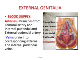
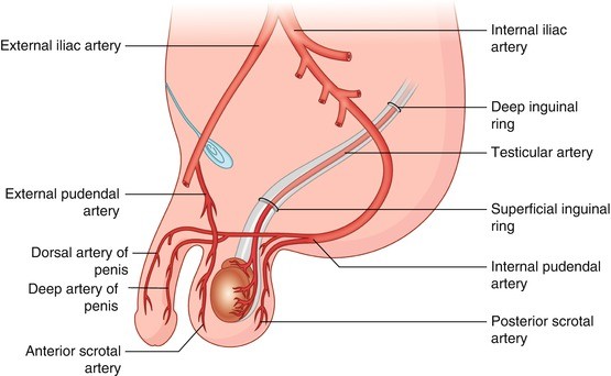
Arterial supply, venous drainage and nervous supply
- The arteries are derived from the inferior vesical and middle rectal arteries.
- The veins accompany the arteries.
- Nervous supply is by sympathetic and parasympathetic nerve fibers.
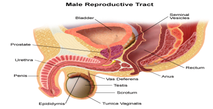
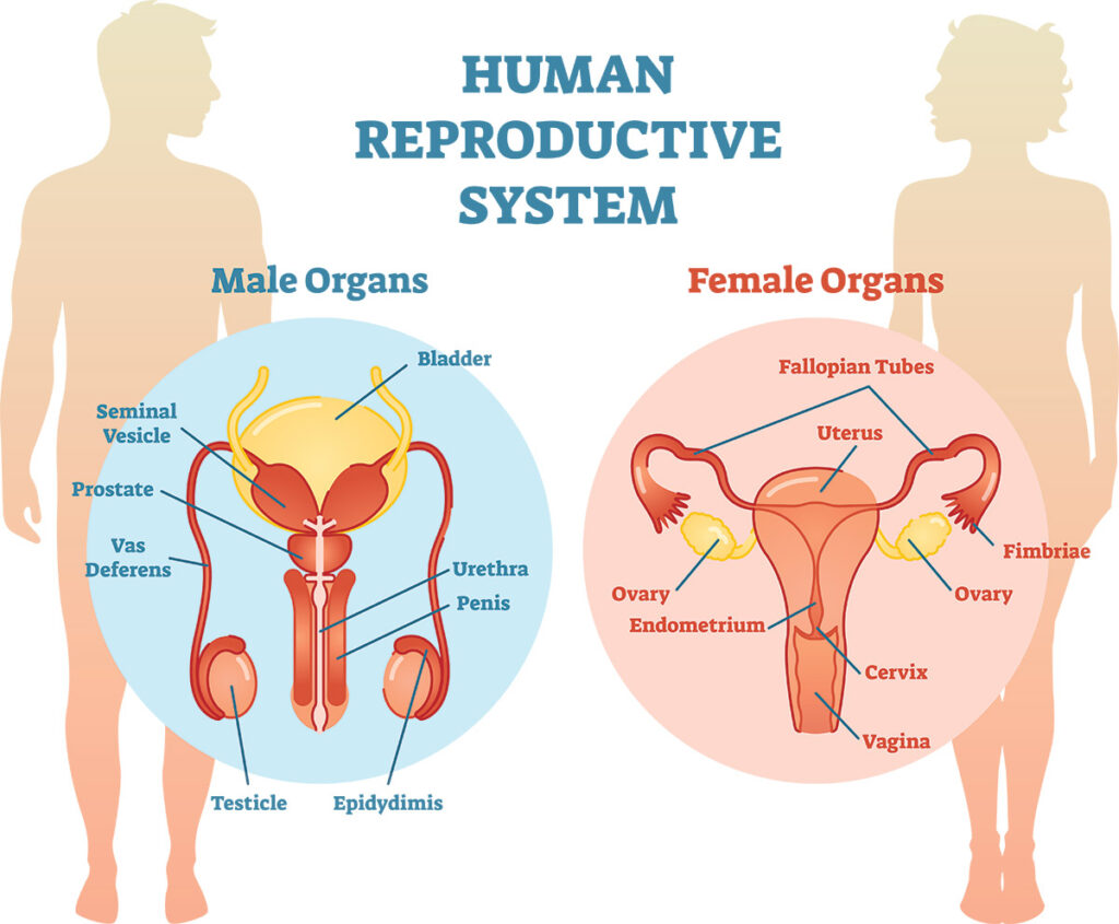
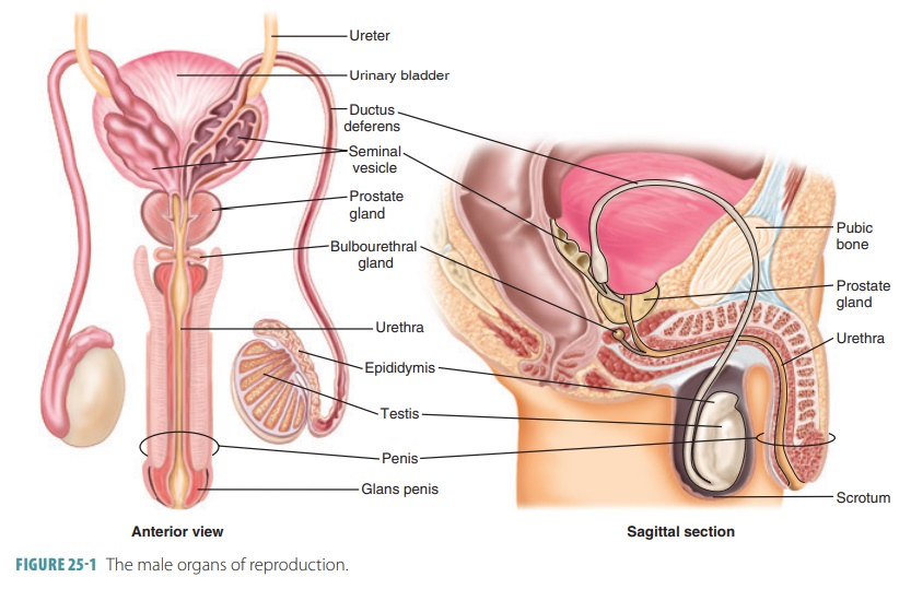
Prostate gland
- This is the largest accessory gland of the male reproductive system.
- It is situated below the bladder.
- The prostate is partly glandular and partly fibromuscular.
- The prostate produces fluid that makes up part of the semen; it helps create a good environment for the sperm in the penile urethra and vagina
- Enables movement of sperm and provides nutrients for the sperm.
Cowper’s gland
- It comprises two small glands situated below the prostate with ducts opening into the urethra.
- Its function is to produce some fluids, which helps create a good environment for the sperm in the penile.
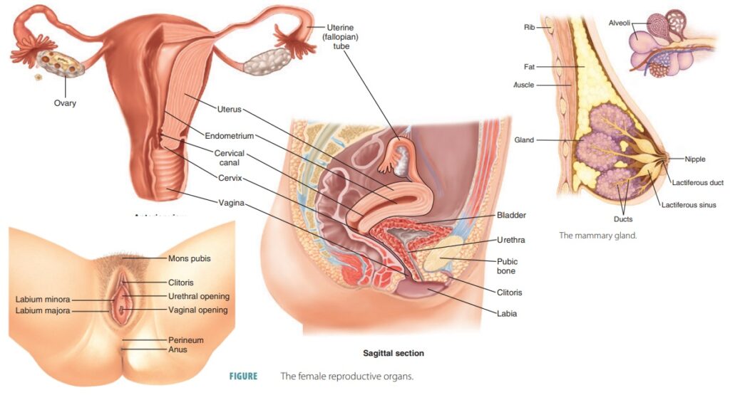
The Female Reproductive System
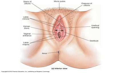
The female external genital organs
- The Mons Pubis
- The Labia Majora
- The labia minora.
- The vestibule of the vagina.
- The External Urethral Orifice
- The Vaginal Orifice
- The Greater Vestibular Glands
- The Lesser Vestibular Glands
- The Clitoris
- The Bulbs of the Vestibule