
Injuries and Trauma: Comprehensive First Aid Guide
Topic 1: Types of Injuries
1.1 Definition of Injury
An injury is defined as damage to the body caused by an external force or agent. This external force can be mechanical (a blow), thermal (heat/fire), chemical (acids), electrical, or radiant energy. Injuries may be intentional (e.g., violence, assault, suicide attempts) or unintentional/accidental (e.g., falls, burns, drowning, occupational accidents).
1.2 Classification of Injuries
To treat an injury correctly, a nurse must rapidly classify it using the following criteria:
(a) By Nature of the Breaking of Skin/Tissue
- Open injuries: The skin is completely broken, and the underlying tissues are exposed to the outside environment. Blood escapes externally, and there is a high risk of bacterial infection. Examples: Abrasions, lacerations, incisions, punctures, avulsions, amputations, and open (compound) fractures.
- Closed injuries: The skin remains intact, meaning the damage is entirely under the surface. Blood and tissue fluid collect internally. Examples: Contusions (bruises), closed simple fractures, dislocations, sprains, strains, internal bleeding, and severe crush injuries.
(b) By Severity
- Minor injuries: Small wounds, superficial (first-degree) burns, mild sprains. These can usually be managed effectively at home or at the basic first aid level.
- Major injuries: Deep/large wounds, severe arterial bleeding, fractures, head/spinal/chest/abdominal injuries, burns covering >10% of the body surface, and crush injuries. These ALWAYS require urgent professional medical attention.
(c) By Mechanism (Cause of Injury)
- Mechanical / Physical: Blunt force (falls, blows from a stick, road crashes) or sharp force (cuts, stabs from a knife).
- Thermal: Burns and scalds from fire/hot water, or extreme cold (frostbite).
- Chemical: Damage from acids, alkalis (like bleach or caustic soda), and irritant gases.
- Electrical: Household electric current, high-voltage power lines, or natural lightning strikes.
- Radiation: Severe sunburn (UV rays), X-ray overexposure, or radioactive sources.
- Asphyxia-type / Environmental: Drowning, choking, and poisoning-related injuries.
(d) By Tissue Involved
- Soft tissue injuries (skin, fat, muscle, ligaments, tendons).
- Bone and joint injuries (fractures, dislocations).
- Head and spinal injuries (central nervous system).
- Chest, abdominal, and pelvic injuries (involving delicate internal organs like the liver or lungs).
- Special sense organ injuries (eye, ear, nose trauma).
📝 Why Classification Matters (The Nurse's Thinking)
- If you see an Open injury, your immediate priority is bleeding control followed by infection prevention (cleaning the wound, applying a sterile dressing, and checking the patient's tetanus vaccination status).
- If you see a Closed injury, you must anticipate the severe danger of hidden internal bleeding and shock. Always suspect that there is much more damage than what you can visually see on the skin surface.
- For a Major injury, you must immediately drop everything and perform a primary survey (DRABC), treat life threats first, immobilise the patient, and arrange an urgent referral.
Topic 2: Wounds (Minor and Major)
2.1 Definition
A wound is a break in the continuity of the skin or mucous membrane, caused by physical means. It opens a doorway for massive blood loss and deadly infections.

2.2 Types of Wounds
Memorize this table to quickly identify the type of wound and its specific risks:
| Type | Description & Appearance | Typical Causes | Bleeding & Infection Risk |
|---|---|---|---|
| Abrasion (Graze) | Superficial scraping of the top layer of skin (epidermis) only. Leaves a raw, weeping surface. | Falls on rough ground, road rash from a boda-boda fall. | Oozes slightly (minor bleeding). Moderate infection risk due to embedded dirt. |
| Incision | A clean, straight, neat cut completely through the tissues. | Knife, razor blade, broken glass, surgical cuts. | Usually moderate to heavy; cuts bleed freely. Lower infection risk due to clean edges. |
| Laceration | Irregular, messy tearing of skin and underlying tissue with jagged, crushed edges. | Machinery accidents, barbed wire, blunt trauma tearing the skin. | Often heavy bleeding. High infection risk because tissue edges are crushed and contaminated. |
| Puncture / Stab | Deep, narrow wound. Has a very small surface opening but a deep, dangerous track inside. | Stepping on a nail, needles, spear, bullet entry, deep thorn. | Often very little external bleeding, but massive deep internal damage. Very high infection risk (especially tetanus, as the deep track lacks oxygen/anaerobic). |
| Avulsion | Tissue is forcibly torn away and left hanging loose (a flap), or is completely detached (amputation). | Heavy machinery, animal bites, explosions. | Often severe and life-threatening bleeding. High infection risk. |
| Contusion (Bruise) | A blunt blow damages small blood vessels under intact skin. (A closed wound). | Blows from a stick, falls, seatbelt trauma. | Internal oozing leads to swelling and discolouration. Low surface infection risk, but watch for deeper organ injury. |
| Gunshot Wound | High-velocity entry (and sometimes exit) wound. Features a deep tract and massive internal tissue cavitation. | Firearms (bullets, shrapnel). | Variable bleeding — but often severe internal organ destruction. Very high infection risk. |
| Bite Wound | A nasty mix of a crush, puncture, and laceration all at once. | Human, dog, or wild animal bites. | Moderate to severe bleeding. Extremely high infection risk (plus massive risk of Rabies from animal saliva). |
2.3 General First Aid for Wounds
The core aims of wound care are: Stop bleeding, prevent infection, prevent shock, and promote healing.
- DRABC first: Treat immediate life threats before focusing on the messy wound.
- Protect yourself: Wash your hands or wear gloves if possible. Standard precautions are vital to protect yourself from blood-borne infections like HIV and Hepatitis B.
- Control bleeding: Apply direct, firm pressure over the wound using a clean dressing or cloth (detailed in Topic 3).
- Clean the wound (For MINOR wounds only): Rinse thoroughly with clean running water or normal saline. Clean the intact skin around the wound with soap and water. Use clean tweezers to pick out visible loose dirt.
DO NOT scrub deep wounds. DO NOT use strong antiseptics like concentrated hydrogen peroxide or undiluted iodine inside the open wound (it destroys exposed healthy tissue and delays healing). - Let it bleed slightly: If it is a very minor wound, leaving it to bleed for a few seconds actually helps flush microscopic germs out from the inside, before you apply pressure.
- Apply a dressing: Use a sterile/clean non-adherent dressing. Bandage it firmly enough to hold it in place, but not so tight that it acts as a tourniquet and cuts off distal circulation.
- Do NOT close deep/dirty wounds: Never attempt home suturing or using superglue on deep cuts. Deep wounds require professional medical assessment, surgical cleaning (debridement), tetanus prophylaxis, and proper sterile stitches.
- Check Tetanus Status: The patient needs a Tetanus Toxoid (TT) booster if they are not fully immunised, or if the wound is dirty/deep and their last dose was more than 5 years ago (per Uganda Clinical Guidelines).
- Raise and rest: Elevate the injured part to reduce throbbing pain and bleeding. Give nothing by mouth if a referral for surgery is likely.
- Monitor for infection: Advise the patient to watch for danger signs over the following days: increasing throbbing pain, expanding redness, unnatural warmth, massive swelling, yellow pus, high fever, or red streaks traveling up the limb toward the heart (lymphangitis).
⚠️ 2.4 Wounds That Must ALWAYS Be Referred (Major Wounds)
Never try to manage these at home. Send immediately to the hospital:
- Deep, gaping wounds, or any wound where yellow fat, muscle, or bone is visible.
- Wounds that will not stop bleeding even after 10 full minutes of hard direct pressure.
- All puncture/stab wounds (due to the hidden deep track and high tetanus risk).
- All bites (human or animal, due to massive infection and rabies risk).
- Wounds situated over major joints, on the face, hands, or genitals (high risk of permanent disability or scarring).
- Wounds with deeply embedded foreign bodies (like glass or knives).
- Amputations: Wrap the severed body part in a clean, slightly moist cloth. Place it inside a sealed, water-tight plastic bag. Put that bag onto ice or in cold water. Never immerse the severed part directly into water or ice, as it destroys the tissue and ruins any chance of surgical replantation!
- Any wound in a casualty who is showing signs of clinical shock (cold, clammy, confused).
Topic 3: Bleeding and Haemorrhage Control
3.1 Definitions
- Haemorrhage: The loss of blood from a ruptured blood vessel. It may be external (visible on the skin) or internal (concealed inside body cavities).
- Mild haemorrhage: Bleeding from injured superficial capillaries; the blood oozes slowly or flows in a steady, manageable stream.
- Severe haemorrhage: Bleeding from a severed artery or large vein; blood exits with massive force (arterial) or in a heavy steady flow (venous). Large volumes of blood are lost very quickly, leading rapidly to Hypovolaemic Shock and death.
3.2 Classification of Haemorrhage
(a) By Time of Occurrence
- Primary haemorrhage: Occurs exactly at the time of the injury.
- Reactionary haemorrhage: Occurs within 24 hours of the injury. When the patient's blood pressure eventually recovers after the initial shock, the rising pressure physically dislodges the weak blood clot that had formed, restarting the bleeding.
- Secondary haemorrhage: Occurs after 48 hours up to 7 days later. It is usually caused by severe bacterial infection (sepsis) that rots and erodes the wall of the injured blood vessel, causing it to burst open again.
(b) By Vessel (Source)
- Arterial bleeding: The blood is bright red (highly oxygenated). It spurts out in violent jets synchronized with each heartbeat. This is the MOST dangerous type due to rapid, high-pressure blood loss.
- Venous bleeding: The blood is dark red/purplish (deoxygenated). It flows in a heavy, steady stream. It can also be rapidly life-threatening if a major deep vein (like the jugular or femoral) is cut.
- Capillary bleeding: Dark red blood that oozes slowly from the entire surface of the wound. It is the most common and is usually self-limiting (stops on its own).
(c) By Site — Internal vs External
- External: Bleeding is clearly visible on the body surface.
- Internal (Concealed): Blood collects silently inside body cavities (chest, abdomen, skull). It is extremely dangerous because it is hidden from the first aider. Blood eventually appears at natural body orifices. As a nurse, memorize these clinical terms:
- Haemoptysis: Bright red, frothy blood coughed up from the lungs/respiratory airways.
- Haematemesis: Blood vomited from the upper GI tract (stomach). If it looks like dark red "coffee-grounds", it means stomach acid has digested a bleeding stomach ulcer.
- Haematuria: Smoky or blood-stained urine, indicating severe kidney or bladder trauma.
- Melaena: Black, tarry, highly offensive-smelling stool. Indicates massive bleeding from the upper bowel (the blood is digested as it travels down).
- Fresh bright red blood per rectum indicates bleeding from the lower bowel or anus.
- Vaginal bleeding: Can indicate a miscarriage, heavy menstruation, injury to the genital tract, or a deadly ectopic pregnancy.
- Thin, watery blood leaking from the ear or nose after a head injury = highly suspicious for a CSF (Cerebrospinal Fluid) leak from a skull fracture.
The Golden Rule: After any severe blunt injury, if the casualty shows signs of clinical shock without any obvious external blood loss — suspect massive internal bleeding immediately.
3.3 The Clotting Mechanism (How the Body Stops Bleeding)
When a vessel is cut, the body triggers a highly complex chemical cascade to save your life. You must understand these physiological steps:
- The injured blood vessel constricts (narrows) immediately to reduce the size of the hole.
- Local blood pressure drops slightly, slowing the flow of blood escaping.
- Platelets rapidly collect at the rough, injured site and stick together to form a temporary "plug" in the gap.
- The damaged platelets and surrounding torn tissues release a chemical enzyme called thrombokinase (thromboplastin).
- Thrombokinase converts the inactive blood protein prothrombin into active thrombin.
- Thrombin then acts as an enzyme to convert soluble fibrinogen (found in blood plasma) into insoluble solid threads called fibrin.
- Fibrin forms a dense, sticky mesh web across the wound. This web traps more platelets and red blood cells, forming a solid clot (scab) as the clear yellowish serum oozes away.
3.4 Signs and Symptoms of Significant Haemorrhage
Whether the bleeding is external or internal, massive blood loss triggers identical systemic shock signs:
- History of severe injury or known medical condition (e.g., bleeding stomach ulcer).
- Pain and severe tenderness over the injury site; rapid swelling; visible deformity.
- Pale face and lips; cold, clammy (sweaty) skin; profuse cold sweating.
- A rapid, very weak (thready) pulse.
- Breathing becomes very shallow, fast, and is often accompanied by "air hunger" (deep sighing or yawning as the brain begs for oxygen).
- Dizziness, blurred vision, roaring in the ears, and fainting.
- Intense, unquenchable thirst; extreme anxiety, restlessness, and a feeling of impending doom.
- Falling blood pressure (a late and very dangerous sign!); lower body temperature; significantly reduced urine output (oliguria) as the kidneys shut down to save fluid.
- In internal bleeding: Blood appearing at orifices + the shock signs above.

3.5 General Management of Haemorrhage
Primary Aim: STOP THE BLEEDING immediately and arrange fast medical aid. The principles of controlling blood loss are to restrict blood flow to the wound and encourage rapid clotting. This is achieved by Direct Pressure and Elevation.
Step-by-Step Management
- Put your gloves on (standard precautions).
- Place the casualty in a suitable, safe position (usually lying down flat to prevent fainting and protect the brain).
- Expose the wound: Remove or cut around as little clothing as possible to clearly see where the blood is coming from.
- Apply DIRECT PRESSURE on the wound:
- Use a clean dressing, thick pad, or cloth and press down extremely hard with your palm.
- Hold the pressure continuously for 10 to 15 minutes without peeking! Lifting the pad to check if it has stopped simply rips off the fragile fibrin clot that is forming, restarting the bleeding.
- If a foreign body (like a knife or glass) is embedded — press firmly AROUND it, never ON it, and build up padding around it.
- If blood soaks completely through your pad — add more dry layers directly on top. NEVER remove the first blood-soaked pad.
- Elevate: Raise the bleeding limb high above heart level and support it. Gravity reduces the hydrostatic blood pressure reaching the wound. (Do not elevate if you suspect a bone fracture in that limb, as movement will cause agony and sever nerves).
- Bandage: Wrap a bandage firmly over the pad to maintain the pressure. Ensure it is not so tight that it cuts off arterial circulation (check that fingers/toes remain warm).
- Immobilise: Keep the injured part perfectly still.
- Treat/prevent shock: Lay them flat, raise their uninjured legs to send blood to the brain, keep them warm with a blanket, reassure them constantly, and give absolutely nothing by mouth.
- Urgent transport: Rush to the hospital — a severe severed artery can drain a human body of blood in under 3 minutes.
Indirect Pressure (Pressure Points)
This is used only if direct pressure fails or is physically impossible. You physically compress the main supplying artery against the underlying bone, located between the heart and the wound.
- For an upper limb bleed: Compress the brachial artery (on the inside of the upper arm).
- For a lower limb bleed: Compress the femoral artery (deep in the groin crease).
- Danger: Prolonged pressure point compression deprives the entire healthy limb of oxygen. Only use it as a temporary bridge while arranging urgent help, and carefully note the time.
🚑 OSCE FOCUS: The Tourniquet (Exams & Real Life)
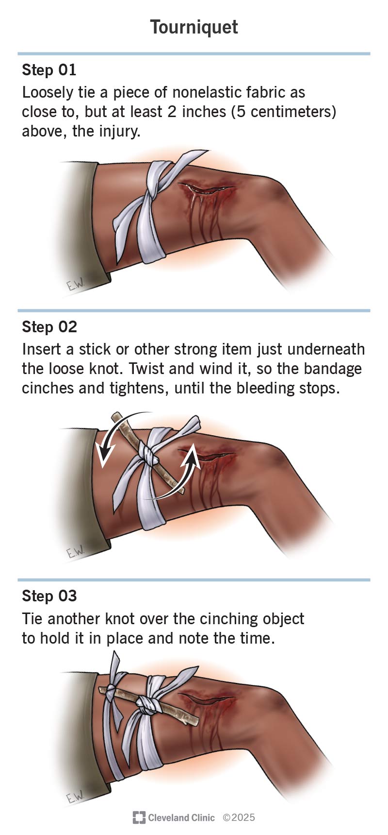
A tourniquet is the absolute last resort for life-threatening limb bleeding that massive direct pressure cannot stop (e.g., traumatic amputation).
- Place it 5 to 7 cm exactly above the wound (between the wound and the heart). Never place it directly over a joint like a knee or elbow.
- Tighten the windlass twisting stick until the bright red bleeding completely stops, and the distal pulse disappears.
- CRITICAL: WRITE THE EXACT TIME OF APPLICATION on the casualty's forehead or on a highly visible tag.
- Never loosen it once applied. Only a surgeon in an operating theatre should remove a tourniquet, to prevent fatal toxins that built up in the dead limb from rushing back into the heart.
- If improvising, use a very wide band of cloth (at least 5cm wide). Never use thin wire or shoe strings, as they will slice straight through the skin and permanently sever nerves.
3.6 Bleeding from Special Sites
(a) Scalp Wounds
- Dangers: The scalp has an incredibly rich blood supply, so even minor cuts bleed profusely and look terrifying. Furthermore, a heavy scalp bleed may hide a depressed skull fracture underneath.
- Signs: Severe pain, massive bleeding, rapid swelling (a "goose egg"). Possible loss of consciousness. If you see blood or clear fluid leaking from the nose or ears, strongly suspect a basal skull fracture.
- Management: Apply direct pressure on the wound. However, press very gently at first—if the skull feels soft or dented inward (depressed fracture), do not press on it! Build a thick ring pad around the dent instead. Bandage firmly. If the casualty is unconscious, keep their head slightly raised (if spinal injury is ruled out), monitor breathing, and refer urgently.

(b) Epistaxis (Nosebleed)
- Bleeding from the delicate vessels inside the nostrils. It may follow a punch to the face, violent nose picking, heavy sneezing, extreme high blood pressure (hypertension), or it may be a deadly sign of a skull fracture (thin watery blood mixed with CSF).
- Aims: Safeguard the airway (prevent blood from being swallowed or inhaled into the lungs) and stop the bleeding.
- Management:
- Sit the casualty down and have them lean SLIGHTLY FORWARD. (Exam Trap: NEVER tilt the head back! Blood will run down the throat, causing violent coughing, choking, or severe vomiting when it hits the stomach).
- Loosen any tight clothing around the neck.
- Advise them to breathe exclusively through their mouth.
- Pinch the soft, fleshy part of the nose (not the hard bony bridge) tightly closed for 10 to 15 uninterrupted minutes.
- Instruct them: No speaking, no swallowing, no coughing, and no spitting during this time. Let any blood in the mouth simply dribble out into a bowl.
- Do not stuff the nose with raw cotton wool (it pulls the clot out when removed).
- After bleeding stops: Clean gently around the nose with lukewarm water. Advise them to have no hot drinks for several hours (heat dilates vessels and restarts bleeding), and avoid blowing or picking the nose for 24 hours.
- Refer if: Bleeding lasts more than 30 minutes, keeps recurring, or follows a significant head injury.
(c) Bleeding from the Mouth / Tooth Socket
- Causes: Severe cuts to the tongue/cheeks, knocked-out teeth, recent tooth extractions, or a fractured jaw.
- Aims: Protect the airway (blood is easily inhaled) and control the bleeding.
- Management:
- Sit the casualty up, with their head tilted slightly toward the bleeding side to let blood drain out.
- Apply direct pressure — use a thick sterile gauze pad over the bleeding tooth socket and ask the casualty to bite down hard on it for 10 to 20 minutes.
- If a thick dark clot has formed in the socket, do not disturb it! Change pads carefully without pulling the clot out.
- No vigorous rinsing or washing of the mouth. No hot drinks for 12 hours (hot liquids dissolve the fragile blood clot). Tell them to gently spit out blood, not swallow it.
- Knocked-out permanent tooth: Handle it only by the white crown (never touch the delicate root). Rinse it very briefly if dirty. Store it submerged in a cup of milk, sterile saline, or have the casualty hold it carefully inside their cheek. Rush to a dentist within one hour for successful replantation.
- Refer if bleeding persists heavily or recurs.
3.7 Complications of Haemorrhage
- Hypovolaemic (Haemorrhagic) Shock: The absolute killer complication.
- Anaemia: A delayed complication due to massive red blood cell loss.
- Asphyxia: If bleeding occurs in the mouth/airway and blood is inhaled into the lungs.
- Unconsciousness and eventual death due to brain hypoxia.
- Hypothermia if bleeding is prolonged and the body loses its warm fluid.
📝 When to Suspect Internal Bleeding (Summary)
If a patient suffers a severe injury (e.g., a femur fracture, a high-speed car crash, a penetrating stab wound, a ruptured spleen from a fall, a bleeding stomach ulcer, or a ruptured ectopic pregnancy) AND they exhibit signs of severe shock (pale, sweating, fast weak pulse) WITHOUT any visible pools of blood on the floor = Treat it as massive internal haemorrhage until proven otherwise by a doctor.
First aid: Lay flat, enforce complete rest, loosen tight clothing, keep them warm, monitor pulse/breathing, save any vomit/urine specimens for the doctor to examine, give absolutely nothing by mouth (they will need emergency surgery), and arrange extremely urgent, gentle transport.
Topic 4: Tissue Injuries (Soft Tissue, Sprains & Strains)
4.1 Soft Tissue Injuries — Overview
This category involves damage to the skin, subcutaneous fat, muscles, tendons, and ligaments. Mechanisms include blunt force (causing contusions or crush injuries) and overstretching/overuse (causing sprains, strains, and tendon tears).
4.2 Contusion (Bruise) — Closed Injury
- A blunt blow violently ruptures tiny blood vessels under intact skin. This leads to throbbing pain, rapid swelling, and a blue-black discolouration (which slowly changes to green/yellow over days as the blood is reabsorbed).
- Management: Apply a cold compress or ice wrapped in a thick cloth to the area for up to 20 minutes to constrict bleeding vessels. Rest the injured part. Elevate it to reduce swelling. (Later, after 48 hours have passed, gentle warmth may help the body absorb the old blood).
- Refer if: There is a huge, expanding swelling (could hide a bone fracture or a much deeper severed artery), if the bruise is over the soft abdomen/chest after severe trauma (internal organ bleeding), or if the casualty is taking prescribed blood-thinning medicines.
4.3 Sprain
- A sprain is the severe stretching or tearing of LIGAMENTS (the tough, inelastic fibrous bands that hold bone firmly to bone around a joint).
- Causes: A joint is violently forced beyond its normal range of motion — e.g., twisting an ankle on uneven ground, twisting a knee during football, or bending a wrist backward during a fall.
- Common sites: Ankle, knee, wrist, and fingers.
- Signs: Intense, immediate pain, rapid and massive swelling, severe bruising appearing shortly after, extreme joint instability, and painful restricted movement. The casualty usually cannot bear any weight on the joint.
4.4 Strain
- A strain is the severe stretching or tearing of MUSCLES or TENDONS (the thick cords attaching muscle to bone).
- Causes: Chronic overuse, sudden unaccustomed violent effort, lifting heavy loads with poor posture, or athletic sprinting without a proper warm-up.
- Common sites: Lower back, thigh (hamstrings), calf, and neck.
- Signs: Deep muscle pain, severe stiffness, profound muscle weakness, painful muscle spasms/cramps, mild swelling. Sometimes a loud "pop" or "snap" is felt or heard at the exact moment of injury.
📝 Sprain vs. Strain — Quick Comparison Table
| Feature | SPRAIN | STRAIN |
|---|---|---|
| Tissue injured | Ligament (Connects Bone to Bone) | Muscle or Tendon (Connects Muscle to Bone) |
| Usual site | Joints (ankle, knee, wrist) | Large muscle bellies (back, thigh, calf) |
| Swelling / Bruising | Marked and rapid | Mild to moderate |
| Movement | Painful, joint feels loose/unstable | Painful, muscle feels extremely weak |
| First aid | RICE / POLICE protocol | RICE / POLICE protocol |
4.6 First Aid: The RICE / POLICE Protocol
For any soft tissue injury, immediately apply this standard protocol:
- P — Protect: Stop the activity immediately. Protect the injured part from further weight-bearing or trauma.
- OL — Optimal Loading: Allow gentle, pain-free movement as tolerated. (Modern sports medicine guidance states that total, prolonged rest is no longer advised, as it causes stiffening. Moving it gently promotes blood flow and healing).
- I — Ice: Apply a cold pack (or ice wrapped inside a wet cloth) to the injury for 15–20 minutes at a time, several times a day. Cold constricts bleeding vessels and numbs pain. (Warning: NEVER put raw ice directly onto bare skin, it causes severe frostbite tissue damage).
- C — Compression: Wrap the joint firmly with an elastic (crepe) bandage to physically squeeze the swelling out. It must be firm, but not tight! Always check that the toes/fingers below the bandage stay warm and pink.
- E — Elevation: Raise the injured limb high above heart level using pillows. Gravity pulls the inflammatory fluid away from the injury, drastically limiting swelling.
- Other actions: Remove rings or tight watches early, before the swelling becomes massive and cuts off circulation. For analgesia, give paracetamol. Avoid giving aspirin early on, as it thins the blood and increases bleeding into the torn tissues.
4.7 When to Refer a Soft Tissue Injury
- If the casualty absolutely cannot bear weight or use the limb at all → treat it as a suspected fracture and refer for an X-ray.
- If there is obvious unnatural deformity, extreme bony tenderness, severe swelling, numbness/tingling, or loss of circulation in the toes/fingers below the injury.
- If there is no improvement after a few days of RICE, or if the pain/swelling worsens.
- The Golden Rule: If you are in any doubt whether an injury is a severe sprain, a strain, or a broken bone — always treat it and splint it as a fracture.
Topic 5: Fractures and Dislocations
5.1 Definitions
- Fracture: A break or crack in the continuity of a bone. It can be a complete snap or a partial crack. (Note: A "broken bone" and a "fracture" mean the exact same thing medically).
- Dislocation: The bones that form a joint are violently forced out of their normal anatomical position, severely tearing or lengthening the surrounding joint capsule and ligaments. (If it pops out and then pops back into place by itself, it is called a subluxation).

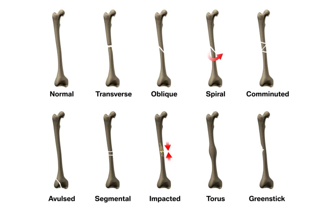
5.2 Types of Fractures
- Closed (Simple) fracture: The bone breaks, but the skin above it remains perfectly intact.
- Open (Compound) fracture: The sharp, broken bone end violently pierces and breaks through the skin, or an external wound leads deep down to the broken bone. This is a massive emergency due to severe bleeding and the extreme risk of deep bone infection (osteomyelitis).
- Complicated fracture: The sharp bone ends damage vital nearby structures (e.g., severing major blood vessels, slicing nerves, or a broken rib puncturing a lung).
- Displaced vs. Non-displaced: Displaced means the bone ends are out of alignment (crooked). Non-displaced means the bone cracked but the pieces remained perfectly aligned.
- Greenstick fracture: The bone bends and cracks only partway down one side, like trying to snap a young, green tree branch. This is extremely common in children because their bones are soft and flexible. It may look like mild swelling only, with no obvious deformity.
- Comminuted fracture: The bone is violently shattered into several small fragments (usually from high-energy injuries like high-speed car crashes or bullet wounds).
- Stress fracture: A tiny hairline crack resulting from repeated, prolonged minor stress (common in long-distance athletes or marching soldiers).
- Colles' fracture: A very specific break of the distal radius bone in the wrist, causing a distinct "dinner fork" upward deformity. Extremely common when people fall and catch themselves on an outstretched hand.
- Pathological fracture: The bone breaks due to an underlying disease (like bone cancer tumours or severe osteoporosis weakening the bone). It often breaks with trivial, everyday force, like simply standing up.
5.3 Signs and Symptoms of Fracture / Dislocation
- Intense pain exactly at the site of the break, massively worsened by any movement or pressure.
- Rapid swelling and severe bruising over the area.
- Deformity / Angulation: The limb looks bent at an unnatural angle, shortened, or the joint looks weirdly "locked" out of place.
- Loss of function: The casualty simply cannot move the limb or bear any weight on it.
- Extreme bony tenderness when touched gently. Abnormal mobility (the limb bends where there is no joint).
- Grating (Crepitus): The horrifying sound or feeling of dry bone ends grinding against each other. (Never intentionally try to feel for this!).
- A loud "snapping" sound heard or felt at the exact moment of injury.
- Numbness, tingling, or a pale, cold limb below the injury (indicating the broken bone has sliced a nerve or artery).
- Signs of profound systemic shock — especially with femur, pelvic, and open fractures. A single closed femur fracture can hide up to 1.5 Liters of internal blood loss; a smashed pelvis can hide even more, causing death by hidden bleeding.
🔍 Fracture vs. Dislocation — Telling Them Apart
| Feature | FRACTURE | DISLOCATION |
|---|---|---|
| What happened | The hard shaft of the bone cracked/snapped. | The joint surfaces were forced entirely apart. |
| Look | Swelling, bruising, bent deformity in the middle of a limb. | Obvious bizarre joint deformity, abnormal joint shape. |
| Movement | Painful or impossible, limb feels floppy. | The joint feels completely locked, stuck, and fixed in place. |
| Common sites | Wrist, ankle, hip, collarbone, shin. | Shoulder, elbow, finger, knee cap. |
| Can occur together? | Yes — always consider both. | Yes — a dislocation may hide a severe fracture inside the joint. |
5.5 General First Aid Management for Fractures
Aims: Prevent any further tissue damage, control bleeding, treat/prevent shock, and arrange safe transport.
- DRABC first. Control any life-threatening bleeding before looking at the broken bone. If it is an open fracture, cover the exposed bone with a large sterile dressing.
- Ask the casualty to keep absolutely still. Do not allow them to try and walk or move the broken limb.
- Do NOT try to straighten it! Never try to push a protruding bone back under the skin, and never try to "pop" a dislocated joint back into place. You will sever nerves and arteries. Immobilise it exactly in the bizarre position you found it.
- Immobilise (Splint) the fracture:
- The golden rule of splinting: Immobilise the joint above AND the joint below the fracture site.
- Use a rigid splint (smooth wood, stiff cardboard, rolled thick newspaper) heavily padded with cloth. Tie broad bandages above and below the fracture site.
- For a broken leg, tie the feet/ankles together securely so the leg cannot rotate outward and grind the bone.
- An injured arm can simply be splinted by tying it firmly to the patient's own body. An injured leg can be tied gently to the uninjured, healthy leg, using it as a biological splint.
- For a fractured collarbone (clavicle) — support the arm on the injured side in a tight sling (e.g., St John elevation sling).
- Check CSM (Circulation–Sensation–Movement): Check the fingers/toes below the injury before and after you tie the splint. If the limb becomes cold, blue, pale, or numb after you tie it, your bandages are too tight! Loosen them immediately and recheck every 15 minutes.
- Apply a cold pack wrapped in cloth to the area to relieve pain and limit swelling.
- Treat for shock: Lay them flat, keep them warm. Remember that femur, pelvic, and open fractures bleed massively internally.
- Suspected spinal fracture: DO NOT move them unless they are in immediate danger of death (like a fire). Support their head and neck firmly in a neutral straight position with your hands. Wait for skilled paramedic help. If you absolutely must move them, use a team to "log-roll" them, keeping the spine perfectly straight.
- Urgent transport: Transport to the hospital with the limb heavily supported. Monitor CSM and vital signs continuously in transit.
5.6 Open Fracture — Extra Emergency Points
- Cover the horrific wound with a large sterile dressing. Do not attempt to wash dirt out deep inside, do not push the sticking-out bone back in, and do not probe the wound with fingers.
- Control bleeding by applying firm pressure around the wound edges, never pressing down directly on the broken bone.
- Immobilise the limb very carefully without disturbing the wound dressing.
- This is an extreme surgical emergency — there is a massive infection risk to the bone marrow; IV antibiotics and urgent tetanus prophylaxis will be needed at the hospital.
🚨 WHAT NOT TO DO (First Aid Errors)
- Never attempt to realign a crooked bone or relocate a dislocated joint (you will slice blood vessels, sever nerves, and crush muscle).
- Never move a casualty with a suspected spinal injury unnecessarily.
- Never let the casualty eat or drink anything (painkillers, water, or food), as they will likely need emergency surgery under general anaesthesia within hours.
- Never bind a splint so tightly that distal circulation is cut off.
Topic 6: Head Injuries
6.1 Definition
A head injury involves any trauma to the scalp, skull, or the brain itself. It ranges from a minor, bloody scalp cut to a rapidly fatal, life-threatening brain injury.
Crucial Concept: The brain can be severely injured even when the hard skull remains perfectly intact. The soft brain is violently shaken and slammed against the hard inside walls of the skull during an impact.
Always suspect a hidden neck (cervical spine) injury whenever there is a significant head injury — especially in falls from a height, road crashes, or heavy blows that throw the head violently backwards.
6.2 Three Key Patterns to Recognise
(a) Scalp Injury
The scalp has a massive, rich blood supply. Even tiny superficial wounds bleed profusely, making the injury look terrifying. This heavy bleeding may hide a severe skull fracture underneath.
Management: Apply direct pressure to stop bleeding, but press gently around the wound first to ensure there is no soft, depressed (caved-in) skull fracture beneath your hand. Bandage firmly and refer.
(b) Concussion (Mild Traumatic Brain Injury)
The brain is violently "shaken" inside the skull, temporarily disrupting its normal electrical function.
- Signs: Brief, temporary unconsciousness (seconds to a few minutes), or no unconsciousness at all. The casualty feels dizzy, confused, nauseated, pale, and sweaty. They complain of a mild, all-over headache. They often suffer from amnesia (they cannot recall the accident or the events leading up to it). They may have blurred vision.
- Prognosis: The casualty usually recovers fully. However, they must be strictly observed for 24–48 hours because deadly bleeding (compression) can develop slowly and delayed over hours.
(c) Cerebral Compression (Serious — Rising Pressure Inside the Skull)
This is a lethal emergency. Bleeding from a torn vessel or massive swelling of brain tissue causes pressure to build up inside the rigid, closed box of the skull. The rising pressure slowly squeezes and crushes the brain downwards toward the spinal cord.
- Signs develop progressively and worsen:
- An intense, severely worsening headache.
- Declining level of consciousness: The patient becomes increasingly drowsy, hard to wake up, and eventually slips into a deep coma. (This is the most critical key sign!).
- Unequal pupils: One pupil becomes huge and blown, while the other remains small.
- Flushed, hot, dry skin with a rapidly raised body temperature.
- A very slow, strong, bounding pulse, accompanied by very slow, noisy, irregular breathing.
- Severe vomiting, and eventually fitting/seizures as the brain is crushed.
- Prognosis: Without urgent neurosurgery to drill a hole and relieve the pressure, it is 100% fatal. Immediate emergency referral is required.
📝 Concussion vs. Compression — Master Table
| Clinical Sign | CONCUSSION (Shaken Brain) | COMPRESSION (Crushed Brain) |
|---|---|---|
| Consciousness | Brief loss, then rapidly recovering and waking up. | Gradually deteriorating, becoming increasingly drowsy and comatose. |
| Skin | Pale, cold, clammy (sweaty). | Flushed red, dry, intensely hot. |
| Pulse | Rapid and weak. | Very slow, full, and bounding. |
| Pupils | Normal, equal, reactive to light. | Unequal (one dilated, one pinpoint), unreactive. |
| Headache | Mild, generalized. | Intense, severe, rapidly worsening. |
| Breathing | Normal, perhaps shallow. | Very slow, noisy, snoring, irregular. |
| Clinical Course | Slowly improves with rest. | Rapidly worsens — a life-threatening surgical emergency. |
6.4 Danger Signs — Emergency Referral Immediately
If a casualty with a head bump shows ANY of these signs, rush them to the hospital:
- Loss of consciousness (even for a second), or rapidly increasing drowsiness / extremely hard to wake up.
- Repeated, forceful vomiting.
- A severe or rapidly worsening headache.
- Unequal pupils, or complaining of double/blurred vision.
- Blood or clear, watery fluid leaking from the nose or ears (CSF leak).
- Sudden seizures, slurred speech, deep confusion, or highly unusual aggressive behaviour.
- Weakness, numbness, or poor coordination/paralysis of their arms or legs.
- A massive soft bump on the head, or dark bruising appearing around both eyes ("raccoon eyes") or dark bruising behind the ears (Battle's sign).
- If the casualty is on prescribed blood-thinning medicines, is over 65 years old, is heavily intoxicated with alcohol (hides symptoms), or lives alone with no one to observe them overnight.
6.5 First Aid Management for Head Injuries
- DRABC: If they are unconscious but breathing normally, place them very carefully into the recovery position (mind the neck — keep the head, neck, and spine perfectly in line while rolling them).
- Support the head and neck: Hold the head firmly in a neutral, straight-ahead position. Do not twist, bend, or tilt the neck. Do not remove a motorcycle helmet unless it is absolutely necessary to maintain an open airway or perform CPR.
- Control scalp bleeding: Use gentle direct pressure around, not on, any depressed/caved-in skull area.
- Conscious casualty: Have them sit or lie down quietly in a very comfortable, dark position. Apply a cold pack wrapped in cloth to the painful bump for up to 20 minutes to reduce swelling.
- Nothing to eat or drink: They may need emergency brain surgery within hours.
- Monitor and record: Track their level of response (AVPU), pulse, breathing, and pupil size every 10 minutes. Watch like a hawk for the danger signs above — deterioration from a slow brain bleed can be delayed for several hours.
- Arrange urgent referral: To the hospital for anything beyond a trivial, tiny bump, and ALWAYS if the mechanism of injury was significant (e.g., fall from a height, road crash, heavy blow with a weapon, or penetration).
6.6 Aftercare Advice (Mild Head Injury, Observed at Home)
If the doctor discharges a mild concussion patient to go home, give this strict advice:
- Strict rest; totally avoid looking at TV/phone screens and avoid any strenuous physical activity for 24–48 hours.
- Do not take aspirin or ibuprofen (these thin the blood and increase the massive risk of a brain bleed) — use only simple paracetamol if pain relief is needed.
- A responsible adult MUST stay with the casualty for 24 hours. The adult must wake the casualty up and check on their responses every few hours during the night.
- Return IMMEDIATELY to the emergency room if: a worsening headache develops, vomiting starts, extreme drowsiness occurs, confusion sets in, pupils become unequal, weakness is felt, or fits/seizures happen.
- Note: Any infant/child who is knocked out cold, or shows any danger signs, must be observed in the hospital, never at home.
Topic 7: Burns and Scalds
7.1 Definitions
- Burn: Severe tissue injury caused by dry heat (open flames, hot metal objects), extreme cold, corrosive chemicals, severe friction, electricity, or radiation.
- Scald: Severe tissue injury caused by wet heat (hot liquids like boiling water, hot cooking oil, hot porridge, soup, and extremely hot steam/vapour).
- Clinical Note: Scalds are usually more superficial because the hot liquid quickly runs off the body, but they tend to cover much larger surface areas. Dry flame burns are usually much deeper and more destructive locally.
7.2 People at High Risk of Burns
As a nurse teaching prevention, focus on these vulnerable groups:
- Children under 5 years: Pulling pots of boiling food/water off tables, or falling into hot bathing basins.
- The elderly: They have very slow reaction times and thin, fragile skin.
- Epileptics & people who faint: Frequently suffer horrific burns after collapsing face-first into cooking fires or hot stoves during a seizure.
- Diabetics & Leprosy patients: Suffer from severe peripheral nerve damage (reduced sensation) — they may rest their feet against a boiling hot object and literally not feel their flesh burning.
- Albinos: Lack protective melanin, suffering extreme sunburns (UV radiation burns) leading to skin cancer.
- Factory workers, petrol station attendants, and commercial cooks.
- People living in crowded slums using charcoal stoves, open fires, or cheap paraffin ("kerosene") stoves.
7.4 Types of Burns
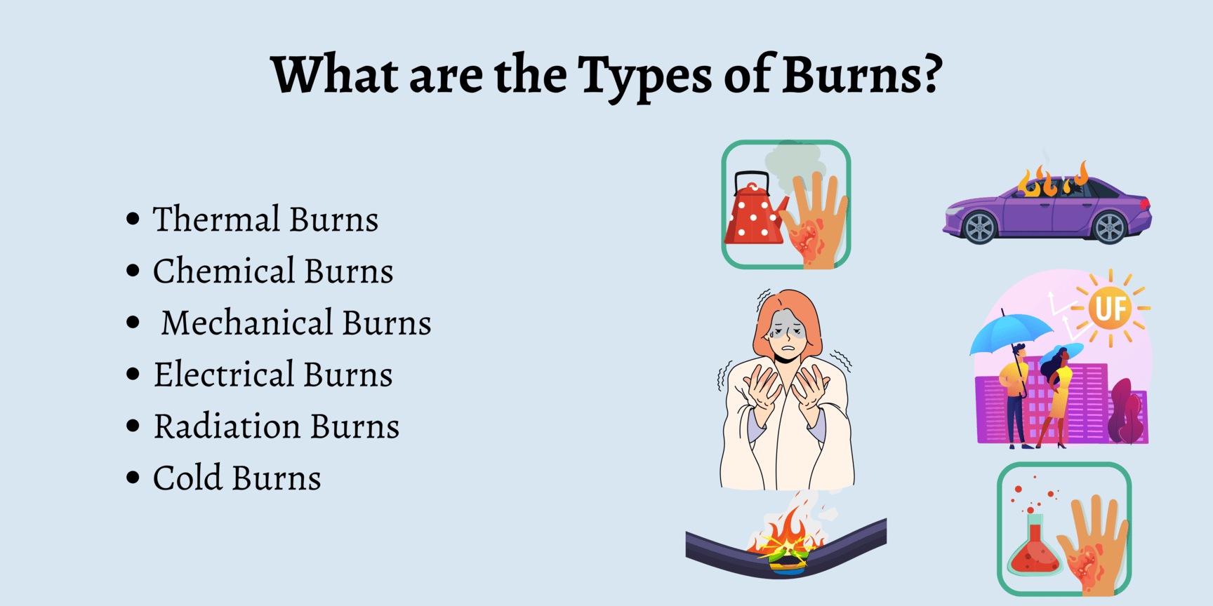
- Dry burns: Direct physical contact with roaring flames, glowing hot objects, or severe friction (road rash).
- Electrical burns: High-voltage current passes violently through the body. Leaves distinctive, deep, charred "entry" and "exit" wounds. The deep internal tissue and muscle damage along the current's path is far worse than the tiny skin wound suggests. Extreme Danger: Immediate cardiac arrest.
- Chemical burns: Strong acids or alkalis (like bleach, caustic soda, battery acid). Tissue destruction signs may develop very slowly (unlike instant heat burns). Alkalis are worse because they penetrate deep into the flesh like butter.
- Radiation burns: Severe UV exposure (sunburn), X-ray overexposure, or "flash burns" to the eyes from industrial welding without a mask.
- Cold injury (Frostbite/Cold burns): Exposure to extreme freezing temperatures or industrial cold liquid gases. The skin becomes numb, pale, waxy, and rock-hard.
7.5 Signs and Symptoms of Burns
- Intense reddening of the skin (erythema), rapid swelling, and massive painful blisters.
- Agonizing pain (paradoxically, pain is most severe in partial-thickness burns because the raw nerve endings are exposed but still alive).
- Peeling of dead skin; dry, charred, black, or leathery white skin in deep burns.
- Extreme restlessness and profound dehydration (fluid rapidly evaporates from the missing skin).
- Airway burns (Inhalation injury): A deadly emergency. Look for singed nasal hairs, black soot around the mouth/nose, a hoarse raspy voice, severe difficulty breathing, and a high-pitched stridor sound. The airway will swell shut in minutes.
- Violent shivering (massive heat loss) — this happens even if the casualty feels hot to the touch.
7.6 Classification by Depth
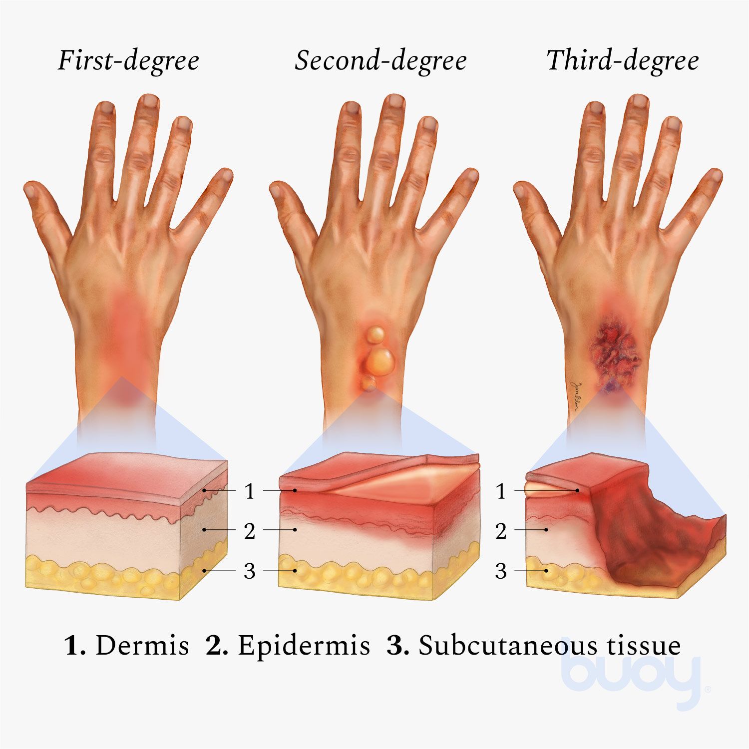
| Depth | Clinical Features & Prognosis |
|---|---|
| Superficial (First-degree) | Damages the top layer (Epidermis) only. Causes intense redness, pain, tenderness, and mild swelling. There are NO blisters. Heals completely and well within days with simple prompt first aid. |
| Partial-thickness (Second-degree) | Damages the Epidermis + the deeper Dermis. Features massive blisters, incredibly severe pain (raw nerve endings exposed), a wet weeping surface, and moderate swelling. The skin often peels off. High risk of infection. Heals in 1–3 weeks, often requiring medical dressings. |
| Full-thickness (Third-degree) | Destroys ALL skin layers (epidermis, dermis, hypodermis) and may reach down to fat, muscle, and tendon. The skin looks absolutely dry, waxy, leathery, white, or charred black. Crucially, there is NO PAIN in the centre because the pain nerves are completely burned away and dead. This painlessness severely MISLEADS bystanders about how fatal the injury is. Always requires urgent, specialized hospital surgical care and skin grafting. |
| Fourth-degree burn | Extends violently straight through to deep muscle, bone, tendons, and ligaments. Threatens the entire limb and function. Extremely life-threatening. |
7.7 Extent of Burns
The larger the surface area burned, the greater the massive fluid loss (plasma weeping from the skin), the greater the heat loss, and the higher the risk of fatal hypovolaemic shock and acute kidney failure.

🔍 The Rule of Nines (Wallace) — For Adults
Used to rapidly estimate the percentage of body surface area burned:
- Head and neck — 9%
- Front of trunk (chest/belly) — 18%
- Back of trunk — 18%
- Each arm — 9% (both arms total = 18%)
- Each leg — 18% (both legs total = 36%)
- Perineum (genitals) — 1%
- Total = 100%
(Note for Children: Use the "Rule of Sevens" or modified Lund & Browder chart, because a baby's head is proportionally massive [approx. 18-28%] and their legs are much smaller).
Approximate Palm Method: The casualty's own flat palm (including their fingers) equals exactly 1% of their body surface. This is highly useful for measuring small, scattered splash burns.
Clinical Danger Zones: If >60% of the body is burned (or just 40% in very young babies or the elderly), fatal kidney failure is highly likely up to 6 weeks later. Any burn covering 30–40% is incredibly severe and requires urgent hospitalization. Any burn >10% requires immediate IV fluid resuscitation.
7.8 First Aid Management of Burns
(a) MINOR Burns (Superficial, small area)
Aims: Relieve pain, halt the burning process, prevent infection, reassure.
- Stop the burning: Pour water immediately on flames. If a person is on fire, wrap them tightly in a heavy blanket and roll them on the ground to smother the fire. Do NOT let a burning person run about in panic (running feeds oxygen to the flames and makes them burn hotter).
- Cool the burn immediately: Flood the area with cool (not freezing ice-cold) running tap water for at least 10 full minutes (up to 20 minutes for chemical burns). The heat continues to burn deeper into the flesh long after the fire is out; water stops this. Never put raw ice directly on burned skin, as the freezing causes further cellular tissue destruction.
- If running water is absent, a clean, cold, soaking wet towel may be applied.
- Leave blisters totally intact: Never pop or burst them! The blister roof is nature's perfect sterile dressing. Bursting it opens a massive doorway for fatal infection.
- Gently pat the area dry. Cover it loosely with a dry, sterile, non-adherent dressing or a clean plastic bag (which won't stick). Never put raw cotton wool directly onto a wet burn; it will stick permanently.
- Absolutely no home remedies: Do not apply butter, cooking oil, toothpaste, raw egg, ash, mud, or aloe vera from unknown sources. They trap the heat inside the skin and cause horrific infections.
- Protect the burn from pressure and rough friction. Reassure the casualty.
- Check their tetanus immunisation status.
- Refer to hospital if: The minor burn involves the delicate face, airway, eyes, hands, feet, genitals, covers a major flexing joint, wraps entirely around a limb (circumferential), is caused by chemicals/electricity, or begins showing signs of yellow pus infection days later.
(b) SUPERFICIAL BUT EXTENSIVE Burns (Large areas)
- Call for professional help immediately.
- Stop the burning process (water/heavy blanket).
- Remove smouldering clothing ONLY if it comes off easily. If melted synthetic clothing is firmly stuck/melted into the flesh, never pull it off! Cut around the stuck pieces carefully with scissors.
- Rapidly remove rings, tight watches, belts, and tight shoes — the burned area will swell massively within minutes, and jewelry will act like a tourniquet, cutting off the limb's blood supply.
- Cooling rules for LARGE burns: If the burn is smaller than the casualty's chest → cool heavily with running water. If the burn is LARGER than the chest → do NOT immerse them in cold water or use cold showers. Because they have lost so much skin, massive cold water will cause fatal systemic hypothermia. Simply cover the vast burn with clean, dry, non-adherent sheets.
- Carefully separate severely burnt fingers/toes with dry sterile dressings so the raw flesh does not heal and fuse together.
- Treat for massive shock: Lay them flat, keep the unburned parts of their body warm with blankets (burnt skin loses body heat extremely fast).
- Urgent hospital transfer. Keep their head still and supported during transit.
🚑 General Referral Criteria for Burns (Exam Gold)
You must refer the patient to a major hospital burn unit if you see ANY of the following:
- Full-thickness (3rd degree) burns of absolutely any size.
- Partial-thickness (2nd degree) burns covering > 5% of the body surface.
- Any burn whatsoever on the face, hands, feet, genitals, perineum, or major joints.
- Circumferential burns (burns wrapping entirely around an arm, leg, or the chest, which act like a shrinking corset and cut off blood/breathing).
- All electrical, chemical, and inhalation (airway/smoke) burns.
- Burns in tiny children, the frail elderly, or people with serious pre-existing illnesses (diabetes, epilepsy).
- Any burn where you highly suspect non-accidental injury (child abuse/torture).
7.9 Complications of Burns
- Immediate: Destruction of vessels, nerves, and tendons; melted foreign bodies embedded in the flesh; profound skin loss and necrosis; deadly airway obstruction/respiratory distress from inhaling superheated smoke.
- Intermediate (Hours/Days later): Severe secondary bacterial infection; massive clinical shock from unbearable pain; profound dehydration and dangerously reduced circulating blood volume (gallons of plasma weep constantly from the raw burn); severe electrolyte imbalance.
- Late (Weeks/Years later): Fatal sepsis; severe contractures (the thick scar tissue shrinks and tightly binds across joints, physically preventing the patient from straightening their arm or neck); massive renal (kidney) failure; ugly unstable/keloid scars; permanent alopecia (hair loss); and Marjolin's ulcer (a highly aggressive squamous cell carcinoma skin cancer that develops inside an old, severe burn scar years later).
7.10 Electrical Burns
Dangers: The massive, hidden danger is Cardiac Arrest (as the raw current passes straight through the heart, destroying its electrical rhythm). It causes severe, extremely deep internal burns (leaving small entry and exit wounds on the skin). The deep muscle and nerve tissue damage along the current's hidden path inside the body is far worse than the tiny skin wound suggests. It causes violent muscle spasms that throw the casualty across the room, causing secondary spine/head fractures.
Note: Wearing damp clothing or wet footwear, or standing on wet ground, drastically increases conduction and makes the electrical damage fatal.
Management of Electrical Injury
- Switch off the current / pull the plug immediately at the wall. NEVER touch the casualty while they are still in contact with the live source. You will be electrocuted instantly and become the second body on the floor.
- If the casualty is lying in a pool of electrified water — stay completely out of it; water conducts electricity perfectly.
- If you absolutely cannot find the switch and they are actively frying on a live wire: forcefully separate them from the wire using a long, completely dry wooden stick (like a broom handle). Stand on a dry, non-conductive surface (a wooden pallet, a thick pile of dry newspapers, or a rubber mat). Wear thick rubber gloves if available.
- High-Voltage Power Lines: If a power pole falls on a car, do not approach within 18 meters! The ground itself is electrified. Wait until the national power company officially confirms the grid is dead.
- Once contact is broken, perform DRABC. Give CPR immediately if they are unresponsive and not breathing (cardiac arrest is extremely common here).
- Flood the visible skin burns with cool water for ≥ 10 minutes.
- Remove jewellery/constrictions before massive swelling occurs. Cover with a sterile non-adherent dressing and bandage loosely.
- Treat for shock. Give oral fluids if they are fully conscious. Monitor and record vital signs carefully.
- URGENT hospital referral: Deep internal heart muscle damage and deadly arrhythmias may suddenly appear hours later. Therefore, ALL electrical injuries must be evaluated in a hospital with an ECG.
7.11 Chemical Burns
Caused by strong industrial acids or alkalis. The intense stinging pain is immediate, but the visible discoloration, massive blistering, peeling, and swelling develop very slowly over time (unlike instant heat burns). Note: Alkalis (like drain cleaner) are far more dangerous than acids because they penetrate deep into the flesh, liquefying fat and tissue.
Management (Aims: Disperse the chemical rapidly, make the area safe)
- Assess scene safety (beware of toxic fumes or gas clouds; wear thick gloves and eye protection to protect yourself).
- Move the casualty far away from the chemical spill zone.
- Flood the affected area with massive amounts of copious running water for at least 20 full minutes. (Water dilutes and washes away the corrosive agent).
- Gently remove contaminated, soaked clothing while you are flooding the area with water. (Use scissors to cut shirts off; do not pull chemical-soaked shirts over the face/head, or you will blind them).
- Do NOT attempt to "neutralise" the burn (e.g., do not pour vinegar on an alkali burn). The chemical reaction will generate explosive thermal heat and boil the flesh. Do not delay washing while searching for a specific chemical antidote. Just use endless water!
- Identify the chemical (grab the bottle or read the hazardous label) and pass this vital information to the hospital doctors.
- Watch the airway closely if fumes were inhaled. Urgent referral.
7.12 Chemical Burn to the Eye — A FIRST AID EMERGENCY
Corrosive chemicals splashed into the delicate eye can scar the clear cornea in seconds, leading to permanent, irreversible blindness. Treatment speed is everything.
- Irrigate immediately: Hold the affected eye under gently running, cold tap water for at least 10 minutes (ideally 15–20 minutes continuously).
- Ensure the water flushes deeply under both sides of the eyelid. This is easier if you pour from a glass/jug, use a tap, or a proper eyewash station.
- The intense pain will cause the eyelid to shut in a violent spasm. You must gently but firmly force the eyelid open with your fingers to let the water inside.
- Ensure the contaminated, toxic rinse water drains away down the side of the face. Do NOT let it flow over the bridge of the nose and splash into the good, uninjured eye!
- Wear gloves. Do not forcibly remove melted contact lenses. The casualty must not rub the eye under any circumstances.
- After 20 minutes of irrigation: cover the eye gently with a sterile eye pad.
- Rush to the hospital immediately. Bring the chemical bottle with you.
Topic 8: Eye, Ear and Nose Injuries
8.1 Eye Injuries
Core Principles for ALL Eye Injuries:
Do not let the patient rub, touch, or press the injured eye. Do not attempt to pull out embedded objects. Do not apply any random ointments or eye drops. Give absolutely NO aspirin or ibuprofen (these thin the blood and will cause massive bleeding inside the delicate eyeball). Rush them to an eye clinic.
Crucial trick: Because human eyes are biologically wired to move together synchronously, you must cover the uninjured, good eye too with a pad. If the good eye is blindfolded, the patient will stop looking around, which perfectly paralyses and rests the injured eye.
(a) Foreign Body on the Eye (Dust, loose eyelash, flying grit, small insect)
- Wash your hands thoroughly.
- Tell the casualty not to rub. Ask them to blink repeatedly and let their natural tears wash the object out.
- Gently pull the upper eyelid out and down over the lower lid (the lower eyelashes act as a brush to sweep the particle out from under the top lid).
- Rinse the eye gently with clean water or sterile saline, pouring from the inner corner (near the nose) outward.
- If it does not wash out easily, or feels scratched → cover the eye with a pad and refer to a clinic.
(b) Embedded / Penetrating Object in the Eye (Metal shard, broken glass, sharp stick)
MASSIVE EMERGENCY. Never attempt removal! Pulling it out will rip the eyeball open, causing all the internal fluids to leak out, leading to instant blindness and massive bleeding.
- Leave the sharp object exactly in place. Do not press on it or touch it.
- Pad around the object: Build a thick ring of sterile gauze (a "doughnut" pad) around the stick/glass, or place a hard plastic paper cup/shield over the entire large object. Tape the shield to the facial bones so that absolutely nothing touches or bumps the embedded object.
- Cover the other, healthy eye with a dark pad to completely stop all eye movement.
- Keep the casualty perfectly still (lying flat on their back if possible) and arrange immediate, smooth hospital transfer.
(c) Blow to the Eye / Black Eye / Blunt Eyeball Injury
Apply a cold compress very gently (for 10 minutes) to the surrounding bruising to reduce swelling. If the severe pain persists, vision becomes blurred or double, or the inside of the eye looks filled with pooling red blood (hyphaema) → refer instantly. There may be a devastating internal blowout fracture or retinal detachment. Never press hard on a swollen, painful eye.
8.2 Ear Injuries
(a) Foreign Body in the Ear (Beans, plastic beads, insects — very common in kids)
Do NOT poke blindly inside with matchsticks, hairpins, or cotton buds! You will push the hard object deeper and permanently rupture the delicate eardrum.
For a live, buzzing insect: Tilt the head sideways and gently pour in a little warm (body-temperature) cooking oil or clean water to drown and float the insect out.
For any other solid object, leave it alone and refer to a health worker with proper suction/forceps.
(b) Bleeding / Clear Fluid from the Ear
If bleeding occurs after a severe head injury, and the blood is thin, watery, or mixed with clear yellowish fluid, this is a CSF (Cerebrospinal Fluid) leak indicating a catastrophic basal skull fracture. EMERGENCY:
- Do NOT plug the ear with tight cotton wool! Plugging it traps the fluid, forcing bacteria backward into the brain, causing fatal meningitis. Let the fluid drain freely.
- Do not clean deep inside. Simply tape a sterile pad loosely over the outer ear to catch the drips.
- Keep the casualty completely still; arrange urgent referral; treat them as having a severe head and possible broken neck injury.
If the ear bleeds without a head injury history (e.g., eardrum rupture from a vicious slap to the side of the head, blast wave, loud noise, or severe middle ear infection): keep the ear totally dry, insert absolutely no ear drops, and refer.
(c) Perforated Eardrum
Signs: The patient had severe ear pain that suddenly felt "relieved" after a pop, followed by hearing loss, loud ringing (tinnitus), and sometimes a little blood trickling out. Keep the ear completely dry (no swimming, place cotton wool loosely when bathing to stop water entering). Apply absolutely no ear drops unless prescribed by a doctor. Refer. Most heal naturally in a few weeks, but infections need antibiotics.
8.3 Nose Injuries
(a) Fractured Nose
Signs: Extreme pain, massive rapid swelling, visible crooked deformity, profuse nosebleed, blocked nasal breathing, and dark black bruising developing under both eyes.
Management: Have them sit leaning forward. Apply a cold compress gently on the bony bridge. Control bleeding by pinching the soft fleshy part. Do not try to forcefully straighten the crooked nose yourself. Encourage mouth-breathing. Urgent referral (doctors get the best cosmetic reduction results if it is fixed within the first few days before the bone sets).
(b) Foreign Body in the Nose (Children pushing beans/beads)
Do not attempt removal at home using tweezers (massive risk of pushing the smooth object backward down the throat into the airway, causing fatal choking). Keep the panicked child calm, tell them to breathe through their mouth, and refer to a clinic.
Topic 9: Bites and Stings
9.1 Human Bites
Human mouths carry incredibly dangerous, aggressive bacteria. There is a massive infection risk. Bites occurring over the knuckles (when someone punches another person in the teeth, known as a "fight bite") infect the deep joint capsule quickly and violently.
Management: Wash the wound intensely and thoroughly with soap and running water. Control bleeding. Cover with a clean dressing. Refer immediately. The casualty will almost certainly need strong prophylactic antibiotics. The doctor will also check their tetanus status and assess the risk of Hepatitis B or HIV transmission. Document carefully and report if it was a violent assault.
9.2 Animal Bites — Dog, Cat, Monkey, Bat (MASSIVE RABIES RISK)
Why it matters in Uganda: Uganda is highly rabies-endemic. Rabies is a terrifying viral disease that attacks the brain. It is 100% fatal once neurological symptoms start (killing ~59,000 people globally per year, mostly children in Africa). However, Post-Exposure Prophylaxis (PEP) given BEFORE symptoms appear is nearly 100% protective and life-saving. Stray dog bites are the main source. Unfortunately, studies in Uganda show that many poor bite victims delay treatment or consult useless traditional healers, and die. The nurse must insist on immediate hospital referral.
First Aid for a Rabid Animal Bite
- Wash the wound IMMEDIATELY and vigorously with soap and running water for at least 15 uninterrupted minutes. This single mechanical step physically washes the fragile rabies virus out of the torn flesh, and combined with PEP, reduces rabies risk by up to 90%.
- If available, apply a strong antiseptic that kills viruses (povidone-iodine or 70% alcohol) directly into the wound after washing.
- Control severe bleeding with direct pressure and cover with a clean dressing.
- Do NOT suture (stitch) the wound closed unless it is absolutely essential to stop fatal arterial bleeding. Stitching physically traps the deadly virus deep inside the tissues. If stitches are essential, doctors will do them loosely, and only after PEP injections have started.
- URGENT referral to a major hospital for Rabies PEP and Tetanus prophylaxis.
Rabies Post-Exposure Prophylaxis (PEP) — WHO Categories
As a nurse, you must know how to categorize the exposure to give the right treatment:
| Category | Type of Contact with Animal | Required Medical Action |
|---|---|---|
| Category I | Touching or feeding the animal; animal licks on perfectly intact, unbroken skin. | No exposure risk. Wash the skin with soap. No vaccine needed. |
| Category II | Animal nibbling on uncovered skin; minor scratches or tiny abrasions without drawing blood. | Vigorous wound washing + immediate Rabies Vaccination. |
| Category III | Single or multiple bites/scratches that break the skin and bleed; or the animal's saliva touches mucous membranes (eyes/mouth) or fresh open wounds. | Vigorous wound washing + Rabies Vaccine + Rabies Immunoglobulin (RIG) injected immediately. |
- The Vaccine: A modern cell-culture vaccine given Intramuscularly on a strict schedule: Days 0, 3, 7, 14, and 28. You must strongly emphasise to the patient that they must return to complete all 5 doses, or they will die.
- Rabies Immunoglobulin (RIG): For severe Category III bites, ready-made antibodies are injected directly into and around the bite wound ASAP to neutralize the virus before it reaches the nerves. Dose is 20 IU/kg (human) or 40 IU/kg (equine). Any remaining volume is injected IM at a distant site.
- Observe the biting animal for 10 days (by vet authorities) if possible to see if it dies of rabies — but never delay starting the PEP injections while waiting for the dog to die!
9.3 Snake Bites
Uganda is home to highly venomous snakes (Cobras, Mambas, Vipers, Puff Adders). Most bites occur on the bare legs, feet, and hands of farmers working in bushes or reaching into dark holes. Many victims die or suffer horrific amputations because they visit traditional healers who cut the wound, delaying modern medical care. Antivenom is the ONLY definitive treatment that saves lives.
First Aid (What TO do)
- Move the casualty safely away from the snake. Do not act brave and try to catch or kill it (the severed head of a snake can still bite and inject venom reflexively hours after death!). A quick photo from a safe distance helps doctors choose the right antivenom.
- Keep the casualty completely STILL and CALM. Extreme panic, a racing heartbeat, and running around wildly pumps the deadly venom rapidly through their lymphatic system into the heart and brain. Lay them down. Splint the bitten limb heavily to completely stop muscle movement. Keep the limb roughly at heart level (or slightly below if you suspect a neurotoxic cobra bite).
- Rapidly remove rings, bracelets, and tight clothing near the bite before massive, crushing swelling begins.
- Wash the bloody bite wound gently with soap and water.
- Use a pen to draw a circle around the leading edge of the swelling and write the exact time next to it. This brilliantly helps doctors track how fast the venom is destroying tissue.
- Arrange incredibly urgent transport to a major facility stocking antivenom. Carry the casualty to the vehicle if possible (walking pumps the venom!).
- Monitor breathing and pulse constantly. Be ready for CPR. If you see weak drooping eyelids, drooping face, or the patient complains of difficulty swallowing or breathing = this is a massive neurotoxic emergency. Their diaphragm is paralysing. Support their breathing with an Ambu bag and drive faster.
🚨 WHAT NOT TO DO (Classic Exam Traps & Deadly Myths)
- Do NOT cut the wound open or make "X" incisions with a razor. It causes fatal bleeding and introduces tetanus.
- Do NOT suck the venom out using your mouth or a pump. It does absolutely nothing to extract venom that has already bonded to tissues, and the bacteria in your mouth will infect the wound.
- Do NOT apply a tight tourniquet that cuts off all arterial blood. It traps the highly destructive venom in the limb, concentrating the acid and guaranteeing the leg will rot and require amputation.
- No ice packs, no electric shocks, no herbal poultices, no traditional "black stones", and no alcohol (which speeds up heart rate).
- Do not give aspirin (snake venom already destroys the blood's ability to clot; aspirin will cause massive internal bleeding).
- Note on bandages: A firm pressure-immobilisation bandage (like wrapping a sprain) is endorsed internationally for neurotoxic elapid bites (cobras/mambas) to slow lymph flow. However, for viper bites (puff adders) which cause massive blistering swelling, a tight bandage acts like a tourniquet and dramatically worsens flesh rotting. Because laypeople cannot identify snakes accurately, the safest universal rule taught in Uganda is: Splint it, keep them totally still, and transport extremely fast.
Signs of Envenomation (For clinical monitoring)
- Local signs: Searing severe pain, massive rapid swelling, dark purple bruising, huge blood blisters, and continuous oozing bleeding from the tiny puncture fangs.
- Haemotoxic (Vipers): Destroys blood clotting. Look for spontaneous bleeding from the gums, nose, blood in urine, or vomiting blood.
- Neurotoxic (Cobras/Mambas): Paralyzes nerves. Look for drooping eyelids (ptosis), blurred/double vision, slurred drunken speech, trouble swallowing saliva, general severe weakness, and finally fatal respiratory muscle paralysis.
- Profound shock, severe nausea, vomiting, and fainting.
9.4 Insect Stings (Bees, Wasps, Ants)
Most stings cause intense local burning pain, redness, and a swollen welt.
Management: Swiftly scrape the stinger off sideways using a fingernail or the hard edge of a credit card. Do not squeeze the stinger with tweezers! Squeezing it acts like a syringe plunger, injecting the remaining venom sac into the skin. Wash the area, apply a cold compress to relieve pain, and observe.
Dangerous situations: Multiple stings (disturbing a swarm of angry bees) or a sting directly inside the mouth/throat (the resulting massive swelling can quickly block the airway and choke the patient to death — give them ice cubes to suck on continuously and refer instantly).
Anaphylaxis (Severe Deadly Allergy): A patient allergic to bee stings will develop sudden facial/tongue swelling, massive hives all over the body, severe wheezing/stridor, a hoarse voice, dizziness, and collapse. Treatment: Lay flat with legs raised (or sit up if struggling to breathe). Administer Adrenaline 0.5 mg IM (1:1000) deep into the anterolateral thigh immediately. Repeat every 5–10 minutes if no response. Use antihistamines + steroids as adjuncts. Urgent hospital referral. Casualties with known severe allergies should carry an adrenaline auto-injector (EpiPen) at all times.
9.5 Tick Bites
Ticks bury their heads in the skin and suck blood, transmitting dangerous fevers.
Management: Grasp the tick's head with fine tweezers as close to the human skin as possible. Pull straight upward with steady, even pressure. Do not twist or jerk the tick (the head will snap off and remain stuck in the flesh). Do not burn it with a match or cover it in petrol/vaseline (this causes the tick to vomit its infected stomach contents into the patient's bloodstream before dying). Clean the site with alcohol. Watch for a strange rash or high fever in the following weeks, and refer to a clinic if illness develops.
Quick Revision Summary (High-Yield Exam Points)
| Injury Type | KEY Action / Management |
|---|---|
| Severe bleeding | Direct pressure 10–15 mins, add layers, elevate, immobilise. |
| Arterial bleed | Bright red, spurting → brutal direct pressure, use pressure points (brachial/femoral). |
| Internal bleeding | Shock signs without visible blood → lay flat, strict NPO (nothing by mouth), urgent transport. |
| Epistaxis (Nosebleed) | Lean FORWARD, pinch the SOFT part of nose 10–15 mins continuously. |
| Sprain / Strain | RICE / POLICE protocol — Protect, Optimal load, Ice, Compression, Elevation. |
| Fracture | Immobilise the joint above + below. NEVER attempt to straighten. Constant CSM checks. |
| Open fracture | Dress the horrific wound, no pushing the bone back, splint as found, give tetanus. |
| Dislocation | Immobilise exactly as found — never try to relocate or "pop" it back. |
| Concussion | Brief knockout. Rest, observe strictly 24–48 h, give no aspirin. |
| Compression (Brain) | Deteriorating consciousness, unequal pupils, slow pulse → SURGICAL EMERGENCY. |
| Head injury | Support the neck constantly, cold pack to bump, monitor AVPU, strict NPO. |
| Burns | Flood with cool water 10–20 mins (chemical 20+ mins), no ice, no bursting blisters, cover with non-adherent dressing. |
| Full-thickness (3rd deg) burn | PAINLESS — this severely misleads bystanders; the flesh is dead. Always refer! |
| Electrical injury | Switch off current first, CPR is priority (cardiac arrest), refer ALL victims for an ECG. |
| Chemical in eye | Irrigate violently with water for 10–20 mins, pad, rush to eye hospital. |
| Embedded eye object | Pad heavily around the object, apply a hard shield, cover BOTH eyes, NEVER remove the object. |
| CSF leak (ear/nose) | Thin bloody water. Never plug it — apply a loose pad, urgent referral (basal skull fracture). |
| Dog bite | Wash with soap + water for 15 mins, NO suturing, rabies PEP urgently (Vaccine days 0,3,7,14,28; +RIG for Cat III). |
| Snake bite | Keep completely still, NO cutting/sucking/tourniquet, splint limb, urgent antivenom transport. |
| Bee sting | Scrape stinger off sideways (don't squeeze), cold pack. Anaphylaxis → Adrenaline IM immediately. |
🧠 THE GOLDEN RULE FOR ALL INJURIES
DRABC first → Control bleeding → Immobilise → Treat/prevent shock → URGENT referral with documentation.