Midwives Revision

Gynaecology

FIBROIDS (FIBROMYOMAS)
Home > Gynaecology & Reproductive Health > Fibroids (Fibromyomas)

Fibroids (Fibromyomas)

Uterine Fibroids are benign (non-cancerous) tumors that originate from the smooth muscle layer (myometrium) of the uterus.

  • Other Medical Names: Uterine leiomyoma, myoma, fibromyoma, and fibroleiomyoma.
  • Demographics: They typically occur after the age of 30 years and are most commonly seen in nulliparous women (women who have never had children).
  • Characteristics: They are composed of dense muscle and fibrous tissue. They can be single or multiple, and range in size from a tiny microscopic pinhead to an enormous mass that distorts the entire abdomen. They are far more likely to arise in the body of the uterus than in the cervix.

Risk Factors for Uterine Fibroids

While the exact primary cause is still being researched, several established clinical risk factors drastically increase a woman's likelihood of developing uterine fibroids:

  • Age: Fibroids are most prevalent and aggressively grow in women between the ages of 30 to 40 years.
  • Parity (Number of Pregnancies): Women who are nulliparous (have never given birth) or have very low parity are at a significantly higher risk. Pregnancy seems to have a protective effect.
  • Race / Ethnicity: Uterine fibroids are disproportionately more prevalent, tend to appear at a younger age, and grow larger in individuals of African descent (Black women) compared to those of Caucasian (White) ethnicity.
  • Family History: Genetics play a strong role. If a woman has close first-degree relatives (such as a mother or sister) with a history of fibroids, her personal risk is highly increased.
  • Hyper-estrogenemia: Elevated levels of the female hormone estrogen directly promote the rapid growth of fibroids. Fibroids are highly estrogen-dependent.
  • Obesity: Being overweight or obese is strongly associated with a higher risk. (Pathophysiology note: Adipose/fat tissue converts androgens into estrogen, leading to higher overall circulating estrogen levels).
  • Early Onset of Menarche: Starting menstruation at a very young age (early exposure to cyclic hormones) is linked to an increased likelihood of fibroid development later in life.
  • Low Level of Vitamin D: Recent clinical studies suggest that a deficiency or insufficient levels of Vitamin D might be associated with a higher risk of fibroid growth.
  • Drugs (Estrogen Replacement Therapy): Long-term use of estrogen replacement therapy, particularly when used without progesterone to balance it, acts as a direct catalyst for fibroid growth.

Classes, Types, and Locations of Uterine Fibroids

Fibroids are classified based on their specific growth patterns and exactly where they are situated within or around the uterus.

1. Types of Uterine Fibroids

The "type" refers to the specific category based on its growth characteristic relative to the uterine layers:

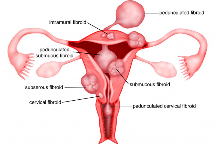
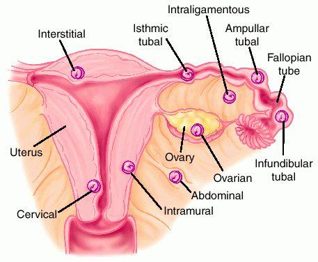
  • Submucosal Fibroids: These grow directly into the uterine cavity, situated just beneath the inner lining (endometrium). Because they disrupt the cavity, they are the main culprits for heavy menstrual bleeding (menorrhagia) and infertility.
  • Intramural (Interstitial) Fibroids: The most common type. They grow centrally within the thick muscular wall of the uterus (myometrium). As they expand, they distort the overall shape of the uterus, leading to severe pain and pressure.
  • Subserosal Fibroids: These grow on the outside surface of the uterus, just under the serosa layer. They project outward into the pelvic cavity, often growing quite large and pressing on nearby organs like the bladder or rectum.
  • Pedunculated Fibroids: These are attached to the uterus by a narrow stalk or pedicle that contains their blood supply. They can be either pedunculated subserosal (hanging outside) or pedunculated submucous (hanging inside the cavity).
  • Cervical Fibroids: Located specifically on the cervix (the lower neck of the uterus). These are relatively rare but can cause severe pain, discomfort, and major complications (like obstructed labour) during childbirth.
  • Fibroid of the Broad Ligament: Develops outward into the broad ligament (the supportive structure holding the uterus in place). They are uncommon and require very specific surgical management to avoid damaging the ureters.
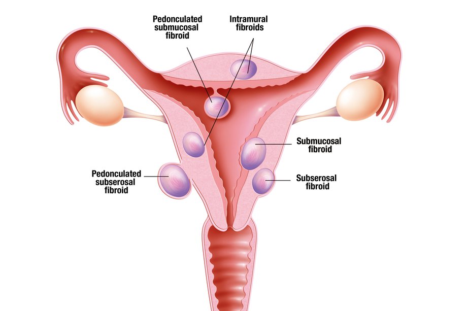
Locations of Uterine Fibroids
Visual classification of fibroids based on their location within the uterine layers.

Understanding the Difference: Types vs. Location

Many students confuse "Types" with "Location." Here is the clinical distinction:

Concept Definition Examples
Types of Fibroids Refers to the different categories or classifications based on their specific growth patterns and characteristics. Submucous, Intramural, Subserosal, Pedunculated, Cervical.
Location of Fibroids Refers to the specific physical site within or around the pelvic organs where the fibroid has situated itself. Location dictates which organs it will press against. Subperitoneal (under peritoneal surface), Bulging into the endometrial cavity, Centrally within the myometrium, At the outer border, On the cervix.

Statistical Frequencies and Specific Positions

  • Intramural / Interstitial: Accounts for the vast majority at 75%. Initially intramural, they may later be pushed outward or inward, but about 70% persist in this central position.
  • Subserous / Subperitoneal: Accounts for 15%. They are pushed outward towards the peritoneal cavity and are either partially or completely covered by the peritoneum.
  • Submucous: Accounts for only 5%. Despite being the least common, it produces the maximum number of symptoms.
  • Cervical Positions: Depending on exactly where they grow on the cervix, they are classified as: Anterior, Posterior, Lateral, or Central cervical fibroids.

Pathology and Microscopic Structure

Understanding the exact cellular structure of a fibroid helps explain its surgical and clinical behavior:

  • Gross Pathology: Fibroids are firm, spherical, or irregularly lobulated. They possess a false capsule (pseudocapsule) made of areolar tissue and compressed normal myometrium. Because the capsule is false, the fibroid can be easily "enucleated" (shelled out) from the surrounding myometrium during surgery.
  • Microscopic Structure: Consists of smooth muscle cells that are spindle-shaped and uniform. They are arranged in a distinct whorled pattern interlaced with varying amounts of fibrous connective tissue.
  • Vascularity: Arteries inside the fibroid are less dense than the surrounding myometrium and lack a regular distribution pattern. Usually, only 1 to 2 major vessels are found at the base or pedicle supplying the entire mass.

Changes in Surrounding Pelvic Organs

  • The Uterus: The shape becomes grossly distorted. Myohyperplasia (overgrowth of muscle cells) is a constant finding. There is massive dilatation and congestion of the myometrial and endometrial venous plexuses. The uterine cavity itself may become elongated and distorted.
  • The Ovaries: Often become enlarged, highly congested, and studded with multiple cysts. Anovulation is commonly associated.
  • The Ureters: Broad ligament fibroids cause severe displacement of normal ureter anatomy, increasing the risk of surgical injury.
  • Associated Conditions: There is a highly increased clinical association with pelvic endometriosis, adenomyosis, and endometrial carcinoma.

Causes of Uterine Fibroids

The exact trigger that causes a single smooth muscle cell to mutate and multiply into a fibroid is not fully understood, but clinical research points to two major driving forces:

  • Hormones (Estrogen and Progesterone): These two hormones, which naturally regulate the menstrual cycle and thicken the endometrium, directly stimulate the growth of fibroids. Fibroids possess more estrogen and progesterone receptors than normal uterine muscle cells. Consequently, fibroids grow rapidly during the reproductive years (especially during pregnancy when hormone levels are massive) and naturally shrink (atrophy) during menopause when hormone production ceases.
  • Genetics: Genetic alterations are frequently found in fibroid cells. A strong familial link exists; women with a mother or sister who had fibroids are genetically predisposed to developing them.
  • Other Contributing Factors: Diet (high in red meat, low in green vegetables), obesity, and race (African descent) act as strong secondary catalysts.

Degenerative Changes in Uterine Fibroids

Because fibroids grow so rapidly, they often outgrow their own blood supply. When this happens, the tissue undergoes "degenerative changes" (alterations in tissue structure):

  • Hyaline Degeneration: The most common type. The firm muscle fibers are gradually replaced by a soft, homogenous, structureless hyaline material. The most common type, occurring in 65% of cases affecting all sizes (except tiny ones). The tumor loses its firm feel and becomes soft and elastic. Naked-eye examination of a cut surface shows irregular, glassy, homogenous areas with a complete loss of the normal "whorl-like" appearance.
  • Cystic Change: This usually follows hyaline degeneration. The soft hyaline tissue liquefies, leaving fluid-filled cavities that make the fibroid look exactly like an ovarian cyst.
  • Red (Carneous) Degeneration: Occurs mainly in the second half of pregnancy and puerperium. Microscopically: shows evidence of severe necrosis. Odor & Color: It emits a distinct "fishy" odor due to fatty acids, and the raw-beef color is due to the presence of haemolysed red blood cells and hemoglobin trapped in the cystic spaces.
  • Fatty Change: The muscle fibers of the fibroid are slowly replaced by fat (adipose) tissue.
  • Atrophy: Following menopause, due to the drastic drop in estrogen and progesterone, the fibroid simply wastes away and shrinks in size.
  • Calcification: Calcium salts are heavily deposited into the fibroid tissue, causing it to harden.
    • Eggshell Fibroid: Calcium deposits form a hard shell strictly on the outside, leaving the inside soft.
    • Womb Stone: The entire fibroid is completely infiltrated with calcium salts, becoming solid and hard like an actual stone.
  • Septic / Infective Degeneration: Infection gains access to the tumor through thinned, sloughed surface epithelium of a submucous fibroid. This usually happens immediately following delivery or an abortion.
  • Sarcomatous (Malignant) Change: The rarest but deadliest change. The benign fibroid undergoes malignant transformation into a leiomyosarcoma. This must be heavily suspected if a fibroid suddenly grows rapidly, especially in post-menopausal women.
  • 🧠 Memory Aid: Degenerative Changes of Fibroids

    Remember the letters: H - C - C - R - F - S - S

    H = Hyaline (Most common, 65%)
    C = Cystic (Liquefaction)
    C = Calcific (Womb stone in menopause)
    R = Red / Carneous (During pregnancy)
    F = Fatty (Post-menopause)
    S = Septic / Infective (Post-delivery/abortion)
    S = Sarcomatous (Malignant transformation)

  • Parasitic Fibroid: Occurs when a pedunculated fibroid twists on its pedicle (torsion), completely cutting off its original blood supply. To survive, it attaches itself to nearby organs (like the omentum or bowel) and establishes a brand new "parasitic" blood supply from them.

⚠️ Attention: Red Degeneration (Obstetric Emergency)

Red Degeneration is an acute complication that almost exclusively occurs during pregnancy. The fibroid grows so fast that its blood supply fails, leading to massive tissue necrosis (cell death) and hemorrhage inside the fibroid. It becomes soft, reddish, and looks like "beefy" raw meat. It causes the pregnant mother sudden, severe, localized abdominal pain, localized tenderness, and low-grade fever.
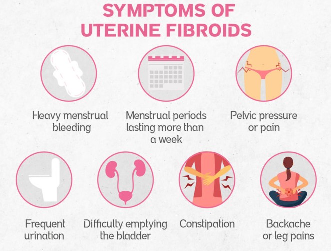
Clinical Presentation (Signs and Symptoms)

Many women with fibroids are asymptomatic. However, depending on the size, number, and location, they can present with severe symptoms:

  • Menstrual Abnormalities:
    • Painful & Prolonged Menstruation: Heavy menstrual bleeding (menorrhagia) and extremely painful periods (dysmenorrhea).
    • Postmenopausal Bleeding: Vaginal bleeding occurring after menopause is highly abnormal and requires immediate investigation.
  • Bulk and Pressure Symptoms:
    • Urinary Issues: Anterior fibroids pressing against the bladder cause increased frequency of urination or difficulty emptying the bladder (retention).
    • Constipation: Posterior fibroids pressing on the rectum block the passage of stool.
    • Pelvic Pressure/Fullness: A constant feeling of heaviness or pressure in the lower abdomen.
    • Abdominal Enlargement: Large or multiple fibroids can cause the abdomen to distend, mimicking the appearance of a pregnancy.
    • Vascular Pressure: Extreme pelvic vein pressure can cause the development of severe hemorrhoids and varicose veins in the legs.
  • Pain and Discomfort:
    • Lower Back and Leg Pain: Caused by large fibroids pressing directly on pelvic nerves.
    • Dyspareunia: Deep pain or discomfort during sexual intercourse.
    • Acute Pain: Sudden, severe pain indicates a complication such as acute red degeneration or torsion (twisting) of a pedunculated fibroid.
  • Reproductive Issues:
    • Infertility: Submucosal fibroids distort the uterine cavity, physically blocking fallopian tubes or preventing the proper implantation of a fertilized egg.

Specific Symptom Triggers

  • Dysmenorrhea Types: Can present as Congestive (due to massive associated pelvic venous congestion or endometriosis) or Spasmodic (associated with the violent uterine contractions attempting to expel a submucous polyp from the cavity).
  • Extreme Urinary Retention: Rarely, a specific posterior fundal tumor can cause an extreme retroflexion of the entire uterus, violently distorting the bladder base and causing acute urinary retention.
  • Bowel Obstruction: A parasitic fibroid that has detached and attached to the bowel can cause direct mechanical bowel obstruction.
  • Cervical Symptoms: Tumors strictly on the cervix heavily provoke foul vaginal discharge, severe bleeding, and dyspareunia.
Diagnosis and Investigations

Diagnosing uterine fibroids involves a combination of thorough clinical examination and advanced imaging.

1. Clinical Examination

  • History Taking: Comprehensive assessment of menstrual patterns, volume of blood loss, reproductive history, and pain levels.
  • Physical Examination: Checking the patient's general health, specifically looking for signs of chronic anemia (extreme pallor of the conjunctiva and palms) resulting from prolonged menorrhagia.
  • Abdominal Examination: Palpating the abdomen to detect large, firm, irregular pelvi-abdominal swellings rising out of the pelvis.
  • Pelvic & Bimanual Examination: A two-handed examination to feel the size, shape, nodularity, and mobility of the uterus. A speculum examination may reveal fibroid polyps protruding directly through the cervical os.

2. Medical Investigations (Labs & Imaging)

  • Pregnancy Test: A mandatory first step to rule out pregnancy as the primary cause of an enlarged uterus or amenorrhea.
  • Full Blood Count (FBC) & Iron Studies: To confirm and quantify the severity of anemia caused by heavy bleeding.
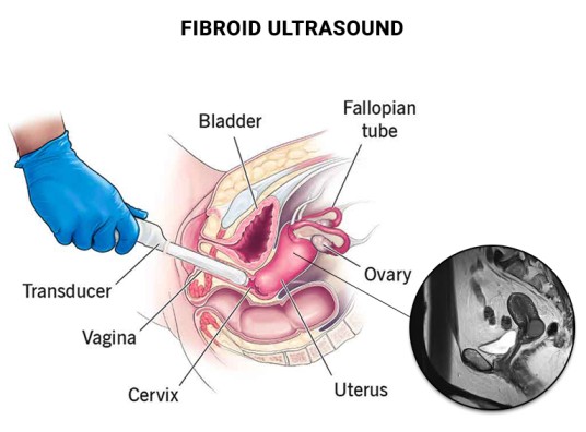
  • Pelvic Ultrasound (U/S): The standard, non-invasive imaging test to clearly visualize the uterus and detect the size and number of fibroids.
  • Transvaginal Ultrasound (TVUSS): Inserting a small probe into the vagina provides a much clearer, high-resolution view of the pelvic organs, which is especially useful in obese patients.
  • Saline Hysterosonography: Injecting sterile saline into the uterus during an ultrasound to sharply outline the cavity and identify hidden submucous fibroids.
  • Hysterosalpingogram (HSG): An X-ray procedure using contrast dye to visualize the exact shape of the uterine cavity and ensure the fallopian tubes are open (crucial for infertility workups).
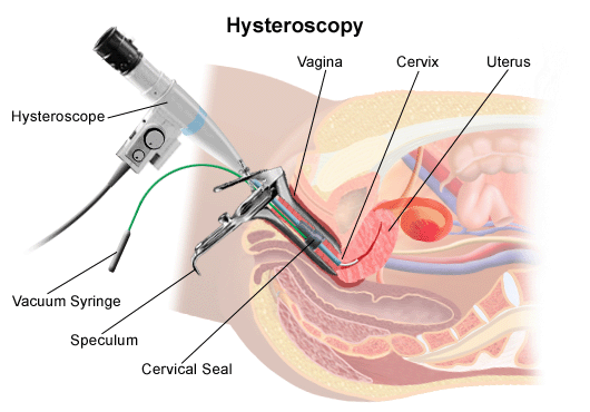
  • Hysteroscopy: Passing a thin, lighted camera (hysteroscope) through the vagina and cervix directly into the uterus to visualize and potentially remove submucosal fibroids.
  • MRI (Magnetic Resonance Imaging): The most highly accurate tool for mapping out the exact size, location, and vascularity of every single fibroid prior to complex surgeries.

Differential Diagnosis (DDx)

Fibroids are usually easily diagnosed, but the midwife/clinician must clinically exclude other conditions that cause similar presentations:

  • Exclude Pregnancy: Always the absolute first step for an enlarged uterus.
  • Exclude Other Pelvic Masses: Ovarian Carcinoma, Tubo-ovarian abscesses, Endometriosis, or Adnexa/omentum/bowel that has adhered to the uterus.
  • Exclude Other Causes of Uterine Enlargement: Adenomyosis, Generalized Myometrial hypertrophy, Congenital anomalies, or Endometrial Carcinoma.
  • Exclude Other Causes of Abnormal Bleeding: Endometrial hyperplasia, Endometrial or tubal Carcinoma, Uterine sarcoma, Polyps, and Dysfunctional Uterine Bleeding (DUB).

⚠️ Attention: Rule Out Malignancy

An endometrial biopsy or Dilation & Curettage (D&C) is strictly essential in the evaluation of any abnormal uterine bleeding to definitively exclude Endometrial Carcinoma before assuming the bleeding is solely from a fibroid.
Management of Uterine Fibroids

Most fibroids are totally asymptomatic and require expectant management (watchful waiting), especially since they shrink naturally after menopause. When treatment is required, the choice depends on: Age, Parity, Size/Location of the fibroids, Desire to preserve the uterus, and the Desire for future pregnancies.

1. Emergency Treatment

  • Blood Transfusion: Immediately given to correct life-threatening severe anemia.
  • Emergency Surgery (Laparotomy): Indicated immediately for an infected/necrotic myoma, acute torsion (twisting) of a pedicle, or severe intestinal obstruction caused by massive fibroid pressure.

2. Medical Management (Pharmacology)

Medical management does not cure fibroids permanently but controls symptoms and temporarily shrinks them prior to surgery.

  • NSAIDs: Nonsteroidal anti-inflammatory drugs (like Ibuprofen) to manage severe pelvic pain and cramping.
  • Antifibrinolytic Agents: Tranexamic acid specifically helps to drastically reduce heavy menstrual bleeding (menorrhagia) by preventing blood clots from breaking down too quickly.
  • Hormonal Control: Low-dose birth control pills or a Levonorgestrel Intrauterine Device (Mirena coil) effectively limit menstrual blood flow and thin the uterine lining with minimal side effects.
  • Haematinics: Aggressive supplementation with Ferrous Sulphate and Folic Acid to rebuild hemoglobin levels in anemic patients.
  • GnRH Agonists (Lupron, Synarel): These drugs put the body into a temporary medical menopause by shutting down estrogen and progesterone production from the ovaries. This stops menstruation and significantly shrinks the fibroids.
  • Mifepristone: (25-50mg twice weekly) acts as a progesterone receptor inhibitor, effectively reducing fibroid size and stopping bleeding.
  • Danazol: A synthetic androgen that interrupts ovulation and shrinks fibroid tissue.

3. Surgical Management

Specific Surgical Criteria

  • Open Myomectomy (Laparotomy): The route of absolute choice for large subserous or intramural fibroids measuring greater than 7 cm, multiple fibroids greater than 5 cm, or when direct entry into the uterine cavity is expected.
  • Hysteroscopic Myomectomy: The route of choice strictly for removing submucous (SM) fibroids measuring greater than 2 cm.
  • Laparoscopic Myomectomy: Mostly done for the subserosal type, removing the mass entirely through a small abdominal keyhole incision.

Advanced Non-Invasive Procedures

  • MRGFUS (Magnetic Resonance Guided Focused Ultrasound Surgery): A non-invasive procedure where focused ultrasound waves are converted into intense heat inside the pathology to destroy it, entirely under MRI guidance.
    Selection Criteria: Fibroid must be 4 to 10 cm, the woman must have completed her family, and she should be perimenopausal.
  • Myomectomy: The surgical removal of the fibroids while leaving the healthy uterus intact.
    Indications: Young women who strongly desire future pregnancies, have a few isolated fibroids, but suffer from heavy bleeding or infertility.
  • Hysterectomy: The complete surgical removal of the entire uterus.
    Indications: Women who have completed their families, are approaching menopause, have massive/numerous fibroids that destroy the uterine structure, or if there is any suspicion of malignant (cancerous) changes.
  • Hysteroscopic Resection: Passing a wire loop through the cervix to shave off and remove submucosal fibroids from inside the cavity without cutting the abdomen.
  • Endometrial Ablation: Surgically destroying the entire inner lining of the uterus using heat or freezing to permanently stop heavy bleeding (only for women who do not want more children).
  • Uterine Artery Embolization (UAE): A radiologist injects tiny polyvinyl particles into the femoral artery, guiding them to block the specific blood vessels feeding the myoma, causing it to starve and shrink.
  • Radiofrequency Ablation: Inserting a needle-like device directly into the fibroid and heating the tissue with radiofrequency energy to destroy it.

Pre and Post-Operative Care for Fibroid Surgery

Whether a patient is undergoing a Myomectomy or a Hysterectomy, meticulous nursing care is required to prevent life-threatening surgical complications.

Pre-Operative Management

  • Admission & History: Obtain personal, medical, social, and detailed gynecological history. Conduct a full physical head-to-toe examination specifically ruling out severe anemia, dehydration, and jaundice. Ensure a general assessment by the gynecologist is documented.
  • Informed Consent: The surgeon must explain the exact reasons for the operation, the benefits, the risks (including the risk of converting a myomectomy to a full hysterectomy if uncontrollable bleeding occurs), and expected results. Involve the partner where culturally or legally appropriate, and secure written consent.
  • Investigations: Ensure all preoperative lab work is back on the file: Urinalysis, Hemoglobin (HB) level, Blood Grouping & Cross-matching (ensure at least 2 units of blood are ready), Abdominal ultrasound, Urea and Electrolytes, INR/PT (clotting profile), and an ECG/ECHO if the patient is older or hypertensive.
  • Patient Education & Counseling: Educate the patient about what to expect when waking up (pain, catheters, IV lines). Provide strong reassurance to relieve surgical anxiety.
  • Physical Preparation: Ensure the patient fasts from food and drinks (NPO) for at least 8 hours prior. Shave and prep the abdominal/pubic area. Arrange for a wide-bore IV line insertion. Insert a urethral Foley catheter to keep the bladder completely empty and safe from surgical injury. Administer prescribed pre-medications.
  • Theatre Transfer: Help the patient change into a clean theatre gown, remove jewelry and dentures, and wheel her to the operating theatre while providing continuous emotional support.

Post-Operative Management

  • Reception & Positioning: Receive the patient from the recovery room into a pre-warmed bed. Keep her lying flat (supine for abdominal surgery) with the head turned to one side to prevent aspiration if she vomits.
  • Intensive Observation: Take vital signs continuously (every 15 minutes for the first hour, then every 30 minutes until fully stable). Closely monitor BP and pulse for early signs of internal hemorrhage or hypovolemic shock. Check the surgical dressing for active bleeding. Monitor the IV infusion rate and continuous blood transfusions.
  • Upon Full Consciousness: Gently welcome the patient, orient her to the ward, and explain that the surgery was successful. Assist with face sponging, provide mouthwash to relieve dry mouth, and change her into a fresh gown.
  • Medical Treatment & Pain Relief: Administer strong analgesics precisely as prescribed (e.g., IM Pethidine 100mg every 8 hours for the first 24 hours, then step down to oral Paracetamol/NSAIDs). Administer broad-spectrum IV antibiotics (e.g., Ampicillin/Gentamicin) to prevent pelvic sepsis. Continue Haematinics and Vitamin C to aid rapid wound healing.
  • Wound Care: Monitor the abdominal wound closely. If minor strike-through bleeding occurs, do not remove the dressing; simply reinforce it with a fresh bandage on top and notify the surgeon.
  • Nursing & Rehabilitative Care: Maintain strict hygiene (bed baths, catheter care). Once bowel sounds return and the doctor permits, start sips of water and progress to a soft diet. Encourage deep chest breathing and early leg exercises while in bed to prevent Deep Vein Thrombosis (DVT) and chest infections. Assist with early ambulation.

Specific Vaginal Surgery Management (If applicable)

  • If a vaginal pack was inserted to control hemorrhage, inspect it frequently for soakage.
  • Once the pack is removed (usually after 24 hours), apply a sterile vulval pad. Swab and clean the vulva at least every 8 hours using aseptic technique to prevent ascending infections.

🧠 Discharge Advice (Crucial for Myomectomy)

For Myomectomy Patients: The uterine wall has been cut and scarred. Advise the mother to strictly avoid conception for at least 2 years to allow the uterine scar to achieve maximum strength. When she does conceive, she must deliver via Elective Cesarean Section to prevent a catastrophic uterine rupture during labour.

For Hysterectomy Patients: Provide deep psychological counseling. Assure her she will no longer have periods and cannot conceive, but her sexual function will remain intact once healed. Recommend abstaining from sexual intercourse for 6 weeks.

Complications of Uterine Fibroids

General Gynecological Complications

  • Severe Menorrhagia: Leading to chronic, debilitating iron-deficiency anemia.
  • Infertility: Due to blocked tubes or a distorted endometrial cavity preventing embryo implantation.
  • Torsion: A pedunculated fibroid twisting on its stalk, causing acute ischemic pain and gangrene.
  • Urinary Tract Diseases: Chronic retention of urine leading to repeated severe urinary tract infections (UTIs) and hydronephrosis (kidney swelling) due to ureter compression.

Complications During Pregnancy and Labour

If a woman with significant fibroids conceives, the pregnancy is automatically classified as High Risk due to the following massive threats:

  • Antepartum Hemorrhage (APH): The presence of fibroids heavily increases the risk of Placenta Previa and Placental Abruption.
  • Abortion/Miscarriage: The distorted cavity lacks enough healthy blood supply to sustain the early placenta.
  • Fetal Growth Restriction (FGR): The fibroids steal the blood supply away from the growing baby, leading to a small, undernourished fetus.
  • Malpresentation: Large fibroids block the baby from turning properly, leading to Breech or Transverse lies.
  • Labor Dystocia & Obstruction: A cervical or lower segment fibroid acts as a literal physical roadblock, causing completely Obstructed Labour.
  • Premature Labor: The uterus becomes overstretched too early by the combined bulk of the baby and the fibroids.
  • Uterine Inertia & PPH: The fibroid-riddled myometrium loses its ability to contract effectively. This leads to weak labour (inertia) and massive Postpartum Hemorrhage (PPH) because the uterus cannot clamp down on the bleeding blood vessels.
  • Subinvolution: During the puerperium, the fibroid prevents the uterus from shrinking back to normal size, causing prolonged, heavy lochia.

Clinical Nursing Care Plan: Uterine Fibroids

Clinical Scenario: Mrs. A.O., a 44-year-old teacher, is admitted for a scheduled total abdominal hysterectomy due to symptomatic fibroids. Complaints: Menorrhagia (bleeding 8-10 days, soaking pad/super tampon hourly), pelvic pain/pressure (5/10), and severe fatigue. Pre-op Hb: 9.8 g/dL. She feels "drained" and is highly anxious about surgery and loss of fertility.

Assessment & Nursing Diagnosis Goals / Expected Outcomes Nursing Interventions & Rationale Evaluation
Subjective: Constant pelvic pain/pressure 5/10. "Heavy ball in stomach."
Objective: Confirmed fibroids, palpable firm enlarged uterus, guarding lower abdomen.

Diagnosis: Chronic Pain related to pressure from enlarged fibroids on pelvic organs/nerves.
- Report pain decrease to manageable level (< 3/10).
- Demonstrate two non-pharmacological relief techniques.
- Rest comfortably.
Interventions:
1. Conduct comprehensive pain assessment.
2. Administer prescribed NSAIDs on a scheduled basis.
3. Teach non-pharmacological comfort (heating pad, guided imagery).
4. Position in semi-Fowler's or side-lying with pillows.

Rationale: Scheduled dosing provides consistent analgesia. Positioning alleviates direct fibroid pressure on structures.
Goals Met. Patient reports pain is 2/10 after scheduled ibuprofen and heating pad. Resting comfortably in semi-Fowler's.
Subjective: "Drained all the time, no energy." Reports menorrhagia.
Objective: Pale conjunctiva, Hb 9.8 g/dL, lethargic, mild tachycardia on exertion.

Diagnosis: Excessive Fatigue burden related to chronic blood loss and anemia.
- Identify relationship between heavy bleeding, anemia, and fatigue.
- Participate in energy conservation.
- Verbalize anemia management plan.
Interventions:
1. Cluster nursing activities; schedule rest periods.
2. Administer iron supplements with Vitamin C.
3. Educate on fatigue causes linking to blood loss.
4. Provide an iron-rich diet (lean meat, spinach).

Rationale: Vitamin C heavily enhances iron absorption. Energy conservation prevents exhaustion while maintaining dignity.
Goals Met. Patient explains, "My heavy periods made my blood count low, which is why I'm tired." Taking iron actively.
Subjective: "Worried about surgery. Feels strange not having a uterus."
Objective: Restless, anxious expression, increased HR/BP when discussing surgery.

Diagnosis: Excessive Anxiety related to impending major surgery, body image changes, and loss of fertility.
- Verbalize specific fears.
- Report noticeable decrease in anxiety.
- Demonstrate effective coping mechanisms (deep breathing).
Interventions:
1. Establish a trusting relationship to express feelings.
2. Provide clear, factual pre/post-op routine info.
3. Teach relaxation (mindfulness, calming music).
4. Acknowledge and validate grief over end of fertility.

Rationale: Validation normalizes emotional response. Factual knowledge dispels myths and reduces fear of the unknown.
Goals Met. Patient reports feeling "less on edge" after talking through fears. Listening to music to relax.
Subjective: Soaking pad and tampon hourly.
Objective: Scheduled for total abdominal hysterectomy. Pre-existing anemia.

Diagnosis: Risk for Bleeding related to underlying condition and impending surgery.
- Remain hemodynamically stable.
- Pre-op prep for blood loss completed.
- Verbalize post-op signs of excessive bleeding to report.
Interventions:
1. Ensure current CBC, blood type, and screen are on chart.
2. Monitor vitals for hypotension/tachycardia.
3. Educate to report soaking >1 pad/hour post-op.
4. Ensure surgical consent (incl. blood transfusion) is signed.
5. Assess/stop anticoagulant use (NSAIDs/fish oil).

Rationale: Type/screen ensures blood is readily available. Stopping NSAIDs prevents coagulation failure during surgery.
Goals Met. Labs complete, consent signed. Patient states two signs of post-op bleeding to report. Hemodynamically stable.

References

  • 1. Myles Textbook for Midwives (Latest Edition) - Anatomy and Physiology of the Female Reproductive System.
  • 2. DC Dutta's Textbook of Gynecology - Benign Lesions of the Uterus.
  • 3. UNMEB Curriculum for Diploma in Midwifery Extension Program - Gynaecology & Reproductive Health (DME 112).
  • 4. Clinical Guidelines for Obstetrics and Gynaecology (Ministry of Health, Uganda).

Quick Quiz

Fibroids Quiz

Gynaecology - mobile-friendly and focused practice.

Privacy: Your details are used only for quiz tracking and certificates.

FIBROIDS (FIBROMYOMAS) Read More »
PELVIC INFLAMMATORY DISEASES (PID)
Home > Gynaecology & Reproductive Health > Pelvic Inflammatory Diseases (PID)

Pelvic Inflammatory Diseases (PID)

Pelvic Inflammatory Diseases (PID) refer to a spectrum of severe, acute, or chronic infections that ascend and affect the upper female genital tract and its surrounding structures.

These affected pelvic organs and structures primarily include:

  • The Cervix (Cervicitis)
  • The Endometrium (Endometritis)
  • The Fallopian Tubes (Salpingitis)
  • The Ovaries (Oophoritis)
  • The Pelvic Peritoneum and Pelvic Vascular System

PID is a major cause of gynecological morbidity, contributing heavily to female infertility, chronic pelvic pain, and ectopic pregnancies.

Causes and Causative Organisms of PID

PID is rarely caused by a single mechanism; it is usually a polymicrobial infection. The infection reaches the sterile upper genital tract through three primary routes of spread:

  • 1. Ascending Infections: This is the most common route. It occurs when pathogenic bacteria from the vagina or cervix travel upwards through the endocervical canal into the normally sterile reproductive organs (uterus, fallopian tubes, and ovaries).
  • 2. Haematogenous Infections: Infections that are spread systemically through the bloodstream to reach the pelvic organs (e.g., Tuberculosis).
  • 3. Direct Spread: Infection spreading from infected neighboring or adjacent organs. For example, a ruptured or highly infected appendix (appendicitis) can directly spill purulent material into the pelvic region, infecting the fallopian tubes.

Specific Causative Organisms

PID is often polymicrobial, involving both aerobic and anaerobic bacteria. The most common culprits include:

  • Neisseria gonorrhoeae: A primary sexually transmitted pathogen responsible for severe acute PID.
  • Chlamydia trachomatis: Another major sexually transmitted pathogen, often causing silent or subclinical but highly destructive PID.
  • Haemophilus influenzae
  • Escherichia coli (E. coli): Common in obstetric PID or when bowel flora cross-contaminates the vagina.
  • Anaerobes: Organisms associated with bacterial vaginosis (e.g., *Gardnerella vaginalis*).

Risk Factors for Developing PID

Certain behavioral, medical, and obstetric factors highly increase a woman's vulnerability to developing Pelvic Inflammatory Disease. These include:

  • History of STIs: Previous or current untreated sexually transmitted infections, especially Gonorrhea and Chlamydia.
  • Bacterial Vaginosis (BV): An overgrowth of harmful bacteria causing an imbalance in normal, protective vaginal flora (Lactobacilli).
  • Sexual Practices: Having multiple sexual partners or a partner who has multiple partners, increasing exposure to pathogens.
  • Douching: This practice vigorously washes away protective vaginal mucus and flora, and can actively push existing lower tract bacteria up into the uterus.
  • Previous History of PID: One episode of PID heavily damages protective mucosal barriers, drastically increasing the risk of recurrence.
  • Use of Intrauterine Devices (IUDs): The risk is slightly elevated immediately after insertion if pre-existing cervical infections are pushed into the uterus.
  • Surgical/Gynecological Procedures: Invasive procedures like Dilation and Curettage (D&C), manual vacuum aspiration, or hysteroscopy can introduce bacteria.
  • Obstetric Causes: Infections following abortion, ectopic rupture, or puerperal sepsis (postpartum infections).

Pathophysiology of PID

The disease process of PID is highly destructive to the delicate reproductive tissues. It typically follows this pattern:

  • The infection typically begins localized in the vagina and cervix (often asymptomatic or presenting as mild cervicitis).
  • The bacteria then actively ascend through the endocervical canal to reach the Fallopian tubes and ovaries. During menstruation, the endocervical canal naturally dilates slightly and the protective cervical mucus plug is lost, facilitating the easy entry of bacteria into the uterus.
  • Once inside the highly vascular reproductive tracts, the bacteria rapidly multiply. The body's immune response triggers massive inflammation.
  • This inflammation destroys the tiny, hair-like cilia inside the fallopian tubes, causing purulent exudate (pus) to build up.
  • The infection can then easily spill out of the open ends (fimbriae) of the fallopian tubes, spreading further to the ovaries, the pelvic peritoneum, and even other abdominal organs, causing severe peritonitis.
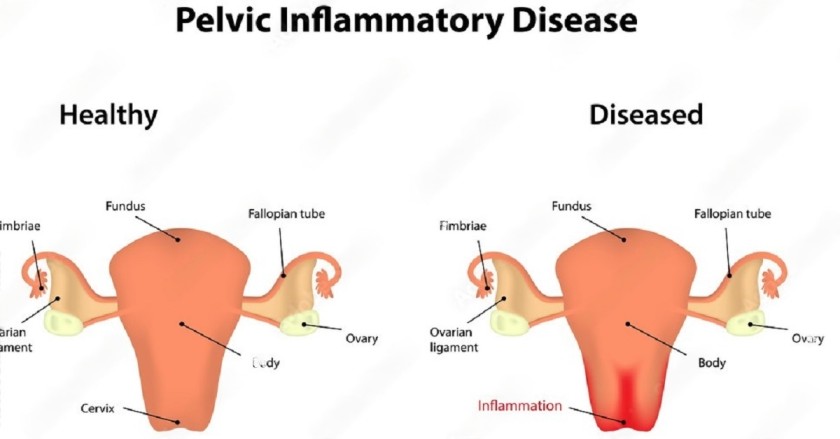
Pathophysiology of Pelvic Inflammatory Disease
Visual comparison: Normal female reproductive anatomy versus ascending infection showing severe Salpingitis and Oophoritis.

Clinical Manifestations (Signs and Symptoms)

The clinical presentation of PID can vary from mild, vague discomfort to an acute, life-threatening acute abdomen. Diagnosis relies heavily on detailed history-taking and physical examination.

1. On History Taking (What the Patient Reports)

  • Severe Lower Abdominal Pain: Approximately 70% of individuals with PID experience intense, continuous pain in the lower abdomen. It can be localized (one side) or diffuse (spread out). It heavily worsens during movement, walking, or sexual activity (dyspareunia).
  • Acute Fever: Around 40% of PID cases present with a high fever (often >38°C) accompanied by chills, responding to the massive internal infection.
  • Purulent Vaginal Discharge: About 90% of PID patients complain of a heavy, purulent (pus-like) vaginal discharge. It is often thick, yellowish or greenish, and foul-smelling.
  • Menstrual Changes: Severe inflammation disrupts normal hormonal and uterine function leading to:
    • Dysmenorrhea: Extremely painful menstrual periods.
    • Menorrhagia: Heavy or abnormally prolonged bleeding.
    • Oligomenorrhea: Infrequent, irregular, or scanty periods.

2. On Physical Examination (What the Midwife/Doctor Finds)

  • Signs of Inflammation: The pelvic region may feel unusually warm, swollen, and look reddened externally.
  • Peritoneal Signs: Abdominal palpation reveals peritonitis signs: Guarding (involuntary tensing of abdominal muscles to protect inflamed organs), Abdominal distension, and Rebound tenderness (sharp pain felt when the examiner's pressing hand is quickly released).
  • Vaginal Examination (Speculum & Bimanual):
    • Vaginal Changes: The vaginal walls appear red, highly inflamed, and dry due to ongoing local irritation.
    • Purulent Discharge: Pus is visibly seen escaping from the cervical os.
    • Cervical Excitation (Chandelier Sign): Extreme pain when the cervix is gently moved side to side during a bimanual exam.
    • Tender Fornices: Palpation of the vaginal fornices reveals extreme tenderness and possibly a swollen mass, indicating pyosalpinx (accumulation of pus in the Fallopian tubes).

    Specific Pelvic Infections Breakdown

    PID is an umbrella term. Depending on exactly where the infection settles, it is medically divided into the following specific inflammatory conditions:

    Condition Definition Specific Signs & Symptoms
    Cervicitis Inflammation of the cervix (the lower part of the uterus opening into the vagina). Often caused by Chlamydia/Gonorrhea.
    • Red, swollen cervix.
    • Cervical friability: Slight bleeding on intercourse or contact.
    • Itching/burning sensation in the vagina.
    • Watery, yellowish, or greenish abnormal discharge.
    Endometritis Inflammation of the endometrium (the inner lining of the uterus). Often occurs postpartum, post-abortion, or post-IUD insertion.
    • Enlargement of the uterus: Feels boggy, tender, and larger than normal.
    • Foul-smelling, offensive lochia/discharge.
    • Lower abdominal pain and high fever.
    Salpingitis Inflammation of one or both fallopian tubes. It is a highly destructive complication causing tubal blockage.
    • Severe lower abdominal or lower back pain.
    • Dyspareunia: Deep pain during sexual intercourse.
    • Risk of forming a pus-filled tube (pyosalpinx).
    Oophoritis Inflammation of one or both ovaries. Often occurs in conjunction with salpingitis (Salpingo-oophoritis).
    • Deep pelvic, abdominal, and back pain.
    • Dyspareunia.
    • Palpable, highly tender adnexal mass.

    Hospital Management of PID

    Because PID can permanently damage a woman's reproductive system, aggressive, immediate, and comprehensive management is required.

    1. Main Aims of Management

    • To strictly prevent permanent complications (like infertility and ectopic pregnancy).
    • To quickly relieve the intense pain.
    • To completely eradicate the infection and prevent the disease from spreading to the peritoneum or systemic bloodstream.

    2. Nursing & Supportive Care

    • Admission & Position: Admit the patient to a clean, well-ventilated gynecological ward for strict, complete bed rest. Place the patient in a Semi-Fowler’s position (propped up). Gravity assists in draining the purulent discharge downwards and prevents the pus from flowing upwards into the abdominal cavity (preventing generalized peritonitis).
    • Hydration: The patient is often febrile and dehydrated. Start an I.V. line immediately and administer intravenous fluids (like Normal Saline or Ringer's Lactate). Encourage high oral fluid intake if she can tolerate it without vomiting.
    • Histories & Examination: Take exhaustive medical and sexual histories. Conduct a comprehensive general and abdominal examination.
    • Continuous Observations: Monitor vital signs strictly (Temperature, Pulse, Respiration, Blood Pressure) every 4 hours. Daily, observe and accurately record the color, specific amount, and smell of the vaginal discharge on a fluid chart.
    • Diet & Elimination: Advise a highly nutritious, easily digestible diet. Provide a bedpan or urinal to maintain bed rest. Observe the urine; disinfect urine and feces with a strong hypochlorite solution (like JIK) before disposal to prevent cross-infection.
    • Hygiene & Exercise: Perform bed-making daily, removing wrinkles to prevent bedsores. Once the fever breaks and pain reduces, encourage light physical exercise (like walking around the ward) to prevent DVT.
    • Care of Mind (Psychotherapy): Severe pain and fears of infertility cause high anxiety. Reassure the patient and her relatives constantly. Provide diversional therapies like newspapers, TV, or radios.

    3. Diagnostic Investigations

    Do not delay initial treatment waiting for results, but perform these urgently:

    • High Vaginal Swab (HVS) / Endocervical Swab: Sent for Culture and Sensitivity (C&S) to exactly identify the causative bacteria (e.g., Gonorrhea).
    • Urinalysis: For C&S to rule out a concurrent severe Urinary Tract Infection (UTI).
    • Blood Tests: A blood culture for C&S to check for a hematogenous (bloodstream) source or septicemia. A malaria blood slide (BS) to rule out malaria as a cause of the high fever. Complete Blood Count (CBC) to check for highly elevated White Blood Cells (Leukocytosis).
    • Ultrasound Scan: A pelvic scan is critical to rule out other severe causes of acute abdominal pain (e.g., ruptured ectopic pregnancy, acute appendicitis, or the presence of a massive Tubo-Ovarian Abscess).

    4. Medical Treatment (Pharmacology)

    Treatment is started immediately using powerful broad-spectrum antibiotics before laboratory results return, covering both aerobic and anaerobic bacteria.

    • Initial I.V. Antibiotic Regimens:
      • Ceftriaxone 2g daily IV for 5 days.
      • Chloramphenicol 2g stat, then 1g 6-hourly for 5 days.
      • Gentamicin 160mg Once Daily (OD) for 5 days.
      • Metronidazole 500mg I.V. 8-hourly (TDS) for strict anaerobic coverage.
    • Oral Step-Down Therapy: If the fever subsides and discharge drastically reduces, switch to oral antibiotics based on sensitivity results. Common choices include Azithromycin (1g single dose for Chlamydia), Doxycycline (100mg BD for 14 days), Ciprofloxacin, or Tetracycline.
    • Analgesics: Severe pain requires powerful relief. Administer strong NSAIDs like Diclofenac or Ibuprofen to reduce both pain and local inflammation. In extremely severe cases, short-term narcotics (e.g., Pethidine) may be required. Paracetamol is used to control fever.

    ⚠️ Attention: Severe Complications of PID

    If PID is left untreated, inadequately treated, or if the patient presents too late, the irreversible destruction of pelvic tissues leads to life-altering complications:

    • Infertility: Severe inflammation leaves thick scar tissue blocking the fallopian tubes, preventing the sperm from reaching the egg.
    • Ectopic Pregnancy: The damaged, narrowed tubes may allow sperm up, but trap the larger fertilized embryo, causing it to grow and dangerously rupture the fallopian tube.
    • Pelvic Abscess & Tubo-Ovarian Mass: Massive collections of walled-off pus (abscesses) forming dangerous masses involving the tubes and ovaries.
    • Chronic Pelvic Pain & Adhesions: Thick scar tissue binds the pelvic organs and intestines together like glue, causing permanent, agonizing daily pain.
    • Intestinal Obstruction & Peritonitis: Severe infection spills into the abdominal cavity, paralyzing or blocking the bowels.

    Advice on Discharge

    A comprehensive counseling session must be held before the patient goes home to prevent recurrence:

    • Medication Compliance: Strictly complete the entire 14-day course of oral antibiotics, even if the pain has completely stopped.
    • Partner Treatment: Emphasize that all recent sexual partners MUST be tested and treated simultaneously to prevent the "ping-pong" re-infection cycle.
    • Safe Sex Practices: Strongly advise a reduction in multiple sexual partners. Advocate for the strict, consistent use of barrier methods (condoms).
    • Contraception Review: Advise avoiding Intrauterine Contraceptive Devices (IUDs) if she is at high risk for STIs. Discuss safer alternative methods.
    • Hygiene: Educate on proper perineal hygiene (wiping front to back) and strictly forbid vaginal douching.
    • Follow-up: Instruct the patient to return to the clinic immediately for review if she experiences a relapse of pain, abnormal discharges, itching, or fever.

    💡 Quick Practice Check

    Question: Why is a patient admitted with severe acute PID placed specifically in a Semi-Fowler’s (propped up) position in bed?

    Answer: To utilize gravity to help drain the toxic, purulent discharge downwards and out through the vagina, preventing the pus from tracking upwards and spilling into the abdominal cavity, which would cause a deadly generalized peritonitis.

    Endometritis

    Endometritis is a specific inflammatory condition characterized by the severe inflammation and infection of the endometrium (the innermost mucosal lining of the uterus).

    It can present as an acute infection (sudden and severe) or a chronic condition (long-lasting, often silent). It is one of the most common precursors to widespread Pelvic Inflammatory Disease (PID) if the bacteria are allowed to ascend further into the fallopian tubes.

    Causes and Major Risk Factors

    Endometritis is primarily caused by an ascending bacterial infection from the lower genital tract (vagina or cervix). The highly vascular, nutrient-rich lining of the uterus provides a perfect environment for rapid bacterial multiplication once the protective cervical barrier is breached.

    The condition is most commonly triggered by major obstetric or surgical events that introduce bacteria directly into the uterine cavity:

    • Postpartum (Childbirth): The most common cause. After delivery, the raw, open placental site acts as a massive wound. If bacteria enter during prolonged labour, frequent vaginal examinations, or unhygienic delivery practices, puerperal endometritis develops rapidly.
    • Post-Abortion: Induced or spontaneous abortions (miscarriages), especially if retained products of conception (tissue) are left behind in the uterus, provide a breeding ground for bacteria.
    • IUD Insertion: The physical insertion of an Intrauterine Contraceptive Device (IUD) can accidentally push pre-existing cervical bacteria (like Chlamydia or Gonorrhea) up into the sterile endometrium.
    • Gynecological Procedures: Invasive procedures such as Dilation and Curettage (D&C), hysteroscopy, or endometrial biopsies.

    Clinical Manifestations (Signs and Symptoms)

    A mother or patient presenting with endometritis will typically exhibit a combination of localized pelvic symptoms and systemic signs of infection. The midwife must carefully assess for the following:

    • Acute Fever and Chills: The patient will often present with a suddenly elevated body temperature (typically above 38.0°C or 100.4°F) accompanied by chills and tachycardia (fast heart rate). This is the body's aggressive systemic response to the bacterial infection inside the uterus.
    • Severe Lower Abdominal Pain: A hallmark symptom. The patient will complain of continuous, deep, dull, or cramping pain and extreme discomfort in the lower abdomen/pelvic region due to the severe swelling of the uterine lining.
    • Abnormal Vaginal Discharge: The infection produces heavy, purulent (pus-filled) vaginal discharge. In postpartum women, this manifests as highly offensive, foul-smelling lochia that may appear yellowish, greenish, or dark brown and prolonged.
    • Enlargement of the Uterus (Subinvolution): Upon abdominal palpation or bimanual examination, the uterus will feel extremely tender, "boggy" (soft instead of firm), and larger than expected. The severe inflammation prevents the uterine muscles from effectively contracting and shrinking back to their normal size.
    • General Malaise: The patient will look visibly ill, fatigued, and may experience anorexia (loss of appetite), nausea, or headaches due to the circulating bacterial toxins.

    ⚠️ Attention: The Danger of Untreated Endometritis

    Endometritis is a medical emergency. If the infection is not aggressively treated with strong intravenous broad-spectrum antibiotics, the bacteria will rapidly ascend out of the uterus and destroy the upper reproductive organs, leading to permanent, irreversible complications.

    Severe Complications of Untreated Endometritis

    Poorly managed or delayed treatment of endometritis sets off a chain reaction of destructive pelvic inflammatory diseases. The direct complications include:

    • Salpingitis: The infection spreads directly from the uterine lining into the fallopian tubes, causing severe inflammation and filling the tubes with pus (pyosalpinx).
    • Pelvic Abscess & Tubo-Ovarian Mass: Massive, dangerous pockets of pus form in the pelvic cavity, wrapping around the fallopian tubes and ovaries, often requiring emergency surgery to drain.
    • Infertility: The intense inflammation leaves behind thick, fibrous scar tissue that permanently blocks or damages the fallopian tubes, preventing sperm and egg from meeting.
    • Ectopic Pregnancy: Because the delicate inner hairs (cilia) of the fallopian tubes are destroyed by the infection, a fertilized egg may get stuck in the tube instead of reaching the uterus, leading to a life-threatening ruptured ectopic pregnancy.
    • Chronic Pelvic Pain & Adhesions: Severe scarring causes the pelvic organs (uterus, tubes, ovaries, and bowels) to stick together unnaturally, resulting in debilitating, constant pain that lasts for years.
    • Peritonitis: The infection completely spills out of the reproductive tract and deeply infects the entire lining of the abdominal cavity, a deadly condition.
    • Intestinal Obstruction: As pelvic adhesions and scar tissue form, they can wrap around and strangle the intestines, causing a partial or complete blockage of the bowels.

    💡 Quick Practice Check

    Question: A mother who delivered a baby 4 days ago presents to the clinic with a high fever, a soft/boggy uterus that is larger than expected, and highly offensive, foul-smelling lochia. What specific condition must the midwife suspect immediately?

    Answer: Puerperal Endometritis. The combination of fever, subinvolution (boggy, enlarged uterus), and foul-smelling discharge in the postpartum period are the classic triad of this dangerous uterine infection.

    Endometriosis

    Endometriosis is a chronic, often painful gynecological condition where tissue that behaves exactly like the lining of the uterus (the endometrium) grows outside the uterine cavity.

    Unlike Endometritis (which is an infection/inflammation of the inside lining), Endometriosis involves stray endometrial tissue implanting on organs such as the ovaries, fallopian tubes, the pelvic peritoneum, the Pouch of Douglas, and sometimes the bowel or bladder.

    Because this misplaced tissue responds to monthly ovarian hormones just like the normal uterine lining, it builds up, breaks down, and bleeds every menstrual cycle. However, because this blood has no way to exit the body, it becomes trapped, leading to severe inflammation, excruciating pain, scar tissue, and infertility.

    Causes and Theories of Origin

    The exact, definitive cause of endometriosis remains a medical mystery. However, several highly researched scientific theories explain how this tissue escapes the uterus:

    • Retrograde Menstruation (Sampson’s Theory): This is the most widely accepted theory. It suggests that during menstruation, some of the menstrual blood containing live endometrial cells flows backwards through the fallopian tubes and spills into the pelvic cavity, instead of leaving the body through the vagina. These stray cells then stick to the pelvic walls and organs, where they grow and thicken.
    • Cellular Metaplasia: This theory suggests that cells in the abdomen or pelvis retain their embryonic ability to transform (metaplasia) into endometrial cells under the influence of hormones or immune factors.
    • Lymphatic and Vascular Spread: Just like cancer cells, endometrial cells may travel through the blood vessels or the lymphatic system to reach distant parts of the body (which explains rare cases of endometriosis found in the lungs or brain).
    • Immune System Dysfunction: A normal immune system should recognize and destroy endometrial-like tissue growing outside the uterus. In women with endometriosis, the immune system fails to seek and destroy these rogue cells.
    • Surgical Scar Implantation: After surgeries involving the uterus, such as a Cesarean section (C-section) or a myomectomy, endometrial cells can accidentally attach to the surgical incision site (e.g., the abdominal wall scar).

    Clinical Manifestations (Signs and Symptoms)

    The primary symptom of endometriosis is pelvic pain, often specifically tied to the menstrual cycle. The severity of the pain does not always correlate with the extent of the disease (some women have severe disease but little pain, and vice versa).

    • Severe Dysmenorrhea: Excruciating pelvic pain and cramping that begins before the menstrual period and lasts several days into it. It is often described as feeling far worse than "normal" period cramps.
    • Dyspareunia: Deep pelvic pain during or immediately after sexual intercourse. The thrusting motion hits the tender, inflamed endometrial nodules tethered behind the uterus or in the vaginal fornices.
    • Chronic Pelvic Pain: A continuous, heavy, dull ache in the lower abdomen, lower back, and pelvis that happens even when not menstruating.
    • Dyschezia and Dysuria: Intense pain during defecation (passing stool) or urination, particularly during the menstrual period. This occurs when endometrial tissue implants on the bowel or bladder.
    • Menorrhagia and Menometrorrhagia: Experiencing excessively heavy menstrual periods or bleeding irregularly between periods.
    • Infertility: Endometriosis is diagnosed in up to 50% of women seeking treatment for infertility. The thick scar tissue (adhesions) distorts the pelvic anatomy, blocking the fallopian tubes and preventing the egg and sperm from uniting.

    ⚠️ Attention: The "Chocolate Cyst"

    When endometrial tissue implants deeply into an ovary, it bleeds into itself every month. Over time, this old, trapped, oxidized blood turns thick and dark brown, forming an Endometrioma, classically referred to in gynecology as a "Chocolate Cyst." If a chocolate cyst ruptures, it causes an acute, surgical abdominal emergency.

    Diagnosis of Endometriosis

    Because the symptoms heavily mimic other pelvic diseases (like PID or Ovarian Cysts), a definitive diagnosis requires specialized investigations:

    • Detailed Pelvic Examination: The healthcare provider may feel cysts on the reproductive organs or thick scars behind the uterus.
    • Transvaginal Ultrasound (TVUS): Highly effective for identifying specific endometrial cysts (endometriomas) on the ovaries, though it may miss smaller, flat implants.
    • Magnetic Resonance Imaging (MRI): Gives a much more detailed image to map out the exact locations and depths of the endometrial implants prior to surgery.
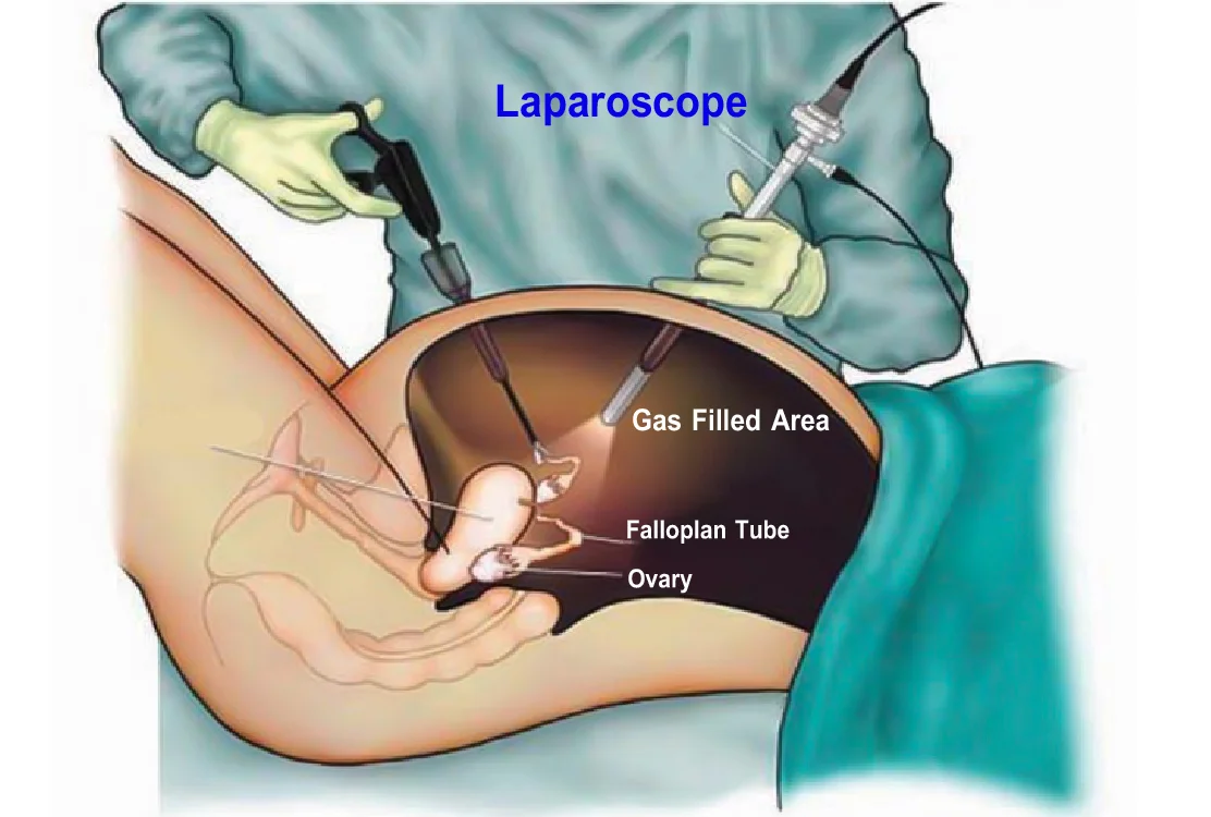
    • Diagnostic Laparoscopy (The Gold Standard): A tiny camera is inserted through a small abdominal incision. This is the only way to 100% definitively diagnose endometriosis. The surgeon can directly visualize the classic "powder-burn" lesions, take a biopsy for histology, and remove the implants simultaneously.

    Management and Treatment

    There is currently no absolute cure for endometriosis. Treatment is highly individualized based on the severity of symptoms, the extent of the disease, and whether the woman desires to have children in the future.

    1. Medical / Pharmacological Management

    The goal is to relieve pain and suppress the menstrual cycle, effectively starving the misplaced tissue of the hormones it needs to bleed and grow.

    • Analgesics: Non-steroidal anti-inflammatory drugs (NSAIDs) like Ibuprofen or Diclofenac are used aggressively to reduce inflammation and severe cramping.
    • Hormonal Contraceptives: Combined oral contraceptive pills (COCs), patches, or vaginal rings are used continuously to stop the hormonal fluctuations, thereby reducing or entirely stopping the bleeding and pain.
    • Progestin Therapy: Progestin-only pills, contraceptive implants, or the Mirena IUD halt the menstrual periods and prevent the growth of endometrial implants.
    • GnRH Agonists (e.g., Danazol, Zoladex): These powerful drugs block the production of ovarian-stimulating hormones, causing a temporary "medical menopause." This stops menstruation entirely, forcing the endometriosis to drastically shrink.

    2. Surgical Management

    • Conservative Surgery (Laparoscopy): For women trying to get pregnant or suffering severe pain. The surgeon carefully excises (cuts out) or burns away (ablates) the endometrial lesions and scar tissue while leaving the healthy uterus and ovaries perfectly intact.
    • Definitive Surgery (Hysterectomy): Used only as a last resort for women who have severe, uncontrollable pain and do not wish to have any more children. It involves the total removal of the uterus (Total Abdominal Hysterectomy), and frequently the removal of both ovaries and fallopian tubes (Bilateral Salpingo-Oophorectomy) to permanently stop all hormone production.

    3. Nursing Care and Psychotherapy

    • Provide extensive emotional support. Endometriosis causes immense psychological stress, depression, and anxiety due to chronic pain, dyspareunia (which damages relationships), and the devastating fear of infertility.
    • Educate the mother extensively on her medication compliance, especially the side effects of hormone therapies (like weight gain or menopausal flashes from GnRH agonists).
    • Encourage attendance at specialized fertility or chronic pain support groups.

    💡 Quick Practice Check

    Question: A 28-year-old woman is admitted to the gynecology ward complaining of severe, incapacitating pelvic pain exactly three days before her period starts, deep pain during sexual intercourse, and inability to conceive for 3 years. Her ultrasound reveals a large "chocolate cyst" on her left ovary. What is the most likely diagnosis?

    Answer: Endometriosis. The classic triad of severe dysmenorrhea, dyspareunia, and infertility, combined with an endometrioma (chocolate cyst) on the ovary, points definitively to endometriosis.

References

  • Ministry of Health (MOH) Uganda - National Clinical Guidelines for the Management of Common Conditions.
  • Berek, J. S. (Ed.). Berek & Novak's Gynecology. Wolters Kluwer.
  • Syllabus guidelines for Diploma in Midwifery Extension Program, Uganda Nurses and Midwives Council (UNMEB).

Quick Quiz

PID Quiz

Gynaecology - mobile-friendly and focused practice.

Privacy: Your details are used only for quiz tracking and certificates.

PELVIC INFLAMMATORY DISEASES (PID) Read More »

Infertility-Causes-Symptoms-Treatment
Home > DME 112: Gynaecology & Reproductive Health > Infertility

Infertility

Infertility refers to the failure or inability of a couple to conceive (get a child) after one full year of regular, unprotected sexual intercourse (coitus) during the childbearing age (15–49 years) without using any form of contraception.

Types of Infertility:

  • Primary Infertility: The inability to conceive in a couple that has had absolutely no previous pregnancies at any time after 1 year of unprotected sex.
  • Secondary Infertility: Refers to a couple who have previously succeeded in achieving at least one pregnancy (even if it ended in a spontaneous abortion/miscarriage) but are now unable to conceive again despite regular unprotected sex.

⚠️ Attention: Infertility vs. Sterility

The term Sterility should only be used when there is absolutely no possible treatment to enable a couple to conceive (achieve pregnancy). For example, when a man has no testes (anorchia) or a woman lacks a uterus (hysterectomy or congenital absence).

Conditions for Normal Implantation and Conception

For a pregnancy to successfully occur, a very specific chain of events must be perfectly executed. The following conditions must be fulfilled:

  • There should be two partners actively involved and willing.
  • They must engage in regular, unprotected coitus without any form of contraception.
  • They must use the right sexual route (vaginal penetration with ejaculation).
  • The female should be within the active childbearing age (14 to 49 years).
  • The male must release healthy semen containing a normal count of motile and morphologically normal spermatozoa.
  • A normal, healthy ovum (egg) must be released from the female's ovary (ovulation).
  • The ovum must unite with the sperms in the fallopian tube to be fertilized.
  • The fertilized ovum (zygote) must successfully travel to and implant in the healthy endometrium of the uterus.

Causes of Infertility in Males

Male factor infertility accounts for a significant portion of infertility cases. The causes range from psychological to severe physiological and genetic abnormalities:

  • Sperm Abnormalities: The release of immature sperms, structurally abnormal sperms, too few sperms (oligospermia), or sperms that are too slow in movement (asthenospermia).
  • Ejaculatory Issues: Poor ejaculation, premature ejaculation, or complete failure to ejaculate. Retrograde ejaculation is a specific condition where semen is ejaculated backward into the bladder instead of out through the urethra (assessed by urinalysis after ejaculation).
  • Extreme Heat: An increase in the temperature of the testes from a prolonged fever, tight clothing, or occupational exposure to excessive heat can greatly reduce sperm count, reduce the vigor of sperm movement, and increase the number of abnormal sperms in the semen.
  • Hydrocele: Excessive collection of fluid in the scrotum. This extra fluid creates pressure and alters the environment, preventing the adequate production of sperms.
  • Varicocele: Varicose (swollen and enlarged) veins of the scrotum. This abnormality prevents the proper supply and drainage of blood from the testes, thus raising the scrotal temperatures and drastically reducing the rate of sperm production. It also affects the quality of ejaculation.
  • Medications & Drugs: Certain drugs cause failure in erection or spermatogenesis. Examples include amoebicides (for amoebiasis), anti-hypertensives like Aldomet (Methyldopa), and diabetic drugs. Nitrofurantoin (an antibiotic) can cause degenerative changes in sperms.
  • Infections: Diseases like Mumps in adult males can cause Orchiditis (severe inflammation of the testes), leading to permanent testicular damage and sterility.
  • Hormonal Imbalance: Inadequate production of testosterone from the Leydig cells can result in immature sperms and poor libido.
  • Lifestyle Factors: Excessive smoking, chronic alcohol consumption, severe obesity, and extreme depression or stress heavily inhibit spermatogenesis and sexual performance.
  • Toxic Exposure: Chronic exposure to toxic industrial chemicals, heavy metals, or radiations physically damages the germinal epithelium of the testes, halting spermatogenesis.

Genetic Factors in Males

  • Klinefelter’s Syndrome: A chromosomal genetic syndrome where a male has 47 chromosomes with an extra X chromosome (47, XXY instead of the normal 46, XY). The loss of a dominant Y-chromosome effect leads to a feminine body build. It presents with gynaecomastia (enlarged breasts), testicular atrophy (shrinking testes), azoospermia (zero sperms), and complete infertility. The testicular atrophy is a result of peritubular fibrosis commencing in childhood and progressing until all seminiferous tubules are replaced by useless fibrous tissue.

Causes/Factors of Infertility in Females

Female infertility is deeply complex and is clinically best discussed under the following distinct headings:

1. Defective Implantation & Tubal Factors

Tubal blockage is the major cause of infertility, especially in Uganda, contributing to 60 – 70% of cases.

  • Pelvic Inflammatory Disease (PID) & Salpingitis: Infection of the fallopian tubes caused by Gonorrhoea, Chlamydia, Tuberculosis, or pelvic peritonitis (e.g., from a burst appendix) after an abortion or delivery. This damages the delicate ciliated tubal epithelium. In severe cases, it causes total tubal blockage. This mostly occurs at the outer end of the tube where the fimbriae adhere together, but it is also seen in the very narrow, interstitial part of the tube.
  • Ectopic Risks: When the tubes are not completely blocked, fertilization of the ovum may still take place. However, because of the damage to the ciliated epithelium, the fertilized ovum cannot be carried down the tube to the uterus, resulting in a dangerous Ectopic Pregnancy.
  • Uterine Abnormalities: Congenital malformations such as having no uterus, a bicornuate uterus (heart-shaped), or a Didelphys uterus (two completely separate horns and cervixes).
  • Uterine Fibroids: Gross fibroids create an irregular, bumpy implantation surface or physically block the fallopian tube ostia.
  • Uterine Synechiae (Asherman's Syndrome): Severe inflammation of the endometrium (endometritis) or scarring caused by over-curetting of the uterus during a D&C, or from surgeries like myomectomy, leads to the walls of the uterus sticking together (intrauterine adhesions).
  • Endometriosis: A painful condition where patches of endometrial-like tissue develop outside the uterine cavity in abnormal locations (ovaries, fallopian tubes, abdominal cavity). This tissue grows and bleeds with hormonal stimulation, causing massive inflammation, scar tissue, pelvic adhesions, and infertility.
  • Cervical & Vaginal Issues: Stenosed (narrowed) cervix due to trauma/surgery. Acquired or congenital Gynaeatresia (a very small vaginal hole with a blind end), septate vagina, or a completely rigid hymen preventing penetration.
  • Infections & Contraceptives: Severe Trichomonas vaginalis infections (evidence of cause noted in Zambia). Prolonged use of barrier contraceptives (condoms, IUDS, Spermicides) without recognizing underlying fertility delays.

2. Endocrine (Hormonal) Disorders

  • Hypothalamic-Pituitary Inefficiency: Stress, extreme weight loss, or excessive weight gain causes an unstable cerebrum and hypothalamus. This alters dopamine or noradrenaline transmission. Certain drugs (Rawolfia, phenothiazines, Metoclopramide) also suppress hypothalamic function, leading to failure of ovulation.
  • Hyperprolactinaemia: A pituitary tumor (micro or macroadenoma) leading to the production of excessive Prolactin hormone. Elevated prolactin is normal during lactation (causing lactational amenorrhea), but if a woman is not lactating, this hormone suppresses GnRH, causing total anovulation.
  • Thyroid & Adrenal Function: Hyperthyroidism, Hypothyroidism, Cushing's syndrome, or Congenital Adrenal Hyperplasia all cause severe hormonal imbalances resulting in anovulation.
  • Age & Menopause: Fertility steeply declines with advanced age. Postponement of childbearing for careers frequently leads to age-related ovulatory decline.

3. Ovarian Causes

  • Resistant Ovary Syndrome: Absence of FSH receptors in the ovarian follicle leads to the complete failure of the ovary to respond to gonadotrophins.
  • Polycystic Ovarian Syndrome (PCOS): Disturbance in the interaction between FSH and the follicle results in an abnormal enzyme reaction. The follicles fail to rupture (anovulation), and the ovary becomes enlarged and multicystic. Without ovulation, the endometrium lacks secretory changes, and cervical mucus remains thick and impenetrable by sperms.
  • Ovarian Damage/Absence: Congenital lack of eggs/follicles, premature menopause, surgical removal of the ovary by mistake, or destruction of the ovarian substance by deep infections like Mumps.
  • Radiation Exposure: Chronic or excessive exposure to radioactive substances or X-rays physically damages and mutates the delicate ova.

4. Defective Transport & Hostility

  • Cervical Hostility: A condition in which the cervical mucus is totally unreceptive to spermatozoa. The mucus either blocks their progressive advance or actively kills them. This may be due to chronic cervical infection or the presence of specific Sperm Antibodies (an allergic immune response to the man's sperms).
  • Vaginal pH: A highly acidic vaginal pH rapidly destroys the motility of the sperms before they can enter the protective cervix.

5. Physical, Psychological & Systemic Causes

  • Psychological & Sexual Dysfunction: Dyspareunia (severe painful sexual intercourse due to psychological or physical factors) and Vaginismus (involuntary spasms of the pelvic floor muscles preventing penetration).
  • Wrong Timing: Having sexual intercourse exclusively during infertile periods (miscalculating the ovulation window).
  • Systemic Diseases: Chronic, uncontrolled diseases such as Diabetes Mellitus, severe Hypertension, and Renal Failure drastically lower fertility chances.
  • Turner’s Syndrome (Genetic): Caused by the complete deletion or abnormality in one of the female's two X chromosomes (Karyotype 45, XO). These females present with complete ovarian failure (streak ovaries), widely spaced nipples, cardiovascular problems, squints, hypothyroidism, and diabetes mellitus. They face absolute infertility and require estrogen therapy in adult life.

General Investigations & Evaluation

All couples complaining of infertility must be investigated together. The length and depth of the investigations will vary depending on the initial findings.

Evaluation in Women

1. Comprehensive History & Physical Examination

  • Menstrual History: Age of menarche, length, and regularity of the menstrual cycles.
  • Gynaecological History: Previous contraceptive use, history of D&C, salpingectomy, abortions, or highly suggestive symptoms of PID.
  • Obstetric History: Any previous pregnancies and the exact number of children fathered specifically by her current partner.
  • General Health: Extremely thin women face amenorrhoea due to lack of body fat. Very obese women face anovulation because the ovary cannot secrete enough estradiol, converting androgens into excess estrone instead. Age is strictly noted (above 50 is generally menopause).
  • Physical Inspection: Check for visual field deficits (suggestive of a large pituitary tumor pressing on the optic chiasm). Check pubic and body hair distribution (presence of male-pattern hair / virilism suggests excess androgens).
  • Vaginal Examination: Check the normality of the vaginal canal, cervix, and uterus. Confirm pelvic anatomy with an ultrasound.

2. Hormonal & Special Tests

  • Progesterone Levels: In a standard 28-day cycle, blood is drawn on Day 21 (or 1 week before the expected period). A progesterone level of more than 20 mmol/L firmly confirms that ovulation took place.
  • FSH and LH Levels: Checked to identify premature menopause, ovarian failure, or polycystic ovaries.
  • Basal Body Temperature (BBT): The woman takes her oral temperature every morning upon waking, before any physical activity. A sustained rise of about 0.5°C in the last 14 days of the cycle indicates ovulation has occurred. Monitored for 6 months.
  • Cervical Mucus Examination: Mid-cycle ovulatory mucus is clear, copious, and stretches into a fine thread (called Spinnbarkeit). On a glass slide, it dries to form a characteristic fern pattern under the microscope.
  • Post Coital Test (Huhner’s Test): Carried out exactly at ovulation. 2 to 8 hours after unprotected intercourse, cervical mucus is withdrawn using a wire loop/pipette, placed on a warm slide, and examined. Normally, a large number of progressively motile sperms are seen. Absence or dead sperms indicate cervical hostility or sperm antibodies.
  • Prolactin Tests: If prolactin is higher than 800 mU/L, a CT scan of the pituitary fossa is mandatory to exclude a prolactin-producing adenoma.
  • Endometrial Biopsy: Done 10-12 days after ovulation. Histology will prove if the glands have undergone necessary secretory changes to support an embryo.

3. Tubal Patency Tests

  • Laparoscopy & Dye Test: The gold standard. A premenstrual procedure combining visual inspection of the pelvis with the injection of dilute methylene blue dye through the cervix. The uterus distends, and if the tubes are patent (open), the dye freely spills from the fimbrial ends. No spill indicates a block. (Pregnancy must be ruled out first).
  • Hysterosalpingogram (HSG): An opaque radio-aqueous solution is injected through the cervix. An X-ray is taken to visualize the uterine cavity and tubal patency. This is done between day 5 and 10 of the cycle (after bleeding stops but before ovulation).
  • Tubal Insufflation: An older, highly unreliable method where Carbon Dioxide (CO2) gas is passed into the uterus. Patency is assumed if the gas is heard via auscultation or confirmed on X-ray.
  • Transvaginal Ultrasound (TVS): Gives clear images of the ovaries and uterus. Contraindications: Suspected pregnancy, severe cervical erosion, active pelvic infection, DUB, or serious heart/lung diseases. Risks: Embolism or ascending infection.

Evaluation in Men

  • General Exam: Check for severe obesity, diabetes, and hypertension. Evaluate male hair distribution and genital development. Check for surgical scars (e.g., undescended testis operation before puberty).
  • Breast Check: Enlargement (gynaecomastia) indicates abnormally high estrogen levels or Klinefelter's syndrome.
  • Testicular Exam: Assess the exact size, consistency, and situation of the testes in the scrotum.
  • Hormonal Blood Tests: Evaluate FSH, LH, and Testosterone levels to rule out endocrine failure.

🧠 Terminology for Seminal Fluid Analysis

Azoospermia: Absolute lack of sperms in the semen (Zero).
Oligospermia: Little or few sperms (Less than 20 million/ml).
Asthenospermia: Decreased motility (slow-moving sperms).
Teratospermia: Excessive abnormal shapes (monsters/mutations) of the sperms.

Normal Seminal Fluid Analysis Findings

A fresh semen sample is analyzed. Normal parameters must meet or exceed the following:

  • Volume: ≥ 2.0 to 2.5 ml.
  • pH: 7.0 to 8.0 (Slightly alkaline to survive the acidic vagina).
  • Total Sperm Count: More than 20 million per ml.
  • Liquefaction: Must be completely liquefied within 1 hour.
  • Motility: ≥ 50% must have strong, forward, progressive motility.
  • Morphology: ≥ 30% or more must have a perfectly normal shape.

Treatment of Infertility

Treatment is strictly tailored according to the identified underlying cause.

General Preventive Measures & Counseling

  • Stop all smoking and significantly reduce alcohol consumption.
  • Adopt a proper, highly nutritious diet to manage weight (obesity or extreme thinness).
  • Educate the couple on meeting at the exact right time (tracking the fertile ovulation window).
  • Actively reduce psychological stress, marital tension, and provide deep psychological counseling to prevent depression, divorce, or polygamy.

Treatment in Women

1. Chemotherapy for Anovulatory Infertility

  • Clomiphene Citrate (Clomid): Induces ovulation by stimulating the Hypothalamic-Pituitary system.
    • Dosage: 50mg daily for 5 days, starting on the 2nd day of menstruation. If ovulation fails, a second course of 100mg daily for 5 days is given (starting as early as 30 days later). Generally, 3 courses are adequate to assess efficacy.
    • Side Effects: Ovarian Hyperstimulation Syndrome (OHSS), multiple pregnancies (twins/triplets), visual disturbances, hot flushes, heavy abnormal bleeding (menorrhagia), hair loss, depression, dizziness, and nausea.
  • Tamoxifen: 20mg daily on days 2, 3, 4, and 5 of the cycle. Dose may be increased to 40mg then 80mg to stimulate ovulation.
  • Bromocriptine (Parlodel, Dopagon): For hyperprolactinaemic infertility. It inhibits the synthesis and release of prolactin from the pituitary.
    • Dosage: Initially 1.25mg at bedtime, gradually increased to 2.5mg three times a day with food. Max 30mg daily.
    • Side Effects: Severe nausea, hypotension, dizziness, nasal congestion, fatigue, and dry mouth.
    • Interactions: Erythromycin increases its toxicity. Antipsychotics and Metoclopramide antagonize its effect.
  • Luteinizing Hormone (LH) / GnRH Pumps: Given via a subcutaneous/IV syringe pump releasing 10-25 micrograms every 90 minutes.
  • HCG & HMG (Pregnyl / Metrodin): Synthetic Human Chorionic Gonadotrophin or Human Menopausal Gonadotrophin triggers ovulation when Clomiphene fails.

2. Surgical Measures in Women

  • Salpingolysis: Dividing peritubal adhesions around the ampullary ends of the tubes to restore free movement.
  • Salpingostomy: Surgically turning back the fimbriae to create a brand new opening for a blocked tube.
  • Tubal Anastomosis & Repair: Coring out the blocked segment (e.g., at the isthmus) and intricately re-stitching (anastomosing) the healthy ends back together.
  • Myomectomy: Surgical removal of uterine fibroids to clear the implantation space.

Treatment in Men

  • Medical: Human Gonadotrophin Therapy or Clomiphene Citrate to forcibly stimulate sperm production. Testosterone therapy to stimulate sexual desire (libido) — Note: Do not give exogenous testosterone if the primary issue is impaired spermatogenesis, as it can suppress natural sperm production further.
  • Surgical: Repair of inguinal hernias, correction of undescended testis, or Varicocelectomy (surgical ligaturing of the internal spermatic vein to reduce testicular heat and pooling). Relief of reproductive tract obstructions.

Assisted Reproductive Technologies (ART) & Other Options

In Vitro Fertilization (IVF)

Developed in 1978 (Robert Edwards won the Nobel Prize for this). IVF bypasses blocked fallopian tubes by fertilizing the egg outside the body.

  • Process: The woman's ovaries are hyper-stimulated using GnRH agonists and FSH to produce multiple eggs. An HCG "trigger shot" is given. Eggs are retrieved via ultrasound-guided transvaginal aspiration exactly between 34 and 36 hours (just before follicle rupture at 38-40 hrs).
  • Fertilization: The retrieved eggs are mixed with purified, washed sperms (to remove microbes/HIV) in a laboratory dish.
  • Transfer: The healthiest resultant embryos are carefully transferred directly into the mother's uterus. (Caution: Often results in multiple pregnancies, and ovarian hyperstimulation must be monitored).

Intracytoplasmic Sperm Injection (ICSI)

Performed alongside IVF for severe male infertility. If sperms are very low in number, highly abnormal, or unable to penetrate the egg wall, a single healthy sperm is captured in a microscopic needle and injected directly into the cytoplasm of the egg.

Other Alternatives

  • Surrogate Mothers: If a woman lacks a functional uterus, her own IVF embryo (using her egg and husband's sperm) is implanted into another woman's uterus. The surrogate carries the pregnancy and hands the baby over to the genetic parents after birth.
  • Artificial Insemination by Donor (AID): If the male partner is completely sterile (azoospermia), semen from a healthy, anonymous fertile donor is introduced into the woman's reproductive tract during ovulation.
  • Adoption: Couples can legally apply to an adoption center to adopt a child of their choice if medical treatments fail.

Nursing Diagnoses & Care for Infertility

The psychological toll of infertility is massive. The midwife/nurse plays a crucial role in providing holistic care:

  • Anxiety and Fear: Related to unknown invasive diagnostic procedures, complex treatments, and unpredictable outcomes, evidenced by the patient’s constant verbalization of worry. (Intervention: Provide clear, empathetic explanations of all procedures).
  • Low Self-Esteem & Grief: Related to the inability to conceive, evidenced by low mood, crying, feeling "inadequate," and social isolation. (Intervention: Offer therapeutic communication, refer to support groups).
  • Knowledge Deficit (Reproductive Process): Related to the complex process of ovulation, fertile windows, and proper sexual relationship timing, evidenced by inadequate verbalization. (Intervention: Teach BBT charting, fertile period calculations, and sexual health).
  • Knowledge Deficit (Anatomy/Causes): Related to the physical causes of infertility, evidenced by misconceptions. (Intervention: Use anatomical charts to explain tubal blockages, sperm counts, etc.).

💡 Quick Practice Check

Question: A male patient's semen analysis returns with a sperm count of 8 million/ml and highly reduced motility. What are the correct medical terms for these specific findings?

Answer: A count below 20 million/ml is termed Oligospermia, and the decreased motility is termed Asthenospermia.

References

  • Myles, M. (Latest Edition). Textbook for Midwives. Churchill Livingstone Elsevier.
  • Uganda Ministry of Health. (2001). Midwifery Handbook and Guide to Practice (11th ed.). Kampala, Uganda.
  • Stanfield, P., Beladia, & Versluys. (2004). Child Health: A Manual for Medical and Health Workers in Health Centres and Rural Hospitals (2nd ed.). English Press Limited, Nairobi.
  • Local Clinical Guidelines and Training Manuals for the Diploma in Midwifery Extension Program (UNMEB).

Quick Quiz

Infertility Quiz

Gynaecology - mobile-friendly and focused practice.

Privacy: Your details are used only for quiz tracking and certificates.

Infertility Read More »

Ectopic Pregnancy
Home > Midwifery I

Ectopic Pregnancy

Overview: An Ectopic Pregnancy is a life-threatening obstetric emergency where a fertilized egg (ovum) implants and grows completely outside the normal uterine cavity. Because these areas cannot stretch or provide enough blood supply like the uterus, the pregnancy cannot develop properly and often results in a dangerous rupture, causing severe internal bleeding.

1. Definition and Causes

Normally, a fertilized egg travels down the fallopian tube and embeds safely inside the thick lining of the uterus. In an ectopic pregnancy, the fertilized ovum embeds outside the uterine cavity.

Causes and Risk Factors

Any condition that slows down or prevents the fertilized egg from moving smoothly down the fallopian tube can cause an ectopic pregnancy.

  • Pelvic Inflammatory Disease (PID): Infections like salpingitis (inflammation of the tubes) destroy the tiny hair-like structures (cilia) inside the tube. PID also forms sticky scar tissue (adhesions) that stop the normal wave-like squeezing motions (peristalsis) of the tubes.
  • Fallopian Tube Damage & Congenital Issues: Previous infections or surgeries can leave behind thick scars. Some women are also born with abnormally long tubes that easily kink (bend), or congenitally narrow tubes, making it hard for the egg to pass.
  • Previous Ectopic Pregnancy: A woman who has already suffered one ectopic pregnancy is at a much higher risk of having another one because the underlying tube damage is usually still present.
  • Endometriosis: This is the abnormal growth of the uterine lining (endometrium) in places outside the uterus. The fertilized egg may accidentally implant on this misplaced tissue.
  • Repeated Induced Abortions: Multiple scraping procedures can introduce infections or cause scarring that damages the reproductive pathways.
  • Tubal Surgery: Surgical operations on the fallopian tubes (like tying them or attempting to unblock them) create scars inside (intraluminal) or outside (extraluminal) the tubes.
  • Intrauterine Devices (IUDs): While IUDs are great at preventing normal pregnancies inside the uterus, if a pregnancy *does* accidentally happen with an IUD in place, it is highly likely to be ectopic because the device blocks the normal implantation site.
  • Tumors: Large growths, like fibroids or ovarian cysts, can press hard against the outside of the fallopian tube, causing a partial or complete physical blockage.
  • Hormonal Factors: Imbalances in hormones can slow down the movement of the egg and affect how quickly it implants.
  • Reproductive System Abnormalities: Being born with a misshapen uterus (like a bicornuate uterus) or abnormally placed tubes.

2. Sites of Ectopic Pregnancy

An ectopic pregnancy can happen in several places, but the Fallopian Tubes are by far the most common site (accounting for about 95% of cases). Other rare sites include the broad ligament, the ovary, the cervix, and the intraperitoneal abdominal cavity.

Specific Sites for Tubal Pregnancy

  • Ampulla (Most Common): The widest part of the fallopian tube where fertilization usually occurs. The egg often gets stuck here.
  • Isthmus (Most Dangerous): The narrowest part of the tube. Because it is so small and tight, a growing pregnancy here will cause the tube to burst (rupture) very early, sometimes even before the mother misses her period or realizes she is pregnant.
  • Fimbriated End (Infundibulum): The finger-like ends of the tube (rare site).
  • Interstitial Part: The section of the tube that crosses through the thick uterine wall (rare but causes massive bleeding if ruptured).

3. Possible Outcomes of a Tubal Pregnancy

If the egg implants in the tube, the pregnancy cannot last to term. One of four things will eventually happen:

Outcome Clinical Description
1. Tubal Mole The fertilized egg (zygote) dies quietly. It is retained inside the fallopian tube, completely surrounded by a thick blood clot. This may result in a slow-leaking, chronic ectopic pregnancy with mild, lingering pain.
2. Tubal Abortion The zygote completely detaches from the wall of the fallopian tube and is pushed out (expelled) through the open fimbriated end into the abdomen. It may die and be absorbed, or very rarely, it can stick to abdominal organs and become an abdominal pregnancy.
3. Tubal Rupture The growing baby quickly becomes too large for the tiny tube. The tube violently bursts open, tearing major blood vessels. This causes massive internal bleeding straight into the abdominal cavity, leading to sudden, severe hypovolemic shock.
4. Tubal Erosion The aggressive cells of the growing zygote slowly eat through (erode) the wall of the fallopian tube, causing a steady leakage of blood into the abdominal cavity.

⚠️ Clinical Emergency: Tubal Rupture

A ruptured ectopic pregnancy is a top-tier obstetric emergency. The mother can bleed to death internally in a matter of hours. Always suspect an ectopic pregnancy in any woman of childbearing age presenting with abdominal pain and fainting, even if she claims she is not pregnant.

4. Signs and Symptoms (Focusing on Tubal Rupture)

On History Taking

  • Amenorrhea: A clear history of missing her periods for 6 to 10 weeks.
  • General Danger Signs: The patient heavily complains of feeling faint, extreme dizziness, severe thirst (a classic sign of internal bleeding), and vomiting.
  • Acute Pain: She reports sharp, sudden, colicky abdominal pain localized in one side of the lower abdomen (iliac fossa).
  • Referred Shoulder Pain: The mother complains of pain at the tip of her shoulder, especially when she lies down flat. This happens because the free blood pooling in her abdomen irritates the diaphragm muscle and the phrenic nerve, which shares a pain pathway with the shoulder.

On Physical Examination

  • Signs of Pregnancy: Early signs may be visible, such as darkening of the breast areola.
  • Signs of Shock: Because of massive internal bleeding, she will have cold and clammy skin, a rapid but very weak (thready) pulse, dangerously low blood pressure, and a slightly low temperature.
  • Mental State: She appears highly anxious, restless, and terrified (air hunger).
  • Pallor: Extreme whiteness (pallor) of the eyes, gums, and mucous membranes due to blood loss.

On Abdominal Palpation

  • Extreme tenderness, especially on the affected side.
  • The abdominal muscles become hard and rigid like a board because the mother is unconsciously guarding against the severe pain.
  • The abdomen is visibly swollen (distended) due to the large amount of free blood floating in the abdominal cavity.

On Vaginal Examination

  • Bleeding Discrepancy: The small amount of bleeding seen coming from the vagina absolutely does not match the mother's severe state of shock (because the real bleeding is hidden inside the abdomen).
  • Cervical Excitation: Moving the cervix slightly during the exam causes her extreme, jumping pain.
  • A distinct, painful swelling or mass can be felt on one side (lateral fornix) or bulging down behind the uterus in the Pouch of Douglas.
  • Dark brown, old blood may be seen on the examining finger.

5. Investigations & Differential Diagnosis

  • Ultrasound Scan: This is the gold standard. It will confirm the diagnosis by revealing an empty uterus, a mass in the fallopian tube, and a large collection of free fluid/blood in the abdomen.
  • Pregnancy Test: A urine or blood hCG test will be positive, proving she is pregnant.
  • Blood Tests: Check Hb (Hemoglobin level will be very low due to bleeding), Grouping, and Cross-matching (to prepare for immediate blood transfusion).
  • Culdocentesis (Emergency Puncture): If a scan is not available in a rural setting, a doctor may insert a needle through the top of the vagina into the Pouch of Douglas. If fresh, non-clotting blood is drawn out (aspirated), it proves there is internal bleeding from a rupture.

❓ Differential Diagnosis

Before confirming an ectopic pregnancy, a midwife or doctor must rule out other conditions that cause severe abdominal pain in women, including:

  • Salpingitis / PID: Specially if associated with foul discharge and irregular menses.
  • Appendicitis: Right-sided pain, often with a fever, but no missed period.
  • Abortion (Miscarriage): Will have heavy vaginal bleeding matching the pain level.
  • Twisted Ovarian Cyst: Sudden, severe pain, but pregnancy test is usually negative.
  • Urinary Tract Infection (UTI): Pain on urination, usually no signs of severe shock.

6. Management at a Health Centre (Primary Care)

At a lower-level health center (Level III or IV), this is a critical emergency. Every second counts to save the mother's life. Do not delay.

Aims of Care: To prevent deep shock, relieve severe pain, reassure the terrified patient, and transfer her safely and immediately.

  • Admission: Temporarily admit her to the gynecological or emergency bed. Quickly take a brief history and do a rapid physical, abdominal, and vaginal examination to make a diagnosis.
  • Observations: Immediately take and record Temperature, Pulse, Respiration, and Blood Pressure to assess how badly her vital organs are failing.
  • Resuscitation (Treatment):
    • Put up an incredibly fast intravenous (IV) drip of Normal Saline to elevate her dropping blood pressure and treat the shock.
    • Administer strong pain relief as prescribed (e.g., Morphine 15mg intramuscularly) to stop pain from worsening the shock.
    • Raise the foot of the bed (Trendelenburg position) to force whatever blood is left to flow to her vital brain and heart centers.
  • Nursing Care: Swab the vulva quickly and apply a clean pad to monitor exactly how much vaginal blood is lost during transit.
  • Transport and Transfer: Inform the patient and her relatives honestly about the danger and the immediate need for surgery. Send for an ambulance or vehicle immediately. Write a detailed referral note stating the time of arrival, her exact condition, and what drugs/IV fluids you have given. A qualified midwife MUST escort the mother to monitor the IV drip and hand her over safely to the hospital staff.

7. Hospital Management

Upon arrival at a main hospital, this is handled as a top-priority gynecological emergency. All nurses and doctors must work as a rapid-response team to get the patient to the operating theatre as fast as possible.

Aims: To treat anemia, reverse shock, reassure the patient, completely stop the internal bleeding, and prevent further complications.

Admission and Immediate Care

  • Admit the patient into a warm, well-ventilated room. Establish a fast but comforting nurse-patient relationship.
  • Take a collateral history from the relatives if the mother is too weak to speak. Focus on exactly when the pain started, vomiting, and missed periods.
  • Call the doctor or surgeon immediately.
  • Do a rapid head-to-toe examination focusing on signs of extreme dehydration, anemia, and hypovolemic shock.

Doctor's Orders and Resuscitation

  • Investigations Ordered: Hb estimation (to see blood lost and rule out malaria), Blood Group & Crossmatch (transfusion is almost always necessary), Pregnancy test, Ultrasound scan, and Urinalysis.
  • IV Fluids & Fluid Balance: Run Normal Saline rapidly. Start a strict fluid balance chart (input vs. output).
  • Blood Transfusion: Book 1 to 2 units of blood immediately based on the Hb results.
  • Pain Relief: Administer strong analgesics like Morphine or Pethidine.

8. Pre-Operative, Theatre, and Post-Operative Care

Pre-Operative Care (Preparation for Theatre)

  • Explain the life-saving nature of the operation and obtain a signed Informed Consent form from her or her husband.
  • Give a quick bed bath, dress her in a clean theatre gown, and inform the theatre staff to prepare.
  • Ensure the IV line is running perfectly. Swab the vulva to prevent taking infections into theatre.
  • Pass a urethral catheter to empty the bladder (an empty bladder is out of the surgeon's way and prevents accidental injury).
  • Pass a Naso-Gastric (NG) tube to empty stomach contents, or give an antacid (like Magnesium Trisilicate) to make stomach acid safe. This prevents the mother from vomiting and breathing acid into her lungs while unconscious.
  • Give premedication like Atropine to dry up mouth and lung secretions.
  • Take one last set of vital signs, gather all her medical charts, and carefully wheel her to theatre, giving a full verbal handover to the theatre nurse. Stay with her until she is asleep if possible.

🩸 What happens in Theatre? (Auto-transfusion)

The surgeon performs a Laparotomy (opening the abdomen) and a Salpingectomy (cutting out the burst fallopian tube). The bleeding is clamped and tied off. If the bleeding just happened and the blood in her stomach is totally fresh, the surgeon can scoop it up, filter (sieve) it, mix it with an anti-clotting chemical (Sodium Citrate), and give her own blood back to her through her IV line. This amazing life-saving trick is called Auto-transfusion.

Post-Operative Care (On the Ward)

While the mother is in surgery, the ward nurse prepares a special recovery bed equipped with an oxygen machine, drip stand, observation charts, and resuscitation tray.

  • Receiving the Patient: Collect her from the recovery room. Check her Airway, Breathing, and Circulation (ABCs). Check her surgical wound for fresh bleeding. Check if the catheter is draining clear yellow urine.
  • Positioning: Place her flat (recumbent) in the bed with her head turned to one side. This allows saliva to drain out and stops her tongue from falling back and choking her.
  • Monitoring Vitals: Take her BP, Pulse, Respiration, and Temp every 15 minutes, then every 30 minutes, then every hour as she gets stronger. Continue this until she is discharged.
  • Fluid/Hydration: Continue IV Normal Saline (0.9%). Monitor the cannula site for swelling. Stop IVs only when her intestines wake up (bowel sounds are heard) and she can drink safely.

Drug Therapy and Diet

The mother will be on a heavy regimen of drugs to ensure full recovery and prevent deadly infections inside her abdomen.

  • Antibiotics: Ampicillin 500mg (6 hourly for 5 days), Ceftriaxone 2gm (once daily), Metronidazole 500mg (8 hourly), or Gentamycin 160mg (once daily).
  • Painkillers: Pethidine 100mg (8 hourly for early severe pain), Diclofenac 75mg, then switch to oral Panadol 1gm (8 hourly) when she can swallow.
  • Blood Builders: Ferrous sulphate and Folic acid to cure her severe anemia over the coming weeks.
  • Diet: Once bowel sounds are positive, start with sips of water. Upgrade to soft foods rich in Proteins (for tissue repair), Roughage (to stop constipation), and Carbohydrates (for lost energy). Remove the NG tube.
  • Physiotherapy: Force her to take deep breaths to prevent lung collapse (hypostatic pneumonia). Encourage her to wiggle her toes, then stand up and walk around quickly to prevent dangerous blood clots in her legs (Deep Vein Thrombosis).
  • Wound & Hygiene Care: Check the dressing daily. Do not let it soak with blood. Stitches are usually removed on the 7th and 8th day. Give her a bed bath until she can walk to the shower. Remove the catheter in 24-48 hours. Give laxatives (Bisacodyl 5-10mg) if she cannot pass stool.

9. Advice on Discharge & Complications

Advice on Discharge

  • Rest completely at home. Absolutely no heavy lifting or digging, to avoid bursting her healing abdominal muscles.
  • Complete every single antibiotic pill given to her.
  • Bring the husband in for STI treatment if Pelvic Inflammatory Disease (PID) caused the ectopic.
  • She must attend the hospital immediately when she gets pregnant again, to verify the new baby is in the right place.
  • Return to the clinic on her exact appointed review dates.

10. Medical Management of Ectopic Pregnancy (Methotrexate Therapy)

In modern midwifery, surgery is not always required for early, silent ectopic pregnancies. Methotrexate is a highly effective, non-surgical alternative that preserves the mother's fallopian tube, protecting her future fertility.

Mechanism of Action: Methotrexate is a folate antagonist (specifically a dihydrofolate reductase inhibitor). It works by blocking folic acid, which prevents fast-growing cells—like the embryonic trophoblastic tissue—from dividing and replicating their DNA. This causes the ectopic pregnancy to stop growing and naturally dissolve over time.

A. Patient Selection Criteria (Who Can Receive It?)

Not every ectopic pregnancy can be treated medically. The midwife must ensure the patient meets strict criteria before starting Methotrexate therapy:

  • Hemodynamic Stability: The mother must have normal vital signs (stable blood pressure and heart rate) with absolutely no signs of internal bleeding, active rupture, or shock.
  • Ectopic Mass Size: The adnexal mass containing the ectopic pregnancy must be small, typically less than 3.5 cm (35mm) on an ultrasound scan.
  • Fetal Cardiac Activity: There must be no visible fetal heartbeat inside the ectopic sac on the ultrasound.
  • Serum Beta-hCG Levels: Ideally, the baseline hCG level should be low. The absolute limit for medical management is < 5,000 mIU/mL (Success is highest when hCG is < 1,500 mIU/mL; success drops drastically if hCG exceeds 5,000 mIU/mL).
  • Normal Organ Function: The mother must have healthy kidneys and liver. Liver Function Tests (LFTs) and kidney function (creatinine) must be normal. She must not have severe blood disorders (like deep anemia or thrombocytopenia).
  • Patient Compliance: The mother must understand, agree to, and be physically able to return to the clinic for strict, repeated follow-up blood tests over several weeks.

⚠️ Absolute Contraindications to Methotrexate

Never administer Methotrexate to a mother who has: Ruptured ectopic pregnancy, active liver or kidney disease, severe anemia, active pulmonary (lung) disease, a compromised immune system, or who is currently breastfeeding (the drug passes into breast milk and is highly toxic to a baby).

B. Dosages and Administration Regimens

There are three standard clinical protocols used to administer Methotrexate:

  • 1. Single-Dose Protocol (Most Common):

    The patient receives a single intramuscular (IM) injection of Methotrexate at a dose of 50 mg/m² (calculated using the mother's body surface area based on her height and weight). It is injected into the gluteal muscle (buttock) or thigh.

  • 2. Two-Dose Protocol:

    Given as a 50 mg/m² IM injection on Day 1, followed by a second 50 mg/m² IM injection on Day 4. This is often used if the starting hCG levels are moderately high.

  • 3. Multi-Dose Protocol (Fixed Regimen):

    Alternating injections of Methotrexate (1 mg/kg IM) on Days 1, 3, 5, and 7, alternated with Leucovorin (Folinic Acid) 0.1 mg/kg IM on Days 2, 4, 6, and 8. Leucovorin acts as a "rescue drug" to protect the mother's healthy liver and bone marrow from the harsh effects of Methotrexate.

C. Follow-up Protocol: The 15% Rule

The midwife must follow a highly strict monitoring timeline to confirm that the medicine is working successfully:

Day of Protocol Clinical Action & Expected Findings
Day 1 Draw blood for a baseline serum beta-hCG test. Give the first IM injection of Methotrexate. (Give Rhogam injection if the mother has an Rh-negative blood type).
Day 4 Draw blood for a second beta-hCG test.
Note: It is normal for the hCG level to rise or stay flat on Day 4. This happens because the dying pregnancy cells are breaking open (lysing) and dumping their hormones into the bloodstream. Do not panic if the level is higher than Day 1.
Day 7 Draw blood for a third beta-hCG test.
Day 7 Evaluation Compare the hCG levels of Day 4 and Day 7.
There must be a decrease of at least 15% between the Day 4 and Day 7 readings.
Weekly Follow-up If the 15% drop is successful, the patient must return every 7 days for a repeat hCG blood draw. This must continue weekly until the hCG level drops to a non-pregnant level (typically < 5 mIU/mL). This process can take anywhere from 2 to 6 weeks.

🚫 Crucial Patient Care Instructions

While undergoing Methotrexate therapy, the mother must be strongly counseled to:

  • DO NOT take folic acid or prenatal vitamins: Folate blocks the action of the drug and will make the treatment fail.
  • Avoid gas-producing foods: Stomach gas causes sharp abdominal pain, which can be easily confused with a ruptured fallopian tube.
  • Avoid aspirin and NSAIDs (like Ibuprofen): These can react poorly with Methotrexate and cause liver or kidney strain. Use Paracetamol for mild pain.
  • Avoid sunlight: Methotrexate makes the skin highly sensitive to sun, leading to fast, painful sunburns.
  • Avoid vigorous exercise and sexual intercourse: Hard physical pelvic movement can physically trigger the fragile tube to rupture.
  • Use strict birth control for 3 to 6 months: If she gets pregnant immediately after taking Methotrexate, the drug can cause severe, fatal birth defects in the new baby.

D. When Has Medical Management Failed? (Indications for Surgery)

The midwife must immediately take the patient to the operating theatre for emergency surgery (Laparotomy and Salpingectomy) if any of the following signs of treatment failure occur:

  • Worsening Abdominal Pain: Sharp, severe, or worsening lower abdominal pain with rigid, guarded abdominal muscles. This is a primary warning sign that the tube has ruptured and she is bleeding internally.
  • Hemodynamic Instability: Sudden signs of shock, such as a drop in Blood Pressure (hypotension), a rapid thready pulse (tachycardia), and cold, clammy skin.
  • Suboptimal hCG Drop: If the beta-hCG level fails to drop by at least 15% between Day 4 and Day 7:
    • If she is still asymptomatic and completely stable, a second dose of Methotrexate may be administered.
    • If the hCG levels still fail to drop after the second dose, or if she develops pain, she must go straight to surgery.

Complications of Ectopic Pregnancy

Immediate Complications Long-Term Complications
Hypovolemic Shock (from massive blood loss) Chronic Pelvic Sepsis / Infection
Peritonitis (inflammation of the abdominal cavity from old blood) Severe Chronic Anaemia
Severe Dehydration Fibrosis and massive abdominal adhesions
Death (if not operated on rapidly) Recurrence (high risk of another ectopic pregnancy)
Secondary Infertility (struggling to get pregnant again because one tube is gone and the other may be damaged)

Quick Quiz

Ectopic Pregnancy Quiz

Gynaecology - mobile-friendly and focused practice.

Privacy: Your details are used only for quiz tracking and certificates.

Ectopic Pregnancy Read More »

MENSTRUAL DISORDERS
Home > Midwifery I > Menstrual Disorders

Menstrual Disorders

Overview: Menstrual Disorders are abnormalities in menstruation that occur during a woman's reproductive life. These conditions can range from the complete absence of a period (Amenorrhoea) to severely painful cramps (Dysmenorrhoea) or heavy, irregular bleeding. Understanding the exact causes, pathophysiology, and detailed nursing interventions is crucial for providing holistic care and improving the patient's quality of life.

There are several common disorders associated with the menstrual cycle that midwives and nurses frequently encounter in clinical practice. These include:

  • Amenorrhoea (Absence of periods)
  • Dysmenorrhoea (Painful periods)
  • Menorrhagia (Heavy or prolonged bleeding)
  • Metrorrhagia (Irregular bleeding between periods)
  • Polymenorrhoea / Epimenorrhoea (Frequent periods)
  • Dysfunctional Uterine Bleeding (DUB) (Abnormal bleeding with no organic cause)
  • Endometriosis (Misplaced uterine tissue)

1. Amenorrhoea

Amenorrhoea refers to the complete absence of menstruation in a female during her reproductive age.

Types of Amenorrhoea

  • Primary Amenorrhoea: This is the complete failure of menses to start by the age of 16 years. A common example is an imperforate hymen, where the girl actually menstruates internally, but the thick membrane blocks the blood from coming out.
  • Secondary Amenorrhoea: This is the cessation (stopping) of menses in a woman who has previously had normal menstrual cycles. It is officially diagnosed as secondary amenorrhoea when she goes for a period of 6 months or more without seeing her menses.

Causes of Amenorrhoea

The causes can be natural (physiological), anatomical, or hormonal:

  • Physiological Causes: Pregnancy and lactation. During pregnancy, the high levels of Oestrogen and Progesterone ensure the endometrium remains intact, resulting in no menses. During lactation, the hormone Prolactin is secreted in massive quantities by the anterior pituitary gland. This partially suppresses the Luteinizing Hormone (LH), meaning ovarian follicles might grow, but ovulation never occurs.
  • Hypothalamic Dysfunction: These patients have abnormally low levels of Follicle Stimulating Hormone (FSH) and LH. This includes congenital syndromes associated with abnormal hypothalamic-gonadal function. A specific example is Kallmann’s Syndrome (absence of the hypothalamus or congenital hypogonadotrophic hypogonadism), characterized by a lack of secondary sexual characteristics.
  • Pituitary Disorders: Conditions leading to elevated levels of prolactin (Hyperprolactinaemia), or tumors of the pituitary gland.
  • Congenital Abnormalities: Anatomical defects such as an imperforate hymen, vaginal septum, absence of the uterus, presence of a uterus but no endometrial lining, absence of ovaries, or extreme narrowing of the cervix (cervical stenosis).
  • Psychological & Environmental: Extreme fear, severe anxiety, high excitement, or a sudden change of environment or occupation.
  • Pseudoamenorrhoea: Meaning "false" amenorrhoea. A woman psychologically convinces herself she is pregnant when she is not, causing her periods to stop.
  • Medical & Surgical Causes: After a hysterectomy or surgical removal of both ovaries (bilateral oophorectomy). Exposure to full doses of radiation. Use of certain drugs, especially hormonal contraceptives. Debilitating systemic diseases like Tuberculosis (TB), HIV/AIDS, or Diabetes Mellitus (DM).
  • Other Causes: Tumors in the ovaries, uterus, or brain. Early onset of menopause, or it may simply be Idiopathic (having no known cause).

Diagnosis and Investigations

  • Detailed history taking (asking about sudden weight changes, high stress, or the growth of excessive facial/body hair which hints at hormones) and a thorough physical examination.
  • Urine test for HCG to rule out pregnancy.
  • Blood tests for hormone analysis to definitively rule out hormonal imbalance.
  • Ultrasound scans of the pelvis to visually inspect the pelvic cavity and its organs.
  • Computerized Tomography (CT) scans to check the brain (pituitary) and pelvic organs.

Management of Amenorrhoea

The treatment approach heavily depends on the root cause and is divided into Medical, Surgical, Psychological, and Nursing care.

Medical Management

  • Hormone Therapy: Prescribed to regulate levels and restore menstruation, especially in cases like Polycystic Ovary Syndrome (PCOS) or hypothalamic dysfunction.
  • Medications: Progestins or combined oral contraceptives may be used to artificially induce menstruation.
  • Hyperprolactinaemia Treatment: Treated by administering Bromocriptine, an ergot alkaloid drug that directly stops prolactin secretion.
  • Radiotherapy: Reserved strictly for patients with tumors who fail to respond to standard medical therapy.

Surgical Management

Surgery is rare but necessary for structural issues:

  • Hysteroscopic Surgery: Inserting a thin, illuminated tube (hysteroscope) through the vagina to view and cut away abnormalities like polyps or scar tissue (adhesions).
  • Imperforate Hymen: Treated by a simple surgical incision and drainage. Large amounts of trapped, old blood may be released. If the membrane is very thick, a minor plastic surgery operation is required.
  • Tumor or cyst removal in the reproductive organs.

Psychological Management

  • Offering deep psychological counseling or referring the woman to mental health professionals to help her cope with emotional distress.
  • Suggesting support groups so she can connect with other women facing similar challenges.
  • Addressing body image and self-esteem. Reassure the patient that the absence of a period does not define her femininity or her worth as a woman.

Comprehensive Nursing Management

  • Assessment: Conduct a comprehensive evaluation of her medical and menstrual history.
  • Emotional Support: Offer empathetic, gentle, and non-judgmental support.
  • Education: Teach the patient about her own reproductive anatomy, physiology, and how treatments work.
  • Lifestyle Modifications: Encourage regular exercise, balanced nutrition, stress reduction techniques, and plenty of sleep to naturally regulate hormonal balance.
  • Contraception Counseling: Discuss family planning options to prevent unintended pregnancies once her cycle returns.

2. Dysmenorrhoea

Dysmenorrhoea is the medical term used to describe severely painful menstrual cramps that occur just before or during the shedding of the uterine lining. Nearly 50% of all women experience some degree of pain, but about 10% are completely unable to perform their normal daily activities because the pain is so severe.

It can happen at any age but is uncommon in the first 6 months of a girl's first period and uncommon right before menopause. The most frequent sufferers are girls in their late teens and women in their early twenties.

The Core Cause

While the exact cause isn't fully understood, the severe cramping is directly linked to the release of strong chemicals called prostaglandins from the cells lining the uterus. These chemicals cause the muscular walls of the uterus to contract aggressively, leading to painful cramps and reduced blood supply to the muscle tissue.

Types of Dysmenorrhoea

A. Primary Dysmenorrhoea

This refers to painful menstruation that naturally starts a few years after puberty, where no exact underlying disease or pathology can be identified.

Predisposing Factors:
  • Narrow Cervical Os (Stenosis): A very tight cervix makes it difficult for blood to flow out, causing immense tension during muscle contractions.
  • Ischaemia: Reduced blood supply and oxygen to the uterine lining.
  • Hormonal Imbalance: Excess prostaglandins.
  • Retroverted Uterus: When the uterus tilts sharply backwards, causing physical tension on the ligaments.
  • Psychological Factors: High social stress, deep fear, or anxiety about menstruation.
Signs and Symptoms:
  • Lower Abdominal Pain (LAP) that ranges from mild to severe, colicky, and crampy. The pain often radiates to the lower back, thighs, and legs.
  • Severe nausea and vomiting.
  • Bowel changes like constipation or diarrhea.
  • Fainting, heavy headaches, and general body weakness (malaise).
  • Emotional irritability, nervousness, and depression.

💊 Specific Drug Management for Dysmenorrhoea

Start treatment 2 days before the expected period begins and continue until 2 days after the bleeding stops.

  • Mild Analgesics: Ibuprofen 400mg three times a day (tds).
  • Prostaglandin Synthetase Inhibitors: Mefenamic acid (250-500mg tds) or Flufenamic acid (100-200mg tds). They stop the cramps from forming.
  • Oral Contraceptives (COCs): Decreases the thickness of the uterine lining (endometrial proliferation). Given for 4-6 months; many women get permanent pain relief even after stopping.
  • Progesterones: Dydrogesterone 10mg twice a day (b.d) taken from day 5 of the cycle for 20 days to relax the uterine muscle (myometrial relaxation).
  • Antispasmodics: Buscopan to relax smooth muscle spasms.
  • Antiemetics: Phenergan to stop severe nausea and vomiting.
Other Primary Management Strategies:
  • Apply heat therapy (hot water bottles or heating pads) to the abdomen for relief.
  • Surgical widening (dilation) of a narrow cervical canal if cervical stenosis is the main problem.
  • Effective counseling, as pain can have psychological triggers. Avoid creating drug dependence.
  • Encourage proper sleep, hygiene, a balanced diet, and light exercise. (Note: Getting older or delivering a baby often permanently cures the pain by naturally relaxing the uterine muscles and improving blood flow).
  • Alternative therapies like hypnotherapy or acupuncture.
  • Note: Dilatation and Curettage (D&C) removes dead tissue but is highly discouraged as it heavily increases the risk of severe infections.

B. Secondary Dysmenorrhoea

This refers to painful periods that start suddenly, many years after a woman has already had normal, pain-free menstrual cycles. This is pathological; upon medical investigation, a clear disease or cause is usually found.

Causes:
  • Pelvic Inflammatory Disease (PID): Chronic infection of the reproductive organs.
  • Uterine Fibroids: Benign tumors causing partial, painful contractions of the uterus.
  • Endometriosis: The growth of endometrial tissue outside the uterus.
  • Endometritis: Severe inflammation of the endometrium lining.
Additional Signs & Symptoms:
  • Lower Abdominal Pain (LAP) begins much earlier—usually 3 to 4 days or even a full week before menstruation starts.
  • Painful sexual intercourse (Dyspareunia).
  • Inability to conceive (Infertility).
  • Signs of Menorrhagia (very heavy bleeding).

Nursing Care Plan for Dysmenorrhoea

Nursing Diagnosis Nursing Interventions & Rationale
Acute Pain related to increased uterine contractility. - Warm the abdomen to cause vasodilation and reduce spasmodic contractions.
- Massage the painful area; therapeutic touch blocks pain signals.
- Perform light exercises to increase blood flow and improve muscle tone.
- Administer prescribed analgesics (NSAIDs) to block nociceptive pain receptors.
Ineffective Coping related to emotional stress and anxiety. - Assess the patient's understanding; anxiety is reduced by knowledge.
- Provide an opportunity to discuss the pain safely.
- Ensure she gets periods of deep sleep and rest to relax the mind and body.
Inadequate Nutrition (Less than body requirements) related to nausea and vomiting. - Encourage very small, frequent feeds which are easier for the sick stomach to tolerate.
- Administer anti-emetics like promethazine to directly block the brain's emetic (vomiting) centers.

3. Menorrhagia

Menorrhagia is characterized by abnormally heavy bleeding, prolonged menstrual bleeding (lasting longer than 7 days), or a combination of both.

Causes

  • Hormonal Imbalances: Sudden fluctuations in Estrogen and Progesterone disrupt the cycle, causing the lining to build up too much.
  • Uterine Fibroids & Polyps: Noncancerous growths or small benign tissue polyps inside the uterus that bleed heavily.
  • Adenomyosis: A painful condition where the inner lining of the uterus actually grows deep into the thick muscular wall of the uterus.
  • Endometrial Hyperplasia: An abnormal, dangerous thickening of the uterine lining.
  • Inherited Bleeding Disorders: Blood clotting issues like von Willebrand’s disease.
  • Other Factors: Pelvic Inflammatory Disease (PID), a retroverted uterus, or serious reproductive cancers (cervical or endometrial cancer).

Signs and Symptoms

  • Bleeding continuously for longer than 7 days.
  • Soaking completely through one or more heavy sanitary pads every single hour for several hours in a row.
  • Passing extremely large blood clots.
  • Extreme fatigue, tiredness, and disruption of daily activities.
  • Signs of severe anemia: Shortness of breath, rapid heart rate (tachycardia), feeling lightheaded, or dizzy.

Investigations

  • Complete medical history, physical exam, and Transvaginal Ultrasound to evaluate the uterine structure.
  • Blood tests: Complete blood count, iron levels, and hormone analysis.
  • Coagulation Profile: Bleeding time, Prothrombin time, and Clotting time. In cases of bleeding disorders, these results will be distinctly abnormal, showing a lack of available platelets or clotting factors.
  • Endometrial biopsy to rule out cancer, and a Hysteroscopy to directly look inside the lighted uterine cavity.

Management of Menorrhagia

The golden rule is to investigate and treat the exact cause.

  • Medical: Give NSAIDs to reduce pain and slightly reduce bleeding. Prescribe hormonal contraceptives (pills or hormonal IUDs) to tightly regulate cycles. Give Iron supplements to restore lost iron and cure anemia.
  • Surgical: Endometrial ablation (a minimally invasive procedure that destroys the lining of the uterus) or Uterine Artery Embolization (injecting small particles into the blood vessels to drastically reduce blood flow to the bleeding uterus).
  • Nursing Interventions: Provide education on menstrual hygiene. Manage symptoms of pain. Offer deep emotional support for the distress caused by heavy bleeding. Encourage a healthy diet.

🚨 Nursing Diagnosis: Ineffective Tissue Perfusion

Due to massive blood loss, the patient is pale, weak, and tissues are not getting enough oxygen.

  • Action 1: Assess vital signs frequently to get accurate baseline data.
  • Action 2: Lift the foot of the bed. This allows gravity to force blood flow back up to vital centers like the brain, kidneys, heart, and liver.
  • Action 3: Rapidly administer Intravenous (IV) fluids and Whole Blood transfusions to restore circulatory volume.
  • Action 4: Administer Vitamin K as prescribed to forcefully activate blood coagulation factors and stop the bleeding.

4. Metrorrhagia

Metrorrhagia is irregular or abnormal uterine bleeding that happens completely between normal menstrual periods. It is also defined as cyclic bleeding that occurs at normal intervals, but the bleeding is massive in amount (>80 ml) or duration. Metrorrhagia is always a warning symptom of some underlying organic or functional pathology.

Causes

  • Uterine Issues: Fibroids, Adenomyosis, Uterine polyps (polyps have a vast blood supply making them bleed very easily), or a retroverted uterus causing deep pelvic congestion.
  • Pelvic Endometriosis and Chronic tubo-ovarian masses.
  • Cervical Erosions: The presence of a raw wound on the cervix paired with an increased blood supply results in frequent spotting/bleeding.
  • Pregnancy Complications: A chronic threatened abortion, incomplete abortion, or retained pieces of placenta (which physically block the uterus from contracting tightly to seal off blood vessels).
  • Molar Pregnancy (Hydatidiform Mole): An abnormal mass growing after fertilization that is supplied by thousands of tiny, fragile blood capillaries that bleed easily.
  • Cancer: Malignant cancer of the cervix or endometrium.
  • Hormonal: Normal ovulation bleeding or very short cycles (polymenorrhoea).

Management

Diagnosis involves a digital and speculum exam to directly visualize the cervix, pelvic scans, hormone checks, and biopsies. Treatment relies heavily on correcting the underlying cause.

  • Prescribe targeted hormonal therapy (progestin or birth control pills) to regulate the cycle.
  • Surgical interventions to remove polyps, fibroids, or retained placental products.
  • Provide strong supportive nursing care, teaching the patient about hygiene and ensuring she comes back for critical follow-up monitoring.

5. Polymenorrhoea / Epimenorrhoea

Polymenorrhoea is a condition characterized by highly frequent menstrual periods. The periods occur at much shorter intervals than usual (between 14 and 21 days), meaning the woman bleeds every two weeks or less, though the bleeding happens at a regular, predictable pace.

Causes

  • Hormonal & Glandular: Severe fluctuations in Estrogen/Progesterone, or Thyroid disorders (both hyperthyroidism and hypothyroidism affect cycle length).
  • Polycystic Ovary Syndrome (PCOS): Causes hormonal chaos and enlarged ovaries filled with cysts.
  • Lifestyle Factors: Chronic, immense stress, excessively heavy athletic exercise, drastic weight changes, and poor nutrition completely disrupt hormonal balance.
  • Uterine Abnormalities: Fibroids, polyps, or adenomyosis.

Signs, Symptoms & Management

The patient will present with frequent bleeding, intense fatigue from constant blood loss, and severe emotional anxiety. Management involves:

  • Hormone-regulating medications to physically force the cycle to lengthen back to 28 days.
  • If caused by retained tissue or polyps, perform a Dilatation and Curettage (D&C) to scrape and clean the uterus.
  • Strict lifestyle modifications: Ensure the patient engages in stress reduction, gets adequate sleep, and eats a nutrient-dense diet to naturally calm the hormonal system.

6. Dysfunctional Uterine Bleeding (DUB)

Dysfunctional Uterine Bleeding (DUB) is abnormal bleeding that results entirely from hormonal changes rather than from physical trauma, inflammation, pregnancy, or a tumor. It is defined as abnormal bleeding with absolutely no organic cause or underlying medical condition.

Incidence: It has a wide prevalence, but accounts for approximately 10% of patients attending outpatient gynaecology clinics.

⚠️ Clinical Rule for DUB

A diagnosis of Dysfunctional Uterine Bleeding is an official "Diagnosis of Exclusion." This means a doctor or midwife can only diagnose a woman with DUB after every other possible cause of bleeding (like cancer, fibroids, or pregnancy) has been completely tested and ruled out.

Pathophysiology

DUB is triggered by sustained, unbroken levels of Oestrogen. This continuous Oestrogen causes the endometrium to grow incredibly thick. Because the hormones are imbalanced, this thickened lining does not shed cleanly all at once; it sheds incompletely and irregularly over long periods.

Furthermore, there is a disturbance in the delicate endometrial blood vessels, capillaries, and the local coagulation of blood. This occurs due to an alteration in the ratio of endometrial prostaglandins (which normally balance bleeding and clotting) and an incoordination in the Hypothalamo-Pituitary-Ovarian (HPO) axis.

Management Strategies

Treatment is highly customized based on the woman's age, the thickness of her uterine lining, and whether she wants to have children in the future.

  • Acute Medical Stop: Heavy bleeding can be stopped quickly using high-dose oral contraceptives (COC) or Intravenous (IV) Oestrogen, followed by oral progestin. This generally stops the massive bleeding within 12 to 24 hours. Afterward, normal low-dose COCs are given for at least 3 months to stabilize the body.
  • Progestin Only: If a woman cannot take Oestrogen safely, she is given Progestin-only pills for 10-14 days each month to force a clean shed of the lining.
  • Fertility Goals: If the woman desperately wants to become pregnant, a drug called Clomiphene is given orally to forcefully induce ovulation and correct the hormonal axis.
  • Surgical D&C: Used quickly if hormonal therapy fails to stop the bleeding.
  • Total Hysterectomy: Strongly indicated if the woman is over 35 years old, her uterine lining is dangerously thick, it contains abnormal pre-cancerous cells, and she definitively does not want to become pregnant.

7. Endometriosis

Endometriosis is a severe, chronic, and incredibly painful condition where tissue that behaves exactly like the lining of the uterus (the endometrium) grows outside the uterus. This "misplaced" tissue still responds to monthly hormones—it thickens, breaks down, and bleeds inside the body cavity, causing massive inflammation and pain.

Incidence: Affects 10-15% of women between 25 and 45 years old. Alarmingly, it is found in 25-50% of all women suffering from infertility.

Common Sites of Misplaced Tissue

This stray tissue can grow almost anywhere, including: the abdominal organs, ovaries, pelvic ligaments, intestines, ureters, urinary bladder, vagina, vulva, navel, lungs, nose, conjunctiva of the eye, and rarely, on normal skin.

Causes and Predisposing Factors

  • Retrograde Menstruation: The most widely accepted theory. Menstrual blood flows backwards through the fallopian tubes and spills into the open pelvic cavity, where the tissue implants and grows like a weed.
  • Hormonal & Immune: Estrogen promotes the growth, while a weakened or dysfunctional immune system fails to seek out and destroy this abnormal tissue outside the uterus.
  • Genetics & Race: It tends to run heavily in families (especially first-degree relatives like a mother or sister) and is more common in Caucasian women.
  • Surgical Transfer: Previous surgeries involving the uterus (like C-Sections or D&C) can accidentally drag and transplant endometrial cells to surgical scars.
  • Other Factors: Environmental toxins, having a retroverted uterus, or having a first baby very late in life (late primipara over 30 years old).

Signs and Symptoms

  • Lower abdominal pain and extremely painful periods.
  • Painful sexual intercourse (Dyspareunia).
  • Infertility (due to blocked tubes and scarred ovaries).
  • Pain when opening the bowels (defecating).
  • Rectal Bleeding: Bleeding from the anus during menstruation because endometrial tissue has grown inside the rectum and is shedding.
  • Visible bleeding from strange sites (like the nose or a surgical scar) exclusively during menstruation.
  • Palpable masses and the formation of heavy scar tissue that glues organs together (Adhesions).

Diagnosis and Medical Management

Diagnosis requires a Laparoscopy to directly see the tissue, followed by a microscopic biopsy to officially confirm it. Ultrasounds, MRIs, and a blood test for the marker cell CA-125 are also utilized.

  • Hormonal Suppression: The goal is to shut down the ovaries to starve the misplaced tissue. This is done using continuous COCs, heavy Progestin, or powerful GnRH agonists.
  • Surgical Excision: Laparoscopic surgery to physically cut out, burn, or destroy as much of the abnormal growths as possible to restore anatomy and relieve pain.
  • Total Hysterectomy: As a last resort, combined with the removal of both ovaries, when the disease destroys the pelvic organs and all other treatments fail.
  • Fertility Assistance: Recommending In Vitro Fertilization (IVF) for patients desperate to bypass their scarred fallopian tubes and conceive.

⚠️ Severe Complications of Endometriosis

If left untreated, Endometriosis leads to permanent Infertility, crippling chronic daily pain, and massive internal scarring (Adhesions). It also forms dangerous ovarian cysts filled with old, dark menstrual blood, medically referred to as Endometriomas or "Chocolate Cysts."

Quick Quiz

Menstrual Disorders Quiz

Gynaecology - mobile-friendly and focused practice.

Privacy: Your details are used only for quiz tracking and certificates.

MENSTRUAL DISORDERS Read More »

Introduction To Gynaecology

Introduction To Gynaecology

Gynaecology is the study of diseases affecting the female reproductive system.

As the genital tract is closely linked anatomically with the urinary tract and the large bowel, certain disorders of the urethra, bladder and rectum may lead the woman to a gynaecologist.

General causes of gynaecological problems

  • Congenital abnormalities: eg absence of the vagina, ovaries, uterus or divided uterus.
  • EnvironmentThis can cause physical or mental illness.eg stress or anxiety that can lead to absence of menstruation.
  • Pathological agentsIn relation with entry of pathogenic micro-organisms which may lead to infection.eg vaginitis, vulvitis etc.
  • TraumaGenetic organs may be traumatized by instruments leading to fistula.

Clinical methods of assessing a gynaecological patients.

  • History taking

The most important information is always provided by the patient or relatives. History taking tactics are required for it is concerned with discussing intimate matters. Therefore, privacy is essential in order to get reliable information from the patient.

  1. Personal dataThis includes name, age, address, next of kin, occupation, religion, tribe etc.
  2. Presenting complaints: eg Pain onset
  • Where it is felt
  • Intensity
  • Defecation or micturition
  • Dyspareunia

      3. History of presenting complaints.

  1. Obstetric history: Number of pregnancies, abortion, type of delivery, history of trauma, prolonged labour etc.
  2. Menstrual cycle: Menarche, regularity, duration and length of cycles, volume of blood loss etc. 
  3. Social history: Look out for marital status, life style, smoking, alcohol, occupation etc. 
  4. Past medical and surgical history: Has she ever suffered from a serious disease eg. Tuberculosis, had an accident which involved the spine, pelvis and lower limbs or any operation on her pelvic organs.
  5. Gynaecological history: Has she ever had any gynaecological condition like fibroids, rectal vaginal fistula, vesicle vaginal fistula, perennial tears, abortions etc., any operations on the cervix or dilation and curettage. 

        4.  Examination 

  1. General examination

The general examination is important in gynaecology. This is done from head to toe. Note. 

  • General appearance of the patient
  • Behavior
  • Look out for signs of anaemia
  • Examine breasts: Look for signs of pregnancy and any discharge.  Examine the breast to exclude malignancies.
  • Abdomen It is inspected for size and shape and palpated for tumours.
  • Pelvic examination: This is the last examination done to confirm the diagnosis already suspected during history taking.

Note:

  • The patient should consent for the examination, if not married, parents can consent for her because the hymen can be broken.
  • The patients bladder and bowels must be empty.
  • Good light is also needed.

      2. Vaginal examination

Each part of the genital tract should be examined in a logical sequence;

  • Vulva
  • Vagina
  • Cervix (inspect for tears, prolapse etc.)
  • Body of the uterus
  • Pouch of Douglas

NB: The cervix and uterus should be examined for size, shape, position and tenderness.

Special procedures and investigations

These are useful to fill gaps which remain after history taking during clinical assessment.

  1. EvacuationIt refers to removal of the contents of a cavity. It is done when pelvic examination has not been possible. Its disadvantage is that important signs of tenderness are missed out. (examination is done under anaesthesia)
  2. Curettage:  Refers to scrapping of the internal surface of an organ or body cavity by means of a spoon shaped instrument called a curette. It is done to; Remove retained products of conception and to obtain a specimen for diagnostic purposes
  3. Biopsy:  This is the removal of a small piece of leaving tissue from an organ or part of the body for microscopic examination so as to exclude certain diseases. It can be obtained from the cervix, endometrium etc.
  4. Ultra sound scanThe use of ultrasound produces images of structures in the human body using sound waves of high frequency. This is now used widely to detect diseases of the pelvic organs and pregnancy.
  5. HysterosalpingographyRefers to x-ray imaging of the uterus and fallopian tubes. It is useful in diagnosing;
  • Tubal obstruction
  • Peritubal and intrapelvic adhesions
  • Malformations of the uterus
  • Small intracavity tumours
  • Detect the internal os of the cervix causing abortion and premature labour.

      6. LaparoscopyExamination of abdominal structures by means of a laparoscope (type of endoscope). This is passed through a small incision in the wall of the abdomen. Used when;

  • Taking a biopsy
  • Aspirating cysts
  • Dividing adhesions
  • Collecting ova for vitro fertilization

GYNAECOLOGICAL OPERATIONS

  • Hysterectomy: This is surgical removal of the uterus.

    Types of hysterectomy

  1. Wertheim’s hysterectomyIt’s a radical operation performed for cervical cancer involving removal of the entire uterus, the connective tissue and lymph nodes close to it, fallopian tubes, ovaries and the upper part of the vagina.
  2. Subtotal hysterectomySurgical removal of the body of the uterus leaving the neck (cervix) in place.
  3. Total hysterectomySurgical removal of the entire uterus.

 Indications 

  • Fibroids 
  • Cancers
  • Raptured uterus
  • Salpingectomy : Refers to surgical removal of the fallopian tubes.

 Indications 

  • Raptured ectopic pregnancy
  • Chronic salpingitis
  • Vesico-vaginal fistula repair :This is an operation done to repair an abnormal communication between the bladder and vagina.
  • Oophorectomy :This is the surgical removal of the ovary(s)

Indications 

  • Tumours of the ovary
  • Chronic oophoritis
  • Myomectomy : Refers to surgical removal of one or more fibroids from the uterus.
  • Rectal vaginal fistula : An operation done to repair an abnormal communication between the rectum and vagina.
  • Mastectomy : Surgical removal of the breast.

Types

  1. Radical mastectomySurgical removal of the breast with the skin and all lymphatic tissue of the armpit. It is performed when breast cancer has spread to involve the lymph nodes.
  2. Simple mastectomySurgical removal of the breast retaining the skin and if possible the nipple. It is performed for extensive but not necessarily invasive tumours.
  • Tubal ligation :An operation done by tying and cutting of fallopian tubes which is used as a permanent family planning method.
  • VulvectomySurgical removal of the vulva.

 Types

  1. Simple vulvectomyExcision of the labia majora, minora and clitoris to eradicate a non-malignant growth.
  2. Radical vulvectomyExcision of the labia majora, minora, clitoris and all regional lymph nodes on both sides together with the skin covering these areas. It is carried out in malignant growths.
  • Dilatation and curettage

An operation in which the cervix(neck) neck of the uterus is dilated using a dilator (heggar’s dilators) and the endometrium is lightly scrapped off with a manual curette or removed by suction using an aspirator.

Indications

  • Removal of any retained products after abortion
  • Obtaining endometrial biopsy for histological examination.
  • Perineoplasty: An operation done to enlarge the vaginal opening by incising the hymen and part of the perineum.
  • Perineorrhaphy: Surgical repair of a damaged perineum. The damage is usually as a result of a tear sustained during child birth.

Introduction To Gynaecology Read More »

Want notes in PDF? Join our classes!!

Send us a message on WhatsApp
0726113908

Scroll to Top