
Table of Contents
ToggleBACILLARY DYSENTERY (SHIGELLOSIS)
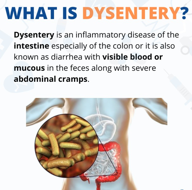
Bacillary dysentery, also known as shigellosis, is an acute diarrheal disease of the intestines characterized by the passage of blood-stained mucoid stool.
It is a local bowel wall infection and does not spread systematically.
It is important not to confuse bacillary dysentery with diarrhea caused by other bacterial infections, as one of the distinguishing characteristics of bacillary dysentery is the presence of blood in the stool, resulting from the invasion of the pathogen into the mucosa.
Cause:
The causative agent of bacillary dysentery is the bacterium Shigella. There are four main species:
- Shigella flexneri
- Shigella sonnei
- Shigella dysenteriae
- Shigella boydii
Shigella dysenteriae is considered the most virulent. These bacteria are strictly pathogenic to humans with no animal reservoir.
Shigella are non-motile, gram-negative bacilli found in the gastrointestinal tract (GIT). They cause disease by invasion and destruction of the colonic mucosa using exotoxins.
Sources of Infection:
- Symptomatic patients: Individuals actively experiencing symptoms of shigellosis.
- Carriers: Individuals who harbor Shigella bacteria but show no symptoms.
Infective Dose:
The infective dose varies. As few as 10 to 100 viable Shigella organisms can cause dysentery, but about 10,000 are required for other species.
Incidence:
Shigellosis affects all age groups, particularly in areas with poor sanitation. It is more prevalent in tropical countries and less developed nations.
Incubation Period:
The incubation period for shigellosis ranges 1-3 days (can extend up to 7 days). Pathology:
All Shigella species have the ability to invade and destroy the epithelial cells of the large intestines. They produce exotoxins with enterotoxic, cytotoxic, and neurotoxic properties.
- Enterotoxins: Produce a secretory effect on the intestine, similar to that caused by cholera toxin, leading to watery (secretory) diarrhea.
- Cytotoxin: Binds to the cell surface and is transported inside cells, inhibiting protein synthesis and causing cell necrosis, which results in dysentery.
- Neurotoxin: May be responsible for neurological complications in children but not adults.
Transmission Routes (Oro-fecal Route):
- Person-to-person contact: Finger-to-mouth transmission is the most important means of transmission, especially among household members where hygienic habits are lacking. The infection is also common where there is ano-oral sexual contact.
- Water-borne: Contamination of water supplies by sewage and excreta containing Shigella leads to outbreaks of shigellosis in communities.
- Food-borne: Contaminated food and milk products are major sources of infection in hospitals and communities. Shigella can survive in various foods for up to 30 days under favorable conditions.
- Flies: Flies can transmit the bacteria by settling on dysenteric stool and then contaminating food or utensils.
Predisposing Factors.
Shigellosis is a bacterial infection that occurs when a person ingests Shigella bacteria, usually through contaminated food or water. The bacteria then multiply in the intestines and invade the lining of the colon, causing inflammation and ulceration.
- Defective sanitation: Poor refuse disposal contributes to the spread.
- Bad hygienic practices: Poor excreta disposal, dirty hands, skin, and clothes, and inadequate cleaning of the anal orifice facilitate transmission.
- Heavy environmental infestation with flies: Flies act as mechanical vectors.
- Wet environment with stagnating water areas: These environments promote the survival of Shigella.
Signs and Symptoms:
The severity of symptoms varies depending on the amount of toxin produced, the infective dose, and the type of Shigella.
- Mild Type (Watery Diarrhea): Onset is gradual, with tenesmus (a painful effort to defecate) that causes straining and painful defecation lasting for a few days after abdominal discomfort and the passage of watery stool.
- Moderate Type: Abrupt onset with abdominal pain, nausea, and vomiting. The passage of blood-stained mucoid stool. Gripping and tenesmus may be severe, with dysuria. High fever and rigor. The face is pinched, and the look is anxious. The patient may become delirious and confused. Marked thirst.
- Fulminating Type (Very Severe): Abrupt onset as described above. Watery diarrhea that later becomes bloody and mucoid. The patient may pass stool as frequently as 10 to 20 times in 24 hours (muco-purulent stools). The motions then decrease, and dysentery symptoms set in. Necrotic sloughs may be passed out. Severe abdominal cramps. Tenesmus. Profound prostration due to fluid loss. Toxaemia occurs from toxin absorption into the circulation. The cheeks are flushed, the expression is anxious, the pulse is rapid, the tongue is coated yellow or dry and furred and brown. Marked dehydration with oliguria, dry, shriveled skin, collapsed veins, low blood pressure. The urine contains albumin. The patient is restless and may die in uraemic coma. Perforation and peritonitis may occur, although rare, with abdominal distension and hiccups.
Other clinical features include;
- Bloody diarrhea: Frequent watery diarrhea, often containing blood and mucus.
- Abdominal cramps: Intense cramping pain in the abdomen. Flatulence
- Fever: High fever, often accompanied by chills.
- Tenesmus: A feeling of incomplete bowel emptying, with frequent straining and urgency to defecate.
- Nausea and vomiting: Can occur, especially in severe cases.
- Headache: General malaise and weakness.
- Dehydration: Significant fluid loss can lead to dehydration, especially in young children and the elderly.
- Electrolyte imbalance: Diarrhea can cause significant electrolyte loss, leading to imbalances that can be life-threatening.
- Rectal prolapse: In severe cases, especially in children, the rectum can protrude from the anus.
Diagnosis/Investigations:
- Stool culture: The most definitive diagnostic test involves culturing stool samples to identify the specific Shigella species present.
- Stool analysis and appearance/rectal swab: Microscopic examination of stool or rectal swab for Shigella bacteria.
- Microscopic examination: Stool examination under a microscope may reveal red blood cells, white blood cells, and bacteria.
- Serological tests: Serological tests can detect antibodies to Shigella bacteria in the blood.
Differential Diagnosis:
- Cholera: Similar symptoms, but cholera is usually more severe.
- Acute diarrhea from food poisoning: Caused by different bacteria or toxins.
- Amoebiasis: Caused by a protozoan parasite.
- Ulcerative colitis: A chronic inflammatory disease of the colon.
- Schistosomiasis from Schistosoma mansoni: Caused by a parasitic worm.
- Carcinoma of the colon and rectum: Cancer of the colon or rectum.
Management:
Aims:
- Prevent the spread of infection: Implement infection control measures.
- Preserve and save the patient’s life: Prioritize life support.
- Support patient recovery (nursing care): Provide supportive care.
- Eliminate the offending bacteria (treatment): Administer antibiotics.
Management depends on the severity of the condition. Severe cases caused by Shigella dysenteriae are considered medical emergencies.
Severe Type of Dysentery (Medical Emergency):
First Aid Treatment:
- The patient is received at the healthcare facility and assessed for airway, breathing, and circulation (ABCs).
- Signs and symptoms of dehydration and anemia are assessed, and appropriate action is taken.
- Brief history is taken, and observations are made. Pulse is checked. A doctor is called, and the patient is reviewed.
- An intravenous (IV) line is established, and fluids such as glucose 50% (30 to 50 ml bolus), normal saline, or Ringer’s lactate (500 to 1000 ml) are connected to control blood pressure and correct electrolyte imbalance.
- The patient and their attendant are reassured about hospital transfer and conditions.
- The patient is referred to the hospital promptly.
Ward Management (Medical Emergency):
- The patient is received and assessed with details from the initial healthcare facility report.
- The patient is admitted in isolation, if possible. If not, high-level hygiene and infection control measures are put in place.
- Disinfection of stool and vomitus with 1% sodium hypochlorite or other disinfectants.
- Strict use of equipment and utensils only for that patient.
- IV fluids are continued as per doctor’s orders.
- Frequent monitoring of the patient (every 4 hours) for vital signs (including temperature, pulse, respiration, and blood pressure), signs and symptoms of dehydration, and signs and symptoms of anemia.
- Immediate investigations are conducted:
- Hemoglobin grouping and cross-matching for blood transfusions if needed.
- Stool for analysis to identify the specific Shigella species.
- Serum electrolytes to assess electrolyte balance.
- Rectal swabs for bacterial culture.
- Full blood count (FBC) and erythrocyte sedimentation rate (ESR).
Continuous Care in the Ward:
- Patient’s personal and environmental hygiene: Similar to the management of cholera or typhoid fever.
- Feeding: During the acute stages, a fluid diet is provided, followed by a soft, balanced, non-irritating, non-spiced, low-residue diet as the stool becomes more solid. Food hygiene is crucial.
- Treatment:
- Antibiotics: Nalidixic acid 1 mg every 6 hours for 5 days or ciprofloxacin 1 mg stat (immediately).
- Pain killers: Paracetamol for children. Bactrim 24 mg/kg for children.
- Vital signs and other assessments: Regular monitoring.
- Nursing care: Provide supportive nursing care, including hygiene, comfort measures, and monitoring.
- Urine and bowel care: Provide regular care and hygiene for the patient’s urinary and bowel functions.
- Terminal disinfection: Thorough disinfection of the patient’s environment after discharge.
IMMEDIATE NURSING CARE:
- Rehydration:
- Intravenous fluids (e.g., Normal saline, Ringer’s lactate)
- Oral rehydration solutions (e.g., ORS)
Hygiene:
- Skin care: Keep the patient’s skin clean and dry.
- Mouth care: Provide oral hygiene to prevent mouth sores.
- Perineal care: Maintain meticulous perineal care to prevent skin breakdown.
Personal protective equipment (PPE):
- Gloves, aprons, and goggles are worn when handling anything from the patient.
Handwashing: Frequent handwashing with soap and water and drying with clean towels.
Safe water and food: Use only treated or boiled water for drinking and cooking.
Disinfection: Dispose of wastes and excreta properly, disinfecting all contaminated items.
Linen treatment: Treat linens as infected material.
Terminal disinfection: Carry out terminal disinfection after the patient’s discharge.
Monitoring: Closely monitor the patient’s condition, particularly abdominal pain, diarrhea, constipation, and any signs of complications. Report any changes promptly.
Diet:
- Initially, provide a clear liquid diet, avoiding dairy products.
- Gradually transition to a bland diet, low in fiber, and avoiding spicy, fatty, and greasy foods.
ADVICE ON DISCHARGE:
- Continue hydration: Maintain adequate fluid intake.
- Handwashing: Continue frequent handwashing.
- Avoid contact: Avoid close contact with others until fully recovered.
- Follow-up: Schedule a follow-up appointment with their healthcare provider.
summary;
- Admission to a medical ward in isolation.
- Strict personal hygiene (barrier nursing) to prevent infecting others.
- Disinfection of the patient’s bed and other items used.
- Proper disposal of fecal matter and vomit into a pit latrine.
- Regular monitoring of temperature, pulse, respiration, blood pressure, hydration levels, and level of consciousness.
- Providing reassurance and support to the patient and relatives.
- Fluid intake maintenance using Oral Rehydration Solution (ORS) or intravenous fluids in severe cases.
- Antibiotic treatment with drugs like nalidixic acid or ciprofloxacin.
- Implementing a BRAT diet (bananas, rice, applesauce, toast) to aid in recovery.
- Use of a nasogastric tube for feeding and medication administration if oral intake is not possible.
- Medications for managing nausea and vomiting, such as metoclopramide (plasil).
- Close monitoring of hydration levels and maintenance of a fluid balance chart.
Prevention:
- Maintain cleanliness in premises and kitchen utensils.
- Proper disposal of rubbish.
- Practice proper hand hygiene before eating or handling food, and after using the toilet or changing diapers.
- Boil or treat drinking water.
- Avoid high-risk foods like shellfish, raw or semi-cooked food.
- Use clean washable aprons and caps during food preparation.
- Thoroughly clean and wash food items, including fruits, in clean water.
- Store perishable food in a well-covered refrigerator.
- Ensure thorough cooking of food before consumption.
- Consume food promptly or refrigerate leftovers and reheat thoroughly before eating.
- Exclude infected individuals and asymptomatic carriers from handling food or providing care to children.
Complications:
- Perforation: A hole in the intestinal wall.
- Hemorrhoids and rectal prolapse: Occur due to over-straining during defecation.
- Hemolytic-uremic syndrome (HUS): A serious complication that can occur with Shigella dysenteriae infection, leading to kidney failure.
- Stricture of the colon: Narrowing of the colon after healing.
- Post-dysenteric colitis (irritable bowel syndrome): Persistent passage of stool after recovery, with colicky abdominal pain, which may clear after 6 months but may be permanent.
- Dehydration: Fluid loss due to diarrhea.
- Renal failure: Kidney dysfunction.
- Shock (Hypovolemic): Low blood pressure due to fluid loss.
- Severe intestinal hemorrhage: Bleeding in the intestines.
Comparison between Bacillary and Amoebic Dysentery:
Feature | Bacillary Dysentery | Amoebic Dysentery |
Occurrence | Epidemic | Endemic |
Severity | “Lying down disease” | “Walking disease” |
Onset | Acute | Gradual |
Fever | Common | Unless complicated |
Tenderness | Whole abdomen, especially sigmoid part | Localized to sigmoid colon |
Quantity of Stool | Scanty but very frequent, bright red, colorless, viscid mucus, jelly-like | Much mingled with blood and mucus, offensive, smelling of decomposing blood, copious |
Tenesmus | Very severe | Present but mild |
Stool Microscopy | Numerous RBCs, WBCs, and macrophages; few bacteria | Many RBCs in clumps; WBCs and macrophages are scanty; large number of mobile protozoa |
Cause | Bacterial | Protozoal |
Complications | Toxic arthritis and eye complications | Hepatic and other abscesses; skin perforations |
Amoebic Dysentery (Amoebiasis)
Amoebic dysentery is a parasitic infection of the gastrointestinal system that is caused by the parasite Entamoeba histolytica.
The infection is most commonly acquired through oral-fecal contamination, which can occur by consuming contaminated food or water, or by coming into contact with contaminated feces and not washing your hands properly.
Symptoms of amoebic dysentery
- Violent diarrhea, often with blood and/or mucus in the stools
- Severe colitis
- Frequent flatulence
- Dehydration
- Abdominal cramps and tenderness
- Slight weight loss
- Moderate anemia
- Moderate fever
- Mild fatigue
- Unrelated symptoms such as liver abscess, lung involvement, amoeboma swelling, and anal ulceration
Diagnosis
The diagnosis of amoebic dysentery is usually made by examining a stool sample under a microscope to look for cysts or motile organisms. Ultrasound scans may also be performed.
Treatment
The treatment of amoebic dysentery involves several steps:
- Correcting any dehydration
- Initiating a 10-day course of the antimicrobial drug metronidazole (Flagyl) or tinidazole to eliminate the infection
- Administering amoebicidal (lumenal) drugs such as diloxanide furoate, paromomycin, or iodoquinol to eradicate any remaining parasites
- Isolating infected individuals to prevent further spread of the infection
- Emphasizing personal hygiene practices
Prevention
To prevent the occurrence and transmission of amoebic dysentery, the following preventive measures should be followed:
- Educate the public about proper handwashing before eating and appropriate fecal disposal practices.
- Ensure the proper management of carriers of the infection.
- Promote the use of clean drinking water and safe food handling practices.
very helpful