Midwives Revision

Uncategorised

Elephantiasis (Bancroftian Filariasis)

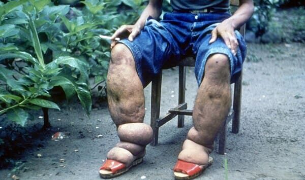
Elephantiasis (Lymphatic Filariasis)

Elephantiasis, also known as lymphatic filariasis, is a chronic parasitic disease caused by filarial worms. 

It results in severe swelling, primarily of the legs, arms, breasts, and genitals, due to blockage of the lymphatic system. The condition gets its name from the enlargement of affected limbs, resembling that of an elephant.

Elephantiasis is a debilitating disease that can significantly impact a person’s physical, social, and economic well-being. The swelling can cause pain, disfigurement, disability, and social stigma. It is a neglected tropical disease (NTD) primarily affecting impoverished communities in tropical and subtropical regions.

Transmission:

Vector: The disease is transmitted by mosquitoes, specifically species of the genera Culex, Anopheles, and Aedes.

Lifecycle:

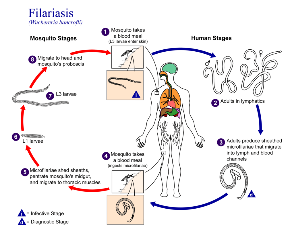
In the Mosquito (Vector):

  • Infected mosquitoes ingest filarial larvae (microfilariae) from an infected person’s blood.
  • The microfilariae develop into mature larvae (L3 larvae) in the mosquito.
  • L3 larvae migrate to the mosquito’s mouthparts, ready for transmission.

In Humans (Host):

  • Infected mosquitoes bite humans, transmitting the L3 larvae into the bloodstream.
  • L3 larvae mature into adult worms in the lymphatic system, typically residing in lymph nodes.
  • Adult worms reproduce, releasing microfilariae into the bloodstream, which can then be ingested by mosquitoes, completing the cycle.

Routes of Transmission:

  • Mosquito Bite: The primary route of transmission is through the bite of an infected mosquito.
  • Blood Transfusion: Transmission is possible through contaminated blood transfusions, although less common.

Causes/Aetiology:

Filarial Worms: The disease is caused by three main species of filarial worms:

  • Wuchereria bancrofti (most common)
  • Brugia malayi
  • Brugia timori

Clinical Features:

1. Early Stage (Lymphangitis):

  • Fever
  • Chills
  • Pain and redness along lymphatic vessels
  • Lymphadenitis (swollen lymph nodes)

2. Subcutaneous Stage (Lymphedema):

  • Swelling in the extremities, particularly legs and arms.
  • Pitting edema (fluid retention that leaves indentations when pressed)
  • Skin thickening and roughness

3. Late Stage (Elephantiasis):

  • Massive swelling of the limbs, breasts, or genitals.
  • Thickened and deformed skin.
  • Lymphatic obstruction leading to fluid buildup.
  • Increased susceptibility to secondary infections.

4. Genital Manifestations:

  • Hydrocele (fluid accumulation in the scrotum).
  • Lymphoedema of the scrotum (scrotal elephantiasis).
  • Lymphoedema of the vulva (vulvar elephantiasis).

5. Other Complications:

  • Chylous ascites (fluid buildup in the abdomen).
  • Chylothorax (fluid buildup in the chest).
  • Chronic kidney disease.

Diagnosis and Investigations:

Clinical Examination: Characteristic swelling and history of mosquito bites in endemic areas.

Microscopic Examination:

  • Blood smear: To detect microfilariae in blood samples.
  • Lymph node aspirate: To detect microfilariae in lymph node fluid.

Serological Tests: Blood tests to detect antibodies against filarial worms.

Imaging Studies:

  • Ultrasound: To visualize the lymphatic system and detect adult worms.
  • CT scan: To evaluate the extent of lymphatic obstruction.
  • MRI: To assess the lymphatic system and surrounding tissues.

Prevention:

Vector Control:

  • Mosquito control measures, such as insecticide spraying, insecticide-treated bed nets, and source reduction (eliminating mosquito breeding sites).
  • Use of repellents.

Mass Drug Administration (MDA):

  • Regular administration of anti-filarial medications (DEC and albendazole) to prevent transmission and reduce disease burden.
  • Large scale treatment/preventive chemotherapy Give annually
  • to all population at risk, for 4-6 years
  • Ivermectin 150-200 mcg/kg plus albendazole 400 mg single dose

Health Education: Educate communities about the disease, transmission, and preventive measures.

Improved Sanitation and Hygiene: Maintain clean surroundings and reduce mosquito breeding sites.

Management:

Aims of Management:

  • To kill adult worms and prevent the spread of infection.
  • To reduce lymphatic obstruction and swelling.
  • To improve quality of life and reduce disability.

Medical Management:

  • Anti-filarial Medications: Diethylcarbamazine (DEC) and Albendazole are the primary drugs for treatment.
  1. Diethylcarbamazine (DEC)
  2. Albendazole
  3. Ivermectin (alternative medication)
  4. Combinations of these drugs may be used.
  • Antibiotics: For treating secondary bacterial infections.

  • Doxycycline 100 mg twice a day for 4-6 weeks (do not administer antiparasitic treatment during an acute attack)

  • Pain Relievers: To manage pain and inflammation.

  • Diuretics: To reduce fluid buildup.

  • Supportive Care:
  1. Elevate affected limbs.
  2. Use compression bandages.
  3. Manage pain and inflammation.
  4. Supportive treatment during an attack (bed rest, limb elevation, analgesics, cooling, hydration)

Continuous Management:

  • Long-term Anti-filarial Therapy: May be required to prevent disease progression.
  • Lymphedema Management:
  1. Regular massage.
  2. Compression therapy.
  3. Skin care and hygiene.
  • Monitoring for Complications: Regular follow-up appointments to monitor for complications, such as secondary infections and kidney damage.

Surgical Management:

  • Chronic case:  Supportive treatment: bandage during the day, elevation of affected limb at rest, analgesics and surgery (hydrocelectomy)
  • Surgery for Complications:
  1. Surgery may be required to treat complications such as hydrocele, lymphoedema, and chylous ascites.
  2. Lymphatic bypass surgery may be considered in some cases.

Nursing Care:

  • Symptom Management: Provide comfort measures for pain, swelling, and discomfort.
  • Skin Care: Promote good skin hygiene to prevent infections.
  • Compression Therapy: Apply compression bandages to reduce swelling and improve lymphatic flow.
  • Lymphedema Management: Teach patients techniques for self-management of lymphoedema.
  • Education: Educate patients about the disease, treatment, and prevention.

Complications:

  • Secondary Infections: Frequent skin infections due to impaired lymphatic drainage.
  • Lymphedema: Progressive swelling of the affected limbs, leading to disfigurement and disability.
  • Elephantiasis: Severe and permanent disfigurement and disability.
  • Hydrocele and Lymphoedema of the Genitals: Can cause pain, discomfort, and reproductive problems.
  • Chronic Kidney Disease: Fluid overload and lymphatic obstruction can strain the kidneys.
  • Social Stigma: Elephantiasis can lead to social isolation and discrimination.
  • Psychological Distress: The disease can have a significant impact on self-esteem and mental health.

Quick Quiz

Elephantiasis Quiz

Tropical Medicine - mobile-friendly and focused practice.

Privacy: Your details are used only for quiz tracking and certificates.

Elephantiasis (Bancroftian Filariasis) Read More »

Schistosomiasis

Schistosomiasis (Snail Fever/bilharzia)

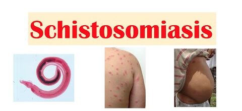
Schistosomiasis, also known as snail fever, is a parasitic disease caused by flatworms of the genus Schistosoma.

Schistosomiasis (also known as bilharzia or snail fever) is a disease of the large intestine and urinary tract caused by parasitic worms of the Schistosoma blood fluke.

It may infect the urinary tract or intestines.

Causes/Aetiology:

Schistosoma species: The disease is caused by five main species of Schistosoma:

  • S. haematobium: Causes urinary schistosomiasis.
  • S. mansoni: Causes intestinal schistosomiasis.
  • S. japonicum: Causes intestinal schistosomiasis.
  • S. intercalatum: Causes intestinal schistosomiasis.
  • S. mekongi: Causes intestinal schistosomiasis.

Snail intermediate host: Snails of specific species are essential for the parasite’s lifecycle.

Transmission:

  • Vector: The disease is transmitted through contact with contaminated freshwater, primarily by snails infected with Schistosoma parasites.
Lifecycle Schistosomiasis

Schistosomiasis  Lifecycle:

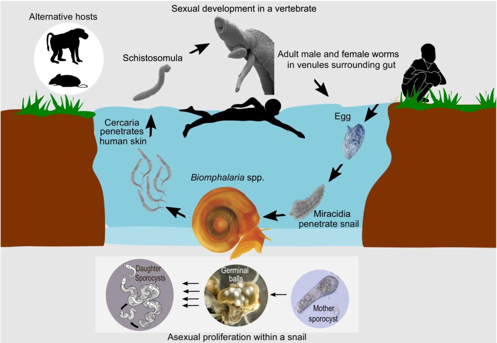
In the Snail (Intermediate Host):

  1. Schistosoma eggs released from an infected human enter freshwater, where they hatch into larvae called miracidia.
  2. Miracidia infect snails.
  3. Within the snail, the miracidia transform into cercariae, which are free-swimming larval forms.

In Humans (Definitive Host):

  1. Cercariae penetrate the skin of humans who come into contact with contaminated water.
  2. Cercariae mature into adult worms within the human body.
  3. Adult worms reside in the blood vessels of the intestines, bladder, or other organs.
  4. Female worms release eggs into the body, and these eggs are excreted in feces or urine.
  5. The cycle repeats as eggs enter freshwater and infect snails.

The lifecycle of Schistosoma worms starts with infected humans. The worms live in the blood vessels of the intestines and produce eggs that are released in feces. These eggs enter freshwater and hatch into microscopic larvae called miracidia.

Miracidia swim until they find a specific type of snail. Inside the snail, they multiply and transform into another type of larvae called cercariae. Cercariae leave the snail and swim in the water.

If a person enters this contaminated water, the cercariae can penetrate their skin. Once inside, they mature into adult worms. The cycle starts again when the worms reproduce and release eggs, which are then expelled in feces and enter the water.

Clinical Features schistosomiasis

Clinical Features:

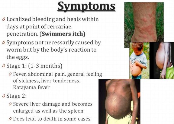
1. Acute Schistosomiasis (Katayama Fever):

  • Fever
  • Chills
  • Headache
  • Muscle aches
  • Rash
  • Cough
  • Abdominal pain
  • Diarrhea
  • Enlarged liver and spleen

2. Chronic Schistosomiasis:

Urinary Schistosomiasis:

  • Blood in the urine (hematuria).
  • Frequent urination, especially at night (nocturia).
  • Painful urination (dysuria).
  • Bladder inflammation (cystitis).
  • In advanced cases, bladder fibrosis and cancer.

Intestinal Schistosomiasis:

  • Abdominal pain and cramping.
  • Diarrhea or constipation.
  • Blood in the stool (hematochezia).
  • Liver enlargement (hepatomegaly).
  • Splenomegaly (enlarged spleen).
  • In advanced cases, portal hypertension, ascites, and esophageal varices.

3. Other Manifestations:

  • Lung involvement (pulmonary schistosomiasis) with cough and shortness of breath.
  • Skin lesions (schistosomal dermatitis) with itching and papules.
  • Brain involvement (neurological schistosomiasis) with seizures, paralysis, and cognitive impairment.

Diagnosis and Investigations:

  • Stool and Urine Examination: Microscopic examination for eggs in stool or urine samples.
  • Serological Tests: Blood tests to detect antibodies against Schistosoma.
  • Imaging Studies:
    • Ultrasound: To visualize the liver, spleen, and other organs.

    • CT scan: To evaluate the bladder and kidneys.

    • MRI: To assess the brain and spinal cord.

  • Biopsy: Biopsy of affected organs (liver, bladder) to confirm the presence of eggs or adult worms.

Prevention:

Safe Water and Sanitation:

  • Provide access to safe drinking water and adequate sanitation facilities.
  • Promote proper waste disposal to prevent contamination of freshwater sources.

Health Education:

  • Educate communities about the disease, transmission, and prevention.
  • Encourage safe bathing and swimming practices in freshwater bodies.

Snail Control:

  • Reduce snail populations by using molluscicides (chemicals that kill snails).
  • Implement environmental interventions to modify snail habitats.

Mass Drug Administration (MDA):

  • Regularly administer praziquantel to all individuals in endemic areas to control the disease.

Management:

Aims of Management:

  • To kill adult worms and prevent egg production.
  • To treat existing symptoms and complications.
  • To prevent further transmission of the disease.

Medical Management:

  • Praziquantel: The primary drug for schistosomiasis, effective in killing both adult worms and reducing egg production.
  • Praziquantel 40 mg/kg single dose
  • OR 2 doses of 20 mg/kg administered 4 hours apart
  • Refer patient if they develop obstruction or bleeding.
  • Other Medications:
  1. Antibiotics: For treating secondary bacterial infections.
  2. Pain relievers: For managing pain and discomfort.
  3. Anti-inflammatory medications: For reducing inflammation.
  • Symptomatic Treatment: Address specific symptoms such as fever, diarrhea, and urinary problems.

Nursing Care:

  • Symptom Management: Provide comfort measures for fever, pain, and diarrhea.
  • Hydration: Encourage fluid intake to prevent dehydration.
  • Hygiene: Promote proper hygiene to prevent infection.
  • Education: Educate patients about the disease, treatment, and prevention.
  • Monitoring: Monitor for signs of complications, including liver problems, bladder dysfunction, and neurological symptoms.

Quick Quiz

Schistosomiasis Quiz

Tropical Medicine - mobile-friendly and focused practice.

Privacy: Your details are used only for quiz tracking and certificates.

Schistosomiasis Read More »

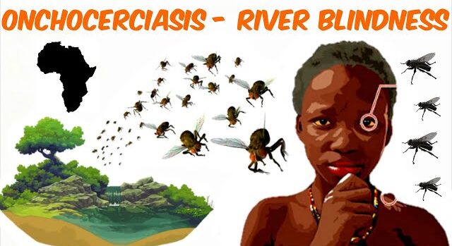
Onchocerciasis (River blindness)

Onchocerciasis (River Blindness)

Onchocerciasis, also known as river blindness, is a chronic parasitic disease caused by the nematode worm Onchocerca volvulus

It is characterized by severe itching, skin lesions, and, in advanced cases, blindness. The disease is primarily found in sub-Saharan Africa, with smaller outbreaks in Central and South America.

Transmission:

Vector: The disease is transmitted through the bite of infected blackflies (genus Simulium) that live near fast-flowing rivers and streams, hence the name “river blindness.”

Lifecycle:

In the Vector (Blackfly):

  1. The female blackfly ingests microfilariae (larval worms) from an infected human during a blood meal.
  2. Inside the blackfly, microfilariae develop into infective larvae.
  3. After approximately 1 to 2 weeks, the larvae mature and migrate to the mouthparts of the blackfly.

In Humans:

  1. An infected blackfly bites a human, injecting the infective larvae into the subcutaneous tissue.
  2. Larvae migrate to the skin, where they mature into adult worms within 1 to 2 years.
  3. Adult worms reside in nodules (onchocercomas) beneath the skin and produce microfilariae.
  4. Microfilariae travel throughout the body, especially to the eyes, skin, and lymph nodes.
  5. The cycle continues as blackflies ingest microfilariae from infected humans.

Clinical Features:

Skin Lesions:

  • Papular, itchy skin lesions (onchocercal dermatitis).
  • Hyperpigmented or depigmented skin patches.
  • Severe itching, leading to secondary bacterial infections.
  • “Elephantine” skin thickening, especially on the legs and scrotum.

Eye Involvement:

  • Microfilariae migrate to the eyes, causing inflammation (onchocercal keratitis).
  • Loss of vision, including blindness, due to corneal scarring, optic atrophy, and retinal detachment.
  • Severe itching and burning in the eyes.

Onchocercomas:

  • Firm, subcutaneous nodules that contain adult worms.
  • Located in various parts of the body, including the head, neck, limbs, and buttocks.
  • Often painful and can cause pressure on nearby nerves or organs.

Lymphatic Involvement:

  • Swelling of lymph nodes and lymphedema.
  • Chronic inflammation of the lymph system.

General Symptoms:

  • Fever
  • Headache
  • Fatigue
  • Joint pain
  • Loss of appetite
  • Weight loss

Diagnosis and Investigations:

  • Skin Biopsy: Examination of skin samples for microfilariae under a microscope.
  • Slit-lamp Examination: Examination of the eyes to detect microfilariae and eye damage.
  • Nodule Biopsy: Biopsy of onchocercomas to confirm the presence of adult worms.
  • ELISA (Enzyme-Linked Immunosorbent Assay): Blood test to detect antibodies against Onchocerca volvulus.

Prevention:

Vector Control:

  • Reducing blackfly populations through insecticides and larvicides.
  • Using insect repellents and protective clothing.
  • Avoiding areas with high blackfly density.

Mass Drug Administration (MDA):

  • Regular administration of antiparasitic drugs (ivermectin) to kill microfilariae.
  • Typically given every 6 months to all individuals in endemic areas.
  • Ivermectin 150 micrograms/kg once yearly for 10-14 years 

Management:

Aims of Management:

  • To eliminate microfilariae and reduce the number of adult worms.
  • To prevent further transmission of the disease.
  • To manage complications and improve quality of life.


Medical Management:

  • Antibiotics: For treating secondary bacterial infections.
  • Doxycycline 100 mg twice a day for 6 weeks followed by;
  • Antiparasitic Drugs:
  • Ivermectin: Kills microfilariae but does not eliminate adult worms.
    • Ivermectin 150 micrograms/kg single dose.

  • Anti-Inflammatory Medications: For managing eye inflammation and other inflammatory conditions.

Surgical Management:

  • Excision of Onchocercomas: Surgical removal of nodules can be considered for symptomatic nodules or those causing pressure on nearby structures.

Nursing Care:

Symptom Management:

  • Provide comfort measures for itching and pain.
  • Administer medications as prescribed.
  • Monitor for side effects of medications.

Eye Care:

  • Teach patients proper eye hygiene and care.
  • Monitor for signs of eye infection and vision loss.

Skin Care:

  • Provide soothing baths and lotions for itching.
  • Encourage the use of insect repellents.

Education:

  • Educate patients about the disease, transmission, and prevention.
  • Encourage adherence to treatment and follow-up appointments.

Quick Quiz

Onchocerciasis Quiz

Tropical Medicine - mobile-friendly and focused practice.

Privacy: Your details are used only for quiz tracking and certificates.

Onchocerciasis (River blindness) Read More »

Helminthic diseases (Intestinal worms)

Helminthiasis/Worm Infestation

Helminthiasis, commonly known as worm infestation, refers to a group of infections caused by parasitic worms living within the human body

These infections are widespread, particularly in tropical and subtropical regions, affecting millions of people globally. 

Helminthiasis:

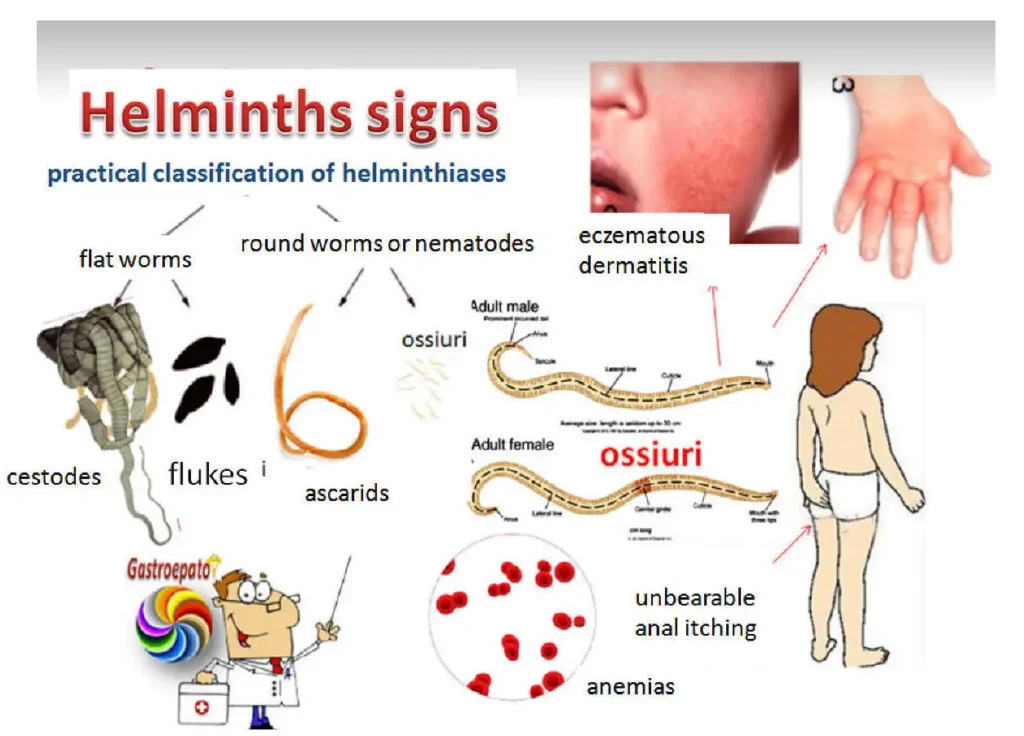
Helminthiasis encompasses infections caused by parasitic worms belonging to three major groups:

1. Nematodes (roundworms): These are cylindrical, unsegmented worms with a pointed tail. Examples include 

  • Ascaris lumbricoides, hookworms (Ancylostoma caninum and Necator americanus), and Trichuris trichiura.

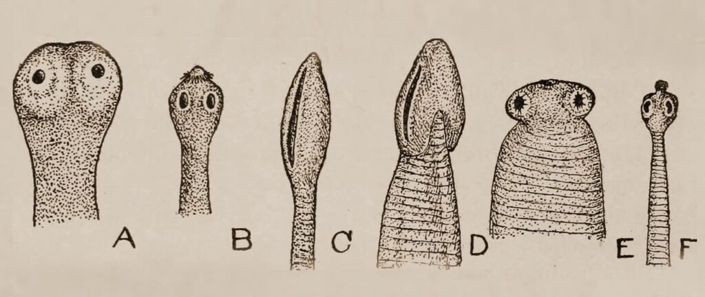
2. Cestodes (tapeworms): These are flat, ribbon-like worms with segments (proglottids). 

  • Taenia saginata, Taenia solium, and Diphyllobothrium latum are some commonly encountered species.

3. Trematodes (flukes): These are flat, leaf-like worms with a complex life cycle involving multiple hosts. 

  • Schistosoma species (blood flukes), Fasciola hepatica (liver fluke), and Clonorchis sinensis (Chinese liver fluke) are examples.
Lifecycles of Helminthiasis

Lifecycles of Helminthiasis

Helminthic infections occur when a host ingests or comes into contact with infectious stages of the parasite. The specific mode of transmission varies depending on the type of worm:

Transmission Routes & Lifecycle:

1. Fecal-Oral Route:

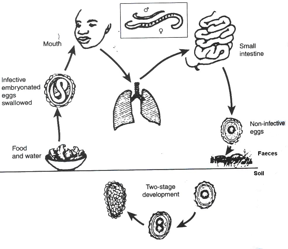
Nematodes like Ascaris lumbricoides, hookworms, and Trichuris trichiura: Human feces containing parasite eggs are released into the environment. These eggs mature and become infective. Humans become infected by ingesting contaminated soil, water, or food containing these eggs.

Lifecycle Example: Ascaris lumbricoides

  • Eggs: Ingested eggs hatch in the small intestine, releasing larvae.
  • Larvae: Larvae penetrate the intestinal wall, enter the bloodstream, and migrate to the lungs.
  • Adult Worms: Larvae mature in the lungs, migrate up the respiratory tract, are swallowed, and reach the small intestine where they mature into adults. Adult worms produce eggs that are passed in feces.

2. Skin Penetration:

Hookworms (Ancylostoma caninum and Necator americanus): Infective larvae present in contaminated soil penetrate the skin, usually through the feet.

Lifecycle Example: Hookworms

  • Larvae: Infective larvae in soil penetrate the skin.
  • Bloodstream Migration: Larvae travel through the bloodstream to the lungs, migrate up the respiratory tract, are swallowed, and reach the small intestine.
  • Adult Worms: Larvae mature into adults in the small intestine, where they attach to the intestinal wall and feed on blood. Eggs are produced and passed in feces.

3. Consumption of Undercooked Meat:

Cestodes like Taenia saginata and Taenia solium: Humans become infected by consuming undercooked meat containing the parasite’s larval stage (cysticercus).

Lifecycle Example: Taenia saginata (beef tapeworm)

  • Ingestion of Cysticercus: Humans ingest undercooked beef containing cysticerci.
  • Adult Worm: Cysticerci mature into adult tapeworms in the small intestine.
  • Eggs: Eggs are released from the adult worm and passed in feces, contaminating the environment.

4. Consumption of Raw or Undercooked Fish:

Cestodes like Diphyllobothrium latum: Humans become infected by consuming raw or undercooked fish containing the parasite’s larval stage (plerocercoid).

Lifecycle Example: Diphyllobothrium latum (broad fish tapeworm)

  • Ingestion of Plerocercoid: Humans ingest raw or undercooked fish containing plerocercoid larvae.
  • Adult Worm: Larvae mature into adult tapeworms in the small intestine.
  • Eggs: Eggs are released from the adult worm and passed in feces, contaminating the environment.
Clinical Features Helminthic diseases (Intestinal worms)

Clinical Features:

The symptoms of helminthiasis vary depending on the type of worm and the intensity of infection. Common features include:

Gastrointestinal Symptoms:

  • Abdominal pain and cramping
  • Diarrhea or constipation
  • Nausea and vomiting
  • Anorexia (loss of appetite)
  • Weight loss

Other Symptoms:

  • Fatigue and weakness
  • Anemia (caused by blood loss due to hookworms)
  • Edema (swelling)
  • Coughing (associated with larval migration in the lungs)
  • Rectal prolapse (particularly in cases of heavy Trichuris trichiura infection)
  • Skin manifestations (rash, itching)
  • Neurologic symptoms (in cases of neurocysticercosis)

Diagnosis & Investigations:

  • History and Physical Examination: Detailed information regarding symptoms, travel history, and potential exposure to contaminated environments is crucial.
  • Stool Examination: This is the primary diagnostic tool for most intestinal helminth infections. Microscopic examination of stool samples can reveal parasite eggs or larvae.
  • Blood Tests: Blood tests can help detect the presence of antibodies against specific parasitic worms (e.g., schistosomiasis).
  • Imaging Studies: Imaging techniques like X-rays, ultrasound, and MRI can be used to detect larval cysts or adult worms in certain tissues (e.g., cysticercosis).

Prevention:

Sanitation and Hygiene:

  • Proper disposal of human feces through toilets or latrines.
  • Washing hands thoroughly with soap and water after using the toilet and before preparing food.
  • Wearing shoes when walking on contaminated soil.

Safe Food Practices:

  • Washing fruits and vegetables thoroughly before consumption.
  • Cooking meat thoroughly to kill any parasitic larvae.
  • Avoiding raw or undercooked fish.

Control of Human Waste:

  • Proper disposal of human waste, including sewage treatment.
  • Education and Awareness: Public education about the risks of helminthiasis, its transmission, and prevention measures is crucial.

Management:

Aims:

  • Elimination of Parasites: The primary aim is to eliminate the parasitic worms from the body.
  • Symptomatic Relief: Addressing the symptoms associated with the infection is vital for patient comfort.
  • Prevention of Complications: Measures are taken to prevent further complications related to the infection.

Management:

  • Supportive Care: Rest, hydration, and proper nutrition are important components of initial management.
  • Anti-Emetic Medications: These can be used to alleviate nausea and vomiting.
  • Anti-Diarrheal Medications: These may be necessary to control diarrhea.
  • Pain Relief: Pain medications can be prescribed for abdominal pain.
  • Iron Supplementation: In cases of anemia caused by hookworm infection, iron supplementation is needed.
  • Anti-Helminthic Medication: Anti-helminthic medications are used to kill or expel the parasitic worms. The specific medication depends on the type of worm causing the infection.
  • Repeat Dosing: Depending on the type of worm and the intensity of infection, repeat doses of anti-helminthic medications may be necessary.

Medical Management:

Drug Therapy: Anti-helminthic medications are the mainstay of treatment for helminthiasis.

Commonly used drugs:

  • Mebendazole: Effective against roundworms (Ascaris lumbricoides, hookworms, Trichuris trichiura) and some tapeworms.
  • Albendazole: Broad-spectrum anti-helminthic agent effective against a wide range of roundworms, tapeworms, and some flukes.
  • Praziquantel: Effective against tapeworms and flukes (e.g., schistosomiasis).
  • Ivermectin: Effective against roundworms (e.g., Onchocerca volvulus, Strongyloides stercoralis) and some other parasites.

Supportive Care:

  • Iron Supplementation: In cases of anemia caused by hookworm infection, iron supplementation is needed.
  • Nutritional Support: Patients with significant weight loss may require nutritional support.
  • Fluid Management: Maintaining adequate hydration is usefu, especially for patients with diarrhea.

Infestation Type

Drug

Dosage

Roundworm, Threadworm, Hookworm, Whipworm

Albendazole

400 mg single dose (200 mg for children under 2 years)

Mebendazole

500 mg single dose (250 mg for children under 2 years)

Ivermectin

150 micrograms/kg single dose

Prevention

  • Practice proper faecal disposal.
  • Maintain personal and food hygiene.
  • Regular deworming for children every 3-6 months.
  • Avoid walking barefoot to prevent skin penetration by larvae.

Complications:

  • Intestinal Obstruction: Large numbers of adult worms (especially Ascaris lumbricoides) can obstruct the intestines, leading to severe abdominal pain, vomiting, and difficulty passing stool.
  • Malnutrition: Chronic helminthic infections can contribute to malnutrition by interfering with nutrient absorption and causing blood loss (e.g., hookworms).
  • Anemia: Hookworms can cause anemia by feeding on blood in the intestines.
  • Rectal Prolapse: Heavy infestations with Trichuris trichiura can lead to rectal prolapse.
  • Cysticercosis: Ingestion of Taenia solium eggs can lead to cysticercosis, where larval cysts develop in various tissues, including the brain.
  • Neurocysticercosis: Cysticercosis in the brain can cause seizures, headaches, and other neurological problems.
  • Schistosomiasisrelated Complications: Schistosoma infection can cause liver damage, urinary tract problems, and other complications.
  • Filariasis: Filariasis, caused by parasitic worms that reside in the lymphatic system, can lead to lymphedema, elephantiasis, and other problems.

Quick Quiz

Helminthiasis Quiz

Tropical Medicine - mobile-friendly and focused practice.

Privacy: Your details are used only for quiz tracking and certificates.

Helminthic diseases (Intestinal worms) Read More »

Trypanosomiasis (sleeping sickness)

African Trypanosomiasis (Sleeping Sickness)

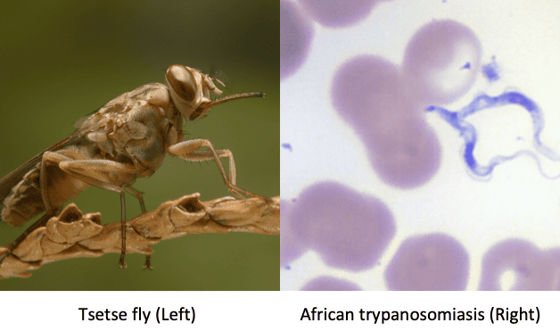
Trypanosomiasis, commonly known as African trypanosomiasis or sleeping sickness, is a parasitic disease caused by protozoa of the genus Trypanosoma.

These parasites are transmitted by the tsetse fly and affect both humans and animals. The disease is endemic to sub-Saharan Africa and can be fatal if left untreated.

Aetiology:

Trypanosomiasis is caused by two main species of Trypanosoma:

  • Trypanosoma brucei rhodesiense (TBr): This species causes the acute form of the disease and is predominantly found in East and Southern Africa.
  • Trypanosoma brucei gambiense (TBg): This species causes the chronic form of the disease and is prevalent in West and Central Africa.

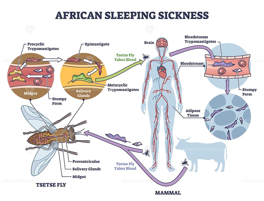
Life Cycle:

Vector (Tsetse Fly):

  1. Infective stage: The tsetse fly ingests trypanosomes in the metacyclic trypomastigote stage from an infected host.
  2. Multiplication: Within the fly’s gut, trypanosomes transform into procyclic trypomastigotes and multiply.
  3. Migration: The trypanosomes migrate to the salivary glands of the fly and differentiate into metacyclic trypomastigotes, the infective stage for humans and animals.
African Trypanosomiasis

Human:

  1. Infection: Metacyclic trypomastigotes are injected into the bloodstream during a fly bite.
  2. Multiplication: The trypanosomes multiply in the bloodstream as bloodstream trypomastigotes.
  3. Spread: They can cross the blood-brain barrier, reaching the CNS and transforming into meningoencephalitic trypomastigotes.

Forms of Transmission:

  • Tsetse fly: This is the primary mode of transmission. The fly acquires trypanosomes by feeding on an infected animal or human. During its subsequent feedings, it injects the parasites into the bloodstream of its new host.
  • Blood transfusion: While rare, the disease can be transmitted through contaminated blood transfusions.
  • Mother to child: Transmission from mother to child can occur during pregnancy or childbirth.

Hosts:

  • TBg: Pigs, dogs, antelopes, cows, sheep, goats, humans.
  • TBr: Antelopes, pigs, and humans (humans are the most common source of infection).

Vectors:

  • Glossina palpalis (Riverine type): Breeds along rivers and lakes and transmits mainly T. b. gambiense.
  • Glossina morsitans (Wounded type): Stays in open, lightly wooded, packed land away from water and transmits T. b. rhodesiense.

Pathogenesis:

Trypanosomiasis arises when humans or animals are infected with trypanosomes through the bite of an infected tsetse fly. The parasite enters the bloodstream and multiplies, eventually reaching the central nervous system (CNS) in the later stages of the disease.

  • Toxins: Trypanosomes produce toxins that damage tissues, causing inflammatory changes at the primary chancre, skeletal and heart muscles.
  • CNS damage: Toxins may destroy ependymal cells lining the brain ventricles, interfering with serotonin release and causing sleep disturbances.
  • Hypersensitivity reactions: The presence of trypanosomes may cause itching (pruritis) and hives (urticaria).

Clinical Presentation:

Onset: Both TBg and TBr follow a similar course, but TBr is often acute and virulent, leading to death within 2-3 years if untreated.

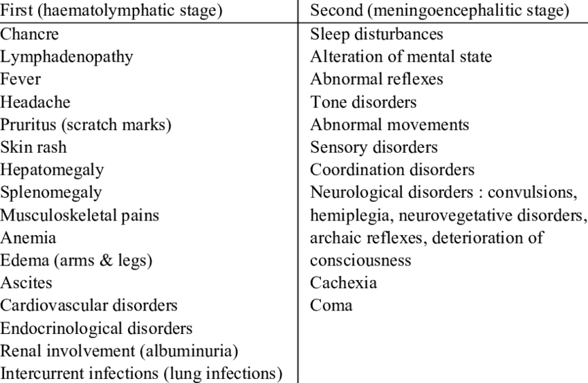
Stage 1: Primary/ Chancre Stage

  • History of bite
  • Local swelling/ nodule at the bite site, possibly hardened and reddened.
  • The chancre may resemble a large boil, but with less pain.
  • This stage can last 1-2 weeks and may resolve.

Stage 2: Blood Stage/ Systemic Stage/Haematolymphatic stage.

  • Fever: Intermittent fever
  • Lymphadenopathy: Swollen spleen and cervical lymph nodes (due to lymphatic spread).
  • Physical weakness: Loss of strength accompanied by fever.
  • Itchy rashes: Skin patches (15-30 cm in diameter) on the chest and back due to hypersensitivity to trypanosomes.
  • Dyspnoea: Shortness of breath due to pericardial effusion and congestive heart failure in chronic cases (TBr).
  • Hepatomegaly: Enlarged liver due to liver damage in chronic cases, with potential jaundice.
  • Pitting oedema: Swelling on the face, lower limbs, eyelids, and abdomen due to cardiac failure or kidney damage.
  • Neurological pains: Muscle cramps are common.
  • Reduced appetite and weight loss: Due to constant sleeping and difficulty eating.
  • Menstrual irregularities: Amenorrhoea in women.
  • Anaemia: Due to the destruction of red blood cells by the trypanosomes.

Stage 3: CNS Stage/ Meningoencephalitis

This stage develops after several months or years of infection, but can occur more rapidly in the acute form (TBr). It’s characterized by involvement of the central nervous system.

Sleep disturbances:

  • Daytime sleepiness, lethargy, and coma.
  • Nighttime insomnia, restlessness, and nightmares.

Behavioral changes:

  • Confusion, apathy, and disorientation.
  • Personality changes, including aggression and irritability.
  • Hallucinations and delusions.

Neurological signs:

  • Motor incoordination: Tremors, jerky movements, paralysis (facial palsy, limb weakness, difficulty swallowing), ataxia (unsteady gait).
  • Sensory disturbances: Numbness and tingling sensations.
  • Headache: Often severe and persistent.

Meningitis:

  • Stiff neck (meningismus)
  • Photophobia (sensitivity to light)
  • Fever
  • Nausea and vomiting

Other Symptoms:

  • Swelling: The face and limbs may become swollen, especially in the late stages.
  • Cardiac complications: Irregular heartbeat, heart failure, and pericarditis can occur.
  • Renal complications: Kidney failure can occur in later stages.

Diagnosis:

Clinical examination: Evaluating the patient’s history, symptoms, and neurological signs.

Laboratory investigations:

Microscopy:

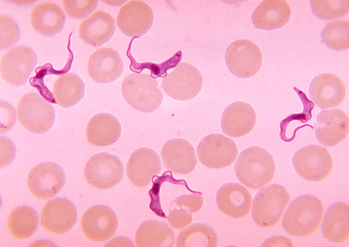
  • Wet blood smear: Examination of a fresh blood sample under a microscope for the presence of trypanosomes.
  • Thick blood smear: A higher concentration of blood is used to improve detection of trypanosomes.
  • Staining: Using Giemsa or Wright’s stain to visualize the trypanosomes.

Serology: Blood tests to detect antibodies against trypanosomes.

Lumbar puncture (LP): A spinal tap to collect cerebrospinal fluid (CSF) for examination.

  • Microscopy: Examining the CSF for trypanosomes, especially in the meningoencephalitic stage.
  • Biochemical analysis: Measuring protein levels and cell counts in the CSF.

Molecular methods: PCR testing can be used to detect trypanosome DNA, especially in the early stages of infection.

Differential Diagnosis:

  • Malaria: Can present with fever, chills, sweating, and headaches.
  • Tuberculosis (TB): Can cause fever, weight loss, and night sweats.
  • Meningitis: Can cause headache, fever, stiff neck, and altered mental status.
  • HIV/AIDS: Can cause fever, weight loss, and neurological symptoms.
  • Other infections: Meningococcal meningitis, encephalitis, and viral infections.

Management:

Early Stages:

  • Suramin: Given intravenously (IV) every 3-5 days for 6-7 doses. Effective against bloodstream trypanosomes, but not CNS involvement.
  • Pentamidine: Given intramuscularly (IM) daily for 7-10 days. Effective against bloodstream trypanosomes.

Late Stages (CNS Involvement):

  • Melarsoprol (MEL.B): The only drug effective against trypanosomes in the CNS. Given intravenously and requires careful monitoring due to potential side effects.
  • Eflornithine: Alternative treatment for late-stage disease, especially in pregnant women.

Supportive Care:

  • Bed rest: To reduce the risk of complications and improve recovery.
  • Hydration: To prevent dehydration.
  • Nutrition: Adequate nutrition is essential to support the body’s fight against infection.
  • Symptom management: Medication for fever, headache, and other symptoms.

Prevention and Control:

  • Vector control: Reducing the tsetse fly population through insecticides, traps, and clearing of vegetation in endemic areas.
  • Early diagnosis and treatment: Active screening and case finding programs are crucial for timely treatment and preventing the spread of the disease.
  • Sleeping sickness awareness: Public health education about the disease, its transmission, and prevention measures.

Prognosis:

  • Untreated: The disease is almost always fatal.
  • Treated: Early treatment is important for a positive prognosis. Late-stage treatment is more challenging and has a higher risk of complications.

Nursing Management:

  • Assessment: Monitor vital signs, neurological status, and symptoms.
  • Medications: Administer medications according to the doctor’s orders and monitor for side effects.
  • Hydration: Ensure adequate fluid intake to prevent dehydration.
  • Nutrition: Provide a balanced diet to support the body’s recovery.
  • Comfort: Provide comfort measures for symptoms like fever, headache, and pain.
  • Patient education: Educate the patient and their family about the disease, its management, and preventive measures.


Early Stage Treatment:

Trypanosoma rhodesiense sleeping sickness:

For both children and adults: Suramin is the drug of choice for early-stage T. rhodesiense infection.

Dosage:

  • A test dose of 5 mg/kg body weight should first be administered intravenously (IV) to check for anaphylactic reactions.
  • If no reaction occurs, five injections of 20 mg/kg body weight are given every 5 days, totaling 100 mg/kg over a 23-day period.
  • The schedule is as follows:
  1. Day 0: 5 mg/kg body weight
  2. Day 3: 20 mg/kg body weight
  3. Day 8: 20 mg/kg body weight
  4. Day 13: 20 mg/kg body weight
  5. Day 18: 20 mg/kg body weight
  6. Day 23: 20 mg/kg body weight
  • Important Note: If anaphylaxis occurs after the test dose, suramin should not be administered.

Trypanosoma gambiense sleeping sickness:

  • For both children and adults: Pentamidine is the preferred treatment for early-stage T. gambiense infection.
  • Dosage: 4 mg/kg body weight daily for 7 days, administered intramuscularly (IM).
  • Important Considerations:
  • Food should be given 1 hour before pentamidine administration to prevent hypoglycemia.
  1. The patient should lie flat (supine position) during administration and for 1 hour afterward to prevent hypotension.

Late Stage Treatment:

Trypanosoma rhodesiense sleeping sickness:

  • For both children and adults: Melarsoprol is the primary treatment for late-stage T. rhodesiense infection.
  • Dosage: 2.2 mg/kg body weight daily for 10 days administered intravenously (IV).

Trypanosoma gambiense sleeping sickness:

Children ≤ 12 years and <35 kg: Eflornithine is the preferred treatment.

  • Dosage: 150 mg/kg body weight every 6 hours for 14 days (total daily dose of 600 mg/kg).
  • Administration: Dilute the 150 mg/kg dose of eflornithine in 100 ml of distilled water and administer the infusion over at least 2 hours.

Children >12 years up to 15 years: Eflornithine is the preferred treatment.

  • Dosage: 100 mg/kg body weight every 6 hours for 14 days (total daily dose of 400 mg/kg).
  • Administration: Dilute the 100 mg/kg dose of eflornithine in 100 ml of distilled water and administer the infusion over at least 2 hours (rate of 20 drops/minute).

Adults >15 years:

  • NECT (Nifurtimox/Eflornithine Combination Therapy): This is the preferred treatment for late-stage T. gambiense infection in adults.
  1. Nifurtimox dosage: 5 mg/kg body weight every 8 hours orally for 10 days (total daily dose of 15 mg/kg).
  2. Eflornithine dosage: 200 mg/kg body weight every 12 hours for 7 days (total daily dose of 400 mg/kg).
  3. Administration: Dilute the 200 mg/kg dose of eflornithine in 250 ml of distilled water and administer the infusion over at least 2 hours (rate of 50 drops/minute).

Relapses: If a relapse occurs, IV melarsoprol at 2.2 mg/kg once daily for 10 days is used.

Important Notes:

Corticosteroids:

  • Corticosteroids should be given to patients with late-stage trypanosomiasis who are receiving melarsoprol, as they may have hypoadrenalism.
  • Corticosteroids can also reduce drug reactions.
  • Hydrocortisone should not be given after day 24, even if the melarsoprol treatment is not yet complete.
  • If prednisolone is used instead of hydrocortisone, the anti-inflammatory action is similar, but the correction of hypoadrenalism will be much less effective.

Suramin:

  • Suramin should not be used for early or late-stage T. gambiense treatment in areas where onchocerciasis (river blindness) is endemic, as it can cause blindness in individuals infected with onchocerciasis by killing the filariae in the eye.

Quick Quiz

Trypanosomiasis Quiz

Tropical Medicine - mobile-friendly and focused practice.

Privacy: Your details are used only for quiz tracking and certificates.

Trypanosomiasis (sleeping sickness) Read More »

Severe Acute Respiratory Syndrome (SARS)

Severe Acute Respiratory Syndrome (SARS)

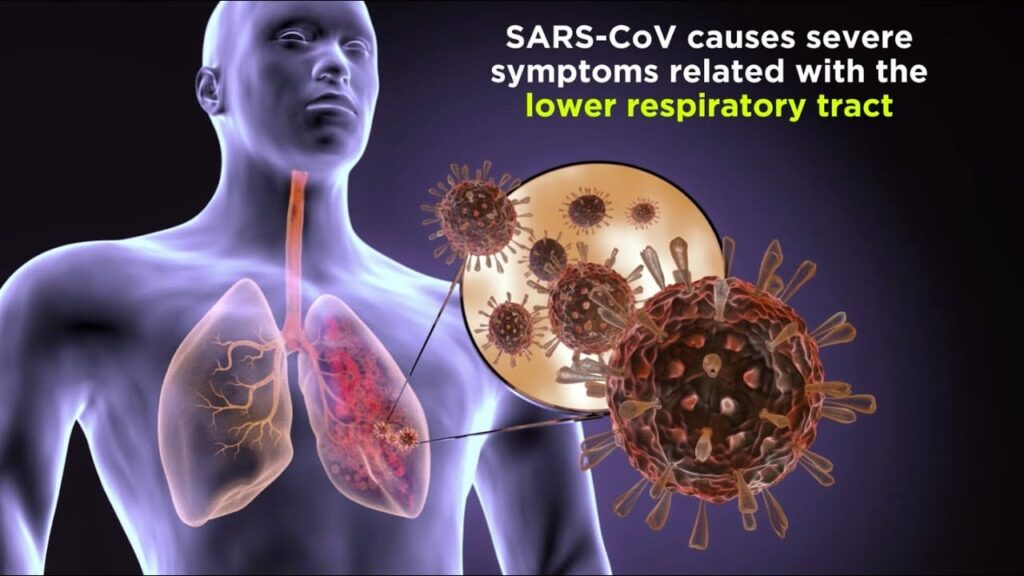
Severe Acute Respiratory Syndrome (SARS) is a viral respiratory illness caused by the SARS coronavirus (SARS-CoV), first identified in 2002. 

It’s characterized by a rapid onset of fever, cough, and shortness of breath, often progressing to pneumonia and acute respiratory distress syndrome (ARDS). 

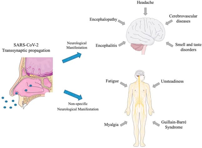
While the 2003 outbreak was effectively contained, the emergence of SARS-CoV-2 (the virus causing COVID-19) highlights the ongoing threat of novel coronaviruses. 

SARS is considered a zoonotic disease, meaning it originated in animals and then spread to humans. 


The exact animal origin remains uncertain, but evidence suggests it may have originated in bats, possibly with an intermediate animal host facilitating transmission to humans.

Forms and Routes of Transmission (1)

Forms and Routes of Transmission:

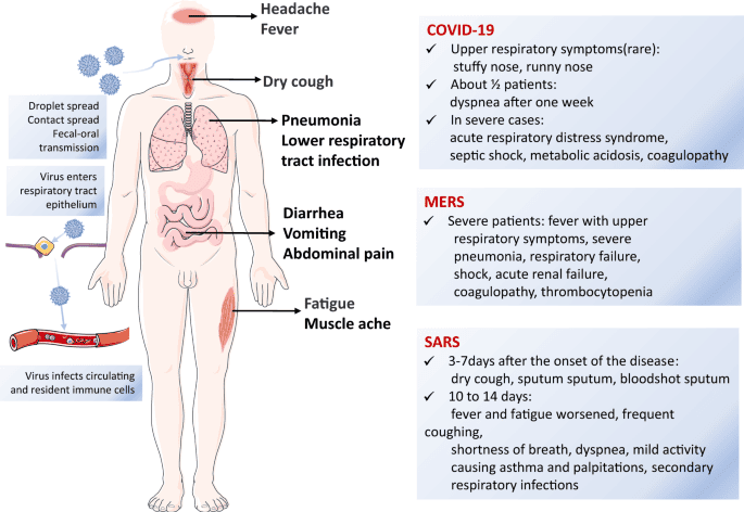
SARS primarily transmits through close contact with an infected individual. There are no known distinct “forms” of SARS like there are for anthrax (cutaneous, inhalation, etc.). The routes of transmission include:

  • Droplet Transmission: The primary route. Large respiratory droplets expelled during coughing, sneezing, or talking can infect individuals within close proximity (generally within 6 feet).

  • Contact Transmission: Touching contaminated surfaces (e.g., doorknobs, handrails) and then touching one’s face (eyes, nose, mouth) can lead to infection.

  • Fecal-Oral Transmission: Although less common, SARS-CoV RNA has been detected in stool samples, suggesting potential fecal-oral transmission, especially in healthcare settings.

Incubation Period:

The incubation period for SARS is usually 2-10 days, with a median of about 5 days. This means that symptoms may not appear until several days after exposure.

Causes/Etiology:

The causative agent is the SARS coronavirus (SARS-CoV), a positive-sense single-stranded RNA virus belonging to the Coronaviridae family.

Clinical Features

SARS presents with a range of symptoms, often starting with a relatively mild prodrome:

  1. High Fever: Typically above 38°C (100.4°F).
  2. Dry Cough: Often a prominent symptom.
  3. Shortness of Breath: Progressing to dyspnea (difficulty breathing).
  4. Myalgia (Muscle Aches): Widespread muscle pain.
  5. Headache: Often reported.
  6. Chills: Feeling cold and shivering.
  7. Fatigue: Significant exhaustion and weakness.
  8. Malaise: A general feeling of illness and discomfort.
  9. Diarrhea: Can occur in some patients.
  10. Sore Throat: May be present.
  11. Pneumonia: Often develops, leading to respiratory distress.
  12. Acute Respiratory Distress Syndrome (ARDS): A life-threatening complication involving severe lung inflammation.

Definitive Diagnosis and Investigations:

Diagnosis is confirmed through laboratory testing:

  • Reverse Transcription-Polymerase Chain Reaction (RT-PCR): Detects the SARS-CoV RNA in respiratory samples (e.g., nasal swabs, sputum). This is the gold standard for diagnosis.
  • Serological Tests: Detect antibodies against SARS-CoV in blood samples. These tests are helpful for retrospective diagnosis but may not be positive early in the course of infection.
  • Chest X-ray or CT Scan: May show characteristic findings of pneumonia. These imaging techniques help assess lung involvement but are not specific for SARS.

Management:

Aims of Management:

  • Supportive care to manage symptoms and complications.
  • Prevention of secondary infections.
  • Prevention of the spread of the virus.

Emergency Management:

Patients with severe respiratory distress (e.g., hypoxia, ARDS) require immediate emergency care including oxygen therapy, mechanical ventilation, and intensive care unit (ICU) admission.

First Aid/Initial Management:

  • Isolate the suspected patient to prevent further spread.
  • Provide supportive care: fluids, rest, fever control (acetaminophen).
  • Seek immediate medical attention.

Medical Management:

  • Antiviral Medications: No specific antiviral treatment proved definitively effective against SARS-CoV during the 2003 outbreak. Research is ongoing. However, supportive care is paramount.
  • Oxygen Therapy: For patients with hypoxia.
  • Mechanical Ventilation: For patients with severe respiratory failure and ARDS.
  • Corticosteroids: May be used in some cases to reduce inflammation, but their benefit is still debated.

Nursing Care:

  • Strict Infection Control: Use appropriate personal protective equipment (PPE) – gowns, gloves, masks, eye protection – to prevent transmission.
  • Respiratory Support: Monitor oxygen saturation, provide oxygen therapy, and assist with mechanical ventilation.
  • Fluid Balance: Monitor fluid intake and output, and administer intravenous fluids as needed.
  • Monitoring Vital Signs: Closely monitor temperature, heart rate, blood pressure, respiratory rate, and oxygen saturation.
  • Psychological Support: Provide emotional support to the patient and their family.
  • Isolation and Ventilation: Admit patients to a well-isolated and ventilated area to prevent further spread.
  • Protective Gear: Wear appropriate personal protective equipment (PPE) like gowns, gloves, masks, and eye protection when caring for SARS patients.
  • History and Physical Examination: Take a thorough history and perform a general physical examination.
  • Vital Signs: Monitor temperature, pulse, respiration, and blood pressure regularly and record the findings.
  • Temperature Management: For high fever, tepid sponging may be used to reduce body temperature.
  • Oxygen Therapy: Provide supplemental oxygen for patients with hypoxemia (low oxygen levels in the blood).
  • Medication Administration: Administer prescribed medications following proper protocols.
  • Hygiene: Practice strict hand hygiene – washing hands before and after caring for the patient.
  • Limit Staff Exposure: Minimize the number of healthcare workers caring for the patient to reduce transmission risk.
  • Reporting: Report suspected cases to the appropriate authorities for effective case management.

Management Up to Discharge:

Patients must meet specific criteria for discharge, including resolution of fever, improvement in respiratory symptoms, and two negative RT-PCR tests.

Advice on Discharge:

  • Continue to monitor for any recurrence of symptoms.
  • Contact a healthcare provider immediately if symptoms worsen.
  • Follow up appointments as scheduled.

Prevention:

  • Infection Control: Strict adherence to infection control measures in healthcare settings and other high-risk environments.

  • Hygiene: Frequent handwashing with soap and water or alcohol-based hand sanitizer.

  • Respiratory Hygiene: Covering coughs and sneezes with a tissue or elbow.

  • Quarantine: Isolation of infected individuals to prevent transmission.

  • Contact Tracing: Identifying and monitoring individuals who have been in contact with infected persons.

  • Early Detection and Case Management: Prompt identification and treatment of infected individuals.

Complications:

  • Pneumonia: A common complication that can be life-threatening.

  • Acute Respiratory Distress Syndrome (ARDS): A severe lung condition leading to respiratory failure.

  • Sepsis: A systemic inflammatory response to infection.

  • Multiple Organ Failure: Can occur in severe cases.

Quick Quiz

SARs Quiz

Tropical Medicine - mobile-friendly and focused practice.

Privacy: Your details are used only for quiz tracking and certificates.

Severe Acute Respiratory Syndrome (SARS) Read More »

Hepatitis B:
Home > Tropical > Hepatitis B

Hepatitis B

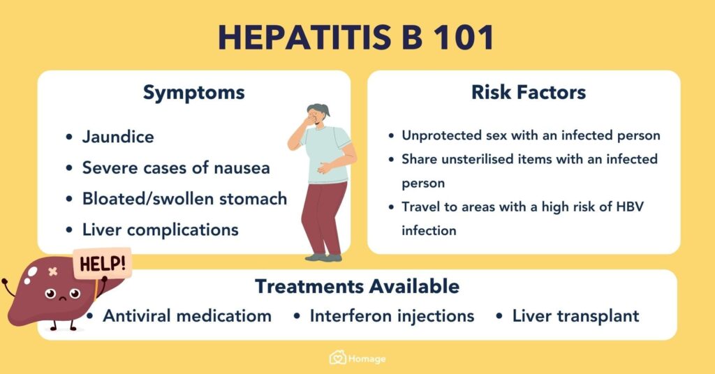
Overview: Hepatitis B is a potentially life-threatening liver infection caused by the Hepatitis B Virus (HBV). It is a major global health problem and a significant occupational hazard for midwives and health workers. In midwifery, our greatest focus is preventing Mother-to-Child Transmission (MTCT) during childbirth, as babies infected at birth have a 90% chance of developing chronic liver disease or liver cancer later in life.

1. Aetiology and Pathophysiology

The Causative Agent

  • The Virus: Hepatitis B is caused by the Hepatitis B Virus (HBV).
  • Virus Family: It belongs to the Hepadnaviridae family.
  • Virus Type: Unlike Hepatitis A or C, Hepatitis B is a DNA virus (it contains double-stranded DNA).
  • Resilience: The virus is extremely tough. It can survive outside the body on dry surfaces (like tables, medical instruments, or dried blood spots) for at least 7 days and remain highly infectious.

How the Virus Damages the Liver (Pathophysiology)

Interestingly, the Hepatitis B virus itself does not directly kill the liver cells (hepatocytes). Instead, the damage is immune-mediated:

  • The virus enters the bloodstream and travels straight to the liver, where it enters the liver cells and begins to multiply.
  • The body's immune system (specifically cytotoxic T-lymphocytes) recognizes that the liver cells are infected.
  • In an attempt to destroy the virus, the immune system aggressively attacks and destroys its own infected liver cells.
  • This massive immune attack causes severe inflammation of the liver (Hepatitis), leading to swelling, tissue death (necrosis), and the leaking of liver enzymes (ALT and AST) into the blood.

2. Modes of Transmission (How It Spreads)

Hepatitis B is highly contagious—it is 50 to 100 times more infectious than HIV. It is transmitted when blood, semen, or other body fluids from a person infected with the virus enter the body of someone who is not infected.

  • Vertical Transmission (Mother-to-Child): This is the most common route of infection in highly endemic areas like Uganda. It mostly happens during childbirth when the baby is exposed to the mother's infected blood and vaginal fluids in the birth canal.
  • Horizontal Transmission (Early Childhood): Spreading from an infected child to an uninfected child during the first 5 years of life (through minor cuts, bites, or sharing toothbrushes/chewed food).
  • Sexual Contact: Unprotected sexual intercourse (vaginal, anal, or oral) with an infected partner. The virus is heavily present in semen and vaginal secretions.
  • Contaminated Needles and Syringes: Sharing injecting equipment (common among drug users), or accidental needle-stick injuries among nurses and midwives.
  • Medical and Traditional Procedures: Receiving unsafe blood transfusions, using unsterilized surgical or dental equipment, traditional scarification, tribal mark cutting, or sharing unsterilized razor blades.
  • Sharing Personal Items: Sharing items that may carry microscopic drops of blood, such as shaving razors, nail clippers, or toothbrushes.

Note: Hepatitis B is NOT spread through breastfeeding, sharing eating utensils, hugging, kissing, holding hands, coughing, sneezing, or through contaminated food or water.
3. Clinical Features and Stages

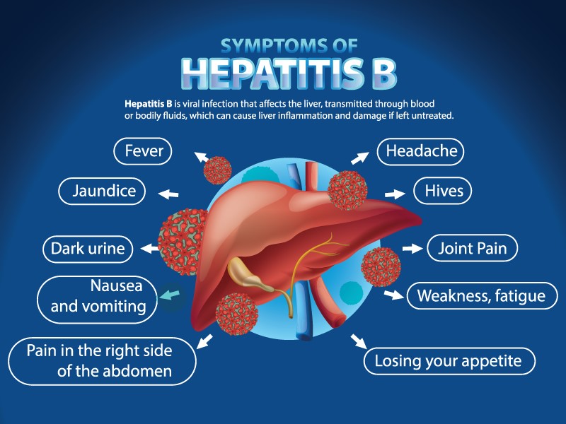
The incubation period (the time from infection to the appearance of symptoms) ranges from 60 to 150 days (average is 90 days). The clinical presentation occurs in distinct phases:

1. Prodromal Phase (Pre-Icteric Phase)

This is the early stage before jaundice appears. Symptoms are vague and look like a bad flu or malaria:

  • Low-grade fever.
  • Severe fatigue, weakness, and general body aches (malaise).
  • Loss of appetite (anorexia), nausea, and vomiting.
  • Mild pain in the upper right side of the abdomen (where the liver is located).
  • Joint pains (arthralgia) and skin rashes.

2. Icteric Phase (The Jaundice Stage)

This phase begins 1 to 2 weeks after the early symptoms. The liver is now actively inflamed and failing to clear waste from the blood.

  • Jaundice: Yellowing of the eyes (sclera) and the skin due to high levels of bilirubin in the blood.
  • Dark Urine: The urine becomes the color of dark tea or Coca-Cola because excess bilirubin is being passed out through the kidneys.
  • Pale Stools: The stool becomes very light, clay-colored, or white because bile is blocked from reaching the intestines.
  • Hepatomegaly: The liver becomes swollen, enlarged, and very tender to touch.
  • Extreme itching of the skin (pruritus) due to bile salts accumulating under the skin.

3. Convalescent Phase (Recovery)

  • The jaundice slowly fades away, and the dark urine clears up.
  • Appetite returns, and the patient slowly regains energy.
  • Complete recovery in healthy adults usually takes 3 to 6 months. In about 90% of healthy adults, the immune system successfully clears the virus, giving them lifelong immunity.

4. Chronic Hepatitis B

  • If the body fails to clear the virus after 6 months, the infection becomes chronic (lifelong).
  • Chronic infection is very common in infants (90% of infected babies become chronic carriers) but rare in adults (only 5% become chronic).
  • Chronic carriers often have no symptoms for decades, but they slowly develop liver scarring (cirrhosis) and are at massive risk of liver cancer.

4. Diagnostic Investigations (Hep B Serology)

Diagnosing Hepatitis B relies heavily on blood tests to look for viral proteins (antigens) and the body's immune defense proteins (antibodies).

Serological MarkerWhat it Means in Simple Terms
HBsAg (Hepatitis B Surface Antigen)This is the outer coat of the virus. If this test is Positive, it means the person is currently infected and can pass the virus to others. (If positive for >6 months, it indicates Chronic Hep B).
Anti-HBs (Hepatitis B Surface Antibody)This is the body's defense against the virus. If this is Positive, it means the person is completely immune and protected. (This happens either because they were successfully vaccinated, or they recovered from a past infection).
HBeAg (Hepatitis B e-Antigen)This indicates active viral replication. If this is Positive, it means the virus is multiplying rapidly in the liver, and the person is highly infectious.
Anti-HBc IgM (Core Antibody IgM)"M" stands for Minute (recent). A positive result means an Acute, very recent infection (usually acquired within the last 6 months).
Anti-HBc IgG (Core Antibody IgG)"G" stands for Gone (past). A positive result means a Chronic or past infection.
HBV DNA (Viral Load)A highly advanced test that counts the exact number of virus particles in the blood. It helps the doctor decide if the patient needs to start taking daily antiviral drugs.

Other Supportive Investigations

  • Liver Function Tests (LFTs): Blood tests to check the levels of liver enzymes (ALT and AST). Very high levels show that the liver cells are currently being destroyed.
  • Abdominal Ultrasound Scan: Done to check the physical size of the liver, look for severe scarring (cirrhosis), or spot early signs of liver cancer tumors.

5. Hepatitis B in Pregnancy & Midwifery Management

Effect of Pregnancy on Hepatitis B

  • Pregnancy generally does not make a chronic Hepatitis B infection worse.
  • However, acute Hepatitis B acquired late in the third trimester can sometimes be severe and increase the risk of maternal death.

Effect of Hepatitis B on Pregnancy

  • High risk of premature labor (preterm birth).
  • Low birth weight babies.
  • Gestational diabetes risk is slightly elevated.
  • High risk of severe bleeding during delivery because a damaged liver cannot produce enough blood-clotting factors (like Fibrinogen).

🚨 Midwifery Alert: PMTCT of Hepatitis B

Prevention of Mother-To-Child Transmission (PMTCT) is the midwife's absolute highest priority. If an HBsAg positive mother delivers, the baby is exposed to massive amounts of infected blood. If the baby is not protected immediately, they will become a chronic carrier.

Midwifery Care & Delivery Management

  • Antenatal Care: Every single pregnant woman MUST be screened for HBsAg during her first ANC visit.
  • Antiviral Therapy in Pregnancy: If the mother's viral load (HBV DNA) is very high, the doctor will start her on Tenofovir (TDF) in the 3rd trimester (usually at 28 weeks). Tenofovir is highly effective and completely safe for the baby. It lowers the viral load to prevent transmission during birth.
  • Conducting the Delivery:
    • Strict Infection Prevention and Control (IPC) must be maintained. The midwife must wear double gloves, goggles, and an apron to prevent accidental blood splashes.
    • Avoid unnecessary traumatic procedures that mix mother and baby's blood (e.g., avoid routine episiotomies, avoid artificial rupture of membranes, and avoid vacuum extractions unless absolutely necessary).
  • Immediate Newborn Care (The First 12 Hours):
    • Immediate Washing: Carefully wipe and wash off all maternal blood and vaginal fluids from the newborn's skin immediately before giving any injections.
    • Birth Dose Vaccine: Administer the single-antigen Hepatitis B birth dose vaccine within 12 to 24 hours of birth.
    • HBIG Administration: Give Hepatitis B Immune Globulin (HBIG) along with the vaccine (in the opposite thigh) within 12 hours. HBIG provides instant, ready-made antibodies to fight off any virus that entered during birth.
  • Breastfeeding: Breastfeeding is 100% encouraged and perfectly safe as long as the baby receives the birth dose vaccine and HBIG. The mother must ensure she has no cracked, bleeding nipples.

6. Medical Management and Treatment

Acute Hepatitis B

There is no specific cure or strong medicine for acute Hepatitis B. Care is strictly supportive:

  • Strict bed rest to reduce the liver's workload.
  • Encourage plenty of oral fluids and fresh fruit juices to provide energy and prevent dehydration from vomiting.
  • Provide a high-carbohydrate, low-fat, and moderate-protein diet. (The liver struggles to digest heavy fats).
  • Avoid all alcohol completely, as it acts as a direct poison to the already damaged liver.
  • Strictly avoid drugs that are processed by the liver (hepatotoxic drugs), such as heavy doses of Paracetamol.

Chronic Hepatitis B

If the infection lasts more than 6 months, the patient needs long-term suppression of the virus.

  • Antiviral Medications: The goal is to completely suppress the virus from multiplying, though it rarely cures it completely. The two most effective, once-daily oral pills used globally and in Uganda are Tenofovir (TDF) and Entecavir.
  • Interferon : Interferon alfa-2b is a synthetic version of a protein that the body naturally produces. Injections of interferon alfa-2b are given to suppress the growth of the HBV virus in the body. It can also help to reduce the intensity of liver damage.
  • Liver transplant: If liver damage has advanced significantly, you or your loved one may need to undergo a liver transplant. This surgical procedure removes the damaged liver and replaces it with a healthy one from either a living or deceased donor. As the liver can regenerate slowly, it is possible to use a part of a healthy liver for the transplant. An unfortunate side effect of a transplant is that the receiver will need to take anti-rejection drugs for as long as they live.
  • Regular Monitoring: Chronic patients must undergo an ultrasound scan every 6 months to catch early signs of liver cancer.

7. Severe Complications of Hepatitis B

If chronic Hepatitis B is left unmonitored and untreated, the constant immune attacks on the liver lead to devastating consequences:

  • Liver Cirrhosis: The healthy, soft liver tissue is completely replaced by hard, useless scar tissue. The liver shrinks and stops working.
  • Hepatocellular Carcinoma (HCC): Primary liver cancer. Hepatitis B is one of the leading causes of cancer deaths worldwide.
  • Fulminant Hepatitis: A rare but deadly acute complication where the liver completely shuts down (acute liver failure) within days of infection, leading to a deep coma and death.
  • Portal Hypertension: Because the scarred liver blocks blood flow, pressure builds up in the abdominal veins, causing massive fluid buildup in the belly (ascites) and swollen, bleeding veins in the throat (esophageal varices).

8. General Prevention and Immunization

1. Universal Vaccination

The Hepatitis B vaccine is highly effective (98-100% protection) and is the ultimate way to prevent the disease.

  • Uganda EPI Schedule: The vaccine is combined into the Pentavalent vaccine (DPT-HepB-Hib). It is given to all infants at 6 weeks, 10 weeks, and 14 weeks of age.
  • Healthcare Workers: Every midwife, nurse, and medical student MUST receive the full 3-dose series of the adult Hepatitis B vaccine (at 0, 1, and 6 months) to protect themselves from needle-stick injuries.

2. Post-Exposure Prophylaxis (PEP)

  • If an unvaccinated midwife accidentally pricks her finger with a needle used on an HBsAg positive patient, she must immediately receive the Hepatitis B Immune Globulin (HBIG) injection within 24 hours, followed by the first dose of the Hepatitis B vaccine.

3. Standard Infection Control

  • Always use strict Universal Precautions (gloves, aprons).
  • Never recap used needles (this is the number one cause of needle-stick injuries). Dispose of them immediately in a puncture-proof sharps box.
  • Properly sterilize all medical, surgical, and dental equipment.
  • Ensure 100% safe screening of all donated blood before transfusions.
  • Promote safe sex practices and the consistent use of condoms.

❓ Quick Review Question

Scenario: You are reviewing the lab results for an antenatal mother. Her serology shows: HBsAg = Negative and Anti-HBs = Positive. What does this mean, and what is your midwifery action?

  • Answer: This means the mother does not have the virus (she is negative for the surface antigen) but she has complete immunity (she is positive for the surface antibody). She is perfectly protected, either from a past infection that healed or from a previous vaccination. No medical action or PMTCT is required for the baby regarding Hepatitis B.

Quick Quiz

Hepatitis B Quiz

Tropical Medicine - mobile-friendly and focused practice.

Privacy: Your details are used only for quiz tracking and certificates.

Hepatitis B Read More »

Scabies
Home >Tropical > Scabies

Scabies: Comprehensive Nursing Guide & Management

Overview: Scabies is a highly contagious, intensely itchy skin infestation caused by the microscopic mite Sarcoptes scabiei. The female mite burrows deep into the outermost layer of the skin to lay her eggs, triggering a severe allergic reaction. It is a massive public health problem globally, especially affecting populations living in crowded environments, refugee camps, prisons, or places with compromised hygiene.

1. Etiology and Pathophysiology

The Causative Agent

  • The causative agent is the Sarcoptes scabiei mite (specifically the variety that affects humans, var. hominis).
  • It is a microscopic, eight-legged parasite that cannot be seen easily with the naked eye.

How It Damages the Skin (Pathophysiology)

  • The Burrowing Process: The pregnant female mite burrows into the stratum corneum (the dead, outermost layer of the epidermis). She tunnels through the skin at a rate of 2 to 3 millimeters per day.
  • Laying Eggs: As she tunnels, she deposits 2 to 3 eggs per day, along with her fecal matter (known clinically as scybala).
  • The Immune Reaction: The intense itching and characteristic rash are not caused by the mite biting. Instead, the host's immune system develops a delayed hypersensitivity (allergic) reaction to the mite's saliva, eggs, and toxic fecal matter left behind in the burrows.
  • The Life Cycle: The eggs hatch into larvae within 3 to 4 days, crawl out to the skin surface, mature into adults, mate, and repeat the cycle.

2. Forms and Routes of Transmission

Scabies is incredibly easy to catch. It is transmitted through two main routes:

  • Direct Skin-to-Skin Contact: This is the primary and most common route. It requires prolonged physical contact. A quick handshake usually will not spread it. It is commonly transmitted during:
    • Sexual activity.
    • Close physical bonding (e.g., mothers cuddling infants, children wrestling).
    • Sleeping in the same bed with an infested person.
  • Indirect Contact (Fomites): Transmission can occur by sharing contaminated inanimate objects like bedding, clothing, or towels. While less frequent than direct contact, the mites can survive off a human host for 48 to 72 hours at room temperature.

Incubation Period

The incubation period is the time between the mites first landing on the skin and the patient actually feeling the itch or seeing a rash.

  • Primary Infestation: If a person has never had scabies before, it takes 4 to 6 weeks for the immune system to build up the allergic reaction. During this silent month, the person is highly contagious but has no idea they are sick.
  • Re-infestation: In individuals previously exposed to scabies, the immune system already recognizes the mite proteins. Symptoms appear much more rapidly, usually within 1 to 4 days.

3. Types of Scabies

Scabies generally presents in two distinct clinical forms depending on the patient's immune system:

FeatureTypical (Classic) ScabiesCrusted (Norwegian) Scabies
Patient ProfileNormal, healthy immune system.Immunocompromised patients (HIV/AIDS, elderly, people on heavy steroids).
Mite LoadLow. Only about 10 to 15 live mites on the entire body.Massive. Thousands to millions of mites multiplying freely.
Clinical AppearanceSmall itchy bumps, linear burrows, scratch marks.Thick, heavy, grey crusts and scales over large areas of the body (looks like severe psoriasis).
Itchiness (Pruritus)Intense, severe, keeps the patient awake at night.Often minimal or absent because the weak immune system does not react to the mites.
ContagiousnessContagious, but usually requires prolonged contact.Extremely contagious. Even brief contact with the patient's clothing or shed skin crusts can spread the massive mite load.

4. Clinical Features (Signs and Symptoms)

  • Intense Nocturnal Pruritus: Severe itching that is classically worse at night. This happens because mites become more active when the skin is warmed under blankets.
  • Papular Rash: Small, red, raised, extremely itchy bumps (papules) that may be clustered in groups or widespread.
  • Linear Burrows: The hallmark sign of scabies. These are thin, slightly raised, grayish-white zigzag lines in the skin measuring 2-10 mm long. They represent the actual tunnel the mite dug.
  • Excoriations: Severe scratch marks and open abrasions resulting from the patient's desperate attempts to relieve the intense itching.
  • Nodules: Small, firm, reddish-brown lumps. (Often found on the male genitalia or groin, and can remain intensely itchy for weeks even after all mites are killed, due to trapped dead mite parts).

Distribution of the Rash

Scabies mites favor areas of the body where the skin is thin, warm, and soft.

  • In Adults/Older Children: Web spaces between the fingers (interdigital webs), the inner wrists, elbows, armpits (axillae), around the umbilicus (belly button), buttocks, female nipples, and male genitalia. (It usually spares the head and face in adults).
  • In Infants/Toddlers: Because their skin is soft everywhere, the rash is widespread and commonly affects the palms of the hands, soles of the feet, scalp, and face.

⚠️ Clinical Alert: Secondary Bacterial Infection

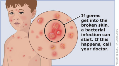
Because the patient scratches their skin open constantly, the protective barrier is broken. Normal skin bacteria (like Staphylococcus aureus or Streptococcus pyogenes) invade the wounds. This causes serious secondary infections like Impetigo (pus-filled sores), Cellulitis, or dangerously, Post-Streptococcal Glomerulonephritis (severe kidney damage triggered by the strep bacteria entering the blood).

Classic presentation of a scabies burrow located in the thin skin web space between the fingers.

5. Definitive Diagnosis and Investigations

The diagnosis of scabies is primarily clinical. A midwife or doctor can usually diagnose it simply by taking a good history (especially asking if others in the house are itching at night) and finding the characteristic rash and burrows.

  • Clinical Identification: Looking for the classic distribution (finger webs, wrists, genitals) and asking about nocturnal itching.
  • Microscopic Skin Scraping: The definitive gold standard. A drop of mineral oil is placed on a burrow. The burrow is scraped firmly with a sterile scalpel blade. The scrapings are placed on a glass slide and viewed under a microscope. Finding the actual mite, its oval eggs, or its brown fecal pellets confirms the diagnosis.
  • The Ink Test (Burrow Ink Test): A drop of dark ink from a pen or marker is rubbed over a suspicious itchy bump. The surface ink is then wiped away with an alcohol swab. If there is a burrow, the ink will remain trapped inside the tunnel, revealing a clear, dark zigzag line.
  • Dermoscopy: Using a special magnifying skin tool to look for the "delta wing" or "jet with condensation trail" sign, which is the shape of the mite's head at the end of the burrow.

6. Management of Scabies

Aims of Management: The primary goals are to absolutely eradicate the living mites and their eggs, relieve the intense itching symptoms, prevent secondary bacterial complications, and halt transmission to the community.

Medical Management (Scabicides)

Topical creams and lotions (scabicides) are the absolute mainstay of treatment. They paralyze and kill the mites.

MedicationApplication & Clinical Notes
Permethrin 5% CreamThe Drug of Choice. Highly effective and safe (even for infants >2 months and pregnant women). Applied neck-down, left on for 8-14 hours (usually overnight), then washed off.
Malathion 0.5% LotionAn alternative if Permethrin fails or is unavailable. Left on the body for 24 hours before washing.
Ivermectin (Oral Tablets)Taken by mouth. Highly effective. Specially reserved for severe Crusted Scabies, institutional outbreaks, or patients who physically cannot apply creams. Usually given as two doses, one week apart.
Lindane 1% LotionRarely used today. It carries a severe risk of neurotoxicity (can cause seizures), especially in young children, premature infants, or if left on the skin too long.
Benzyl Benzoate 25%A cheaper, older alternative commonly used in developing settings. Must be applied for 24 hours, but can cause severe skin irritation.

Step-by-Step Nursing Care & Medication Administration

The success of the treatment depends almost entirely on how well the nurse educates the patient on how to apply the cream. If a single spot is missed, the mites survive.

  • Proper Application Technique:
    • The patient should take a lukewarm shower and dry off completely before applying the cream. (Hot water dilates blood vessels and causes the body to absorb too much medicine into the blood instead of keeping it on the skin).
    • Massage the cream thoroughly into the entire body from the chin down to the toes.
    • Pay special attention to hiding spots: deep between the fingers and toes, under the fingernails (cut nails short!), the groin, and the cleft of the buttocks.
    • For infants and the elderly, the cream must also be applied to the scalp and face (avoiding the eyes and mouth).
  • Duration: Leave the medicine on for the prescribed time (usually overnight, 8 to 14 hours). If the patient washes their hands to eat or use the toilet during the night, they must reapply the cream to their hands immediately.
  • The "Second Dose" Rule: Creams kill live mites but struggle to kill unhatched eggs. A second application is almost always required exactly 7 days later to kill the newly hatched larvae before they can lay more eggs.
  • Treating Contacts: The most important rule in scabies management: All household members and close sexual contacts MUST be treated at the exact same time, even if they have absolutely no itching or rash! (Remember the 4-6 week silent incubation period).

Symptom Management & Monitoring

  • Post-Scabietic Itch: Warn the patient that the intense itching may continue for 2 to 4 weeks even after all the mites are completely dead. This is because the dead mite bodies and feces are still trapped under the skin, keeping the allergy active until the skin naturally sheds.
  • Help manage this itching using soothing cool compresses, calamine lotion, and oral antihistamines (like Chlorpheniramine or Cetirizine) to help them sleep. Mild topical steroid creams (Hydrocortisone) can be prescribed for severe inflammation.
  • Monitoring: Closely examine the patient's skin for signs of secondary bacterial infection (increased redness, massive swelling, severe pain, warm to touch, or yellow pus). If present, systemic antibiotics are required.

7. Prevention and Environmental Control

Because mites can survive off the body, the patient's environment must be aggressively decontaminated.

  • Laundering: All clothing, underwear, towels, and bedsheets used by the infested person within the last 3 to 4 days before treatment must be washed in hot water and dried on the highest heat cycle. Heat kills the mites and eggs instantly.
  • The Plastic Bag Method: Items that cannot be washed (like heavy blankets, stuffed toys, or certain shoes) should be sealed tightly in airtight plastic bags and locked away for 72 hours (3 days). The mites will starve and die without a human host.
  • Good Hygiene: Practice frequent handwashing with soap and water. Never share personal items such as towels, bed sheets, or unwashed clothing.
  • Limit Contact: Avoid direct skin-to-skin contact with individuals known or suspected to have scabies until they have completed their medical treatment.
  • Institutional Control: In hospitals or nursing homes, if a patient is diagnosed with severe Crusted Scabies, they must be placed in strict contact isolation rooms immediately to prevent a massive outbreak among health workers and other patients.

❓ Quick Clinical Review

Scenario: A mother brings her 4-year-old child to the clinic. You successfully treat the child with Permethrin. One week later, the mother returns angrily, stating the medicine didn't work because her child is still scratching severely. What is your nursing explanation?

  • Answer: You must kindly educate the mother about "Post-Scabietic Pruritus." Explain that the medicine successfully killed the mites, but the child's body is still having an allergic reaction to the dead bugs and waste trapped under the skin. The itching can last for up to 4 weeks while the skin naturally sheds. Prescribe antihistamines to comfort the child.

8. References

  • Uganda Ministry of Health (MoH) Clinical Guidelines for the Management of Common Conditions.
  • World Health Organization (WHO). Fact Sheet on Scabies and Ectoparasites.
  • UNMEB Curriculum for Diploma in Midwifery/Nursing - Dermatology and Parasitic Infections.

Quick Quiz

Scabies Quiz

Tropical Medicine - mobile-friendly and focused practice.

Privacy: Your details are used only for quiz tracking and certificates.

Scabies Read More »

Rabies

RABIES

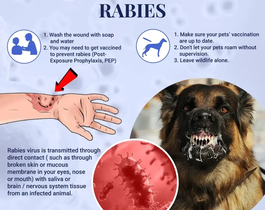
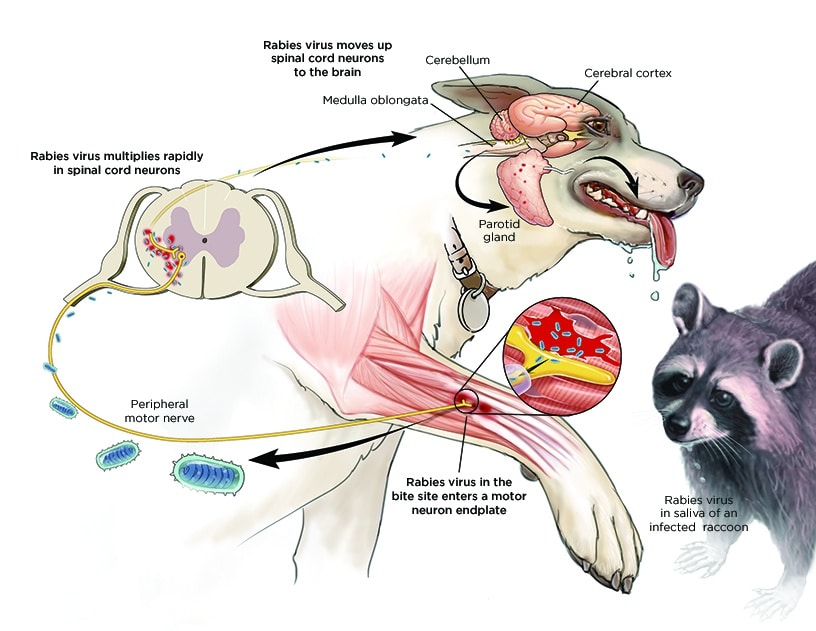
Rabies, also known as hydrophobia, is a fatal viral infection of the central nervous system (CNS) characterized by inflammation and acute encephalitis. It’s caused by contact with the saliva of an infected animal.

Causes:

Rabies is caused by the rabies virus, a single-stranded RNA virus with a bullet-shaped morphology (130-300 nm). It belongs to the Lyssavirus genus of the Rhabdoviridae family and possesses an external envelope with short projections.

Source/Reservoir:

The primary reservoirs are infected wild animals, particularly those in the Canidae (dogs, foxes, etc.) and Felidae (cats, leopards, lions, etc.) families. Domestic dogs also serve as significant reservoirs, especially in areas with limited rabies control programs. Humans are accidental hosts.

Transmission:

Transmission primarily occurs through the saliva of a rabid animal, typically via a bite. Other less common routes include:

  • Direct inoculation: A bite from a rabid animal directly introduces the virus into tissues.
  • Mucosal contact: Saliva contact with mucous membranes (eyes, nose, mouth), particularly on broken skin.
  • Aerosolization (rare): Inhalation of aerosolized saliva, primarily in bat caves or during close contact with infected animals.
Routes of Transmission: The routes highlight the direct entry of the virus into the body: Neural route: The virus travels along peripheral nerves to the central nervous system (CNS). This is the primary route and explains the neurotropic nature of rabies. Hematogenous route (less common): The virus can enter the bloodstream and spread throughout the body, though neural spread is the predominant mechanism. Incubation Period: The incubation period varies (2 weeks to 1 year, averaging 2-342 days), depending on: Bite site: Bites closer to the CNS (head, neck) have shorter incubation periods. Tissue penetration: Deeper wounds allow for faster viral dissemination. Viral load: Higher viral loads result in shorter incubation periods. Site of the bite: Bites on the head and neck tend to have shorter incubation periods than bites on the extremities due to proximity to the brain. Pathology: Following inoculation, the virus initially replicates at the bite site for approximately 96 hours. It then spreads via peripheral nerves to the spinal cord and brain, primarily replicating in the gray matter. The virus subsequently disseminates through autonomic nerves to various organs (salivary glands, adrenal medulla, kidneys, lungs, liver, skeletal muscles, and skin). At this stage, the patient's saliva and secretions become infectious.

Routes of Transmission:

The routes highlight the direct entry of the virus into the body:

  • Neural route: The virus travels along peripheral nerves to the central nervous system (CNS). This is the primary route and explains the neurotropic nature of rabies.
  • Hematogenous route (less common): The virus can enter the bloodstream and spread throughout the body, though neural spread is the predominant mechanism.

Incubation Period:

The incubation period varies (2 weeks to 1 year, averaging 2-342 days), depending on:

  • Bite site: Bites closer to the CNS (head, neck) have shorter incubation periods.
  • Tissue penetration: Deeper wounds allow for faster viral dissemination.
  • Viral load: Higher viral loads result in shorter incubation periods.
  • Site of the bite: Bites on the head and neck tend to have shorter incubation periods than bites on the extremities due to proximity to the brain.

Pathology:

Following inoculation, the virus initially replicates at the bite site for approximately 96 hours. It then spreads via peripheral nerves to the spinal cord and brain, primarily replicating in the gray matter. The virus subsequently disseminates through autonomic nerves to various organs (salivary glands, adrenal medulla, kidneys, lungs, liver, skeletal muscles, and skin). At this stage, the patient’s saliva and secretions become infectious.

Clinical Presentations:

Rabies progresses through distinct stages:

Prodromal Stage(pre-encephalitic): This initial phase is characterized by nonspecific symptoms:

  • Pain at the bite site
  • Headache
  • Fever
  • Malaise
  • Weakness
  • Anorexia
  • Vomiting
  • Sore throat
  • Non-productive cough

Encephalitic Phase

Furious (Excitory) Stage: Neurological symptoms become prominent:

  • Excessive motor activity, agitation, excitation
  • Confusion, anxiety, hallucinations
  • Muscle spasms
  • Aggression
  • Seizures
  • Hypersensitivity to light, noise, touch, temperature
  • Dilated pupils, lacrimation (excessive tearing), drooling, sweating
  • Hydrophobia (fear of water)
  • Aerophobia (fear of drafts)

Paralytic Stage:  Progressive paralysis sets in:

  • Pharyngeal spasms (difficulty swallowing), dysphagia, odynophagia
  • Weakness spreading from the bite site, leading to constipation, urinary retention, respiratory failure
  • Coma
  • Death

Survival beyond a week after the onset of encephalitic symptoms is uncommon.

Management:

Medical management of developed rabies is largely supportive and focuses on alleviating symptoms and maintaining vital organ function. Unfortunately, there is no specific treatment that cures rabies once clinical symptoms are evident.

Aims:

  • Prevent the progression of rabies to the encephalitic stage.
  • Provide supportive care to maintain vital functions.
  • Prevent further transmission of rabies if the patient is rabid.

First Aid Management:

Immediate wound care is crucial to minimize viral load:

  1. Thoroughly wash the wound with soap and water for at least 15 minutes. Scrub the wound gently.
  2. Rinse thoroughly with copious amounts of clean water.
  3. Leave the wound open (do not suture).

Hospital Management:

  • Admission: Isolate the patient in a barrier room to prevent transmission.
  • Treatment:
  1. Antibiotics: Systemic antibiotics (e.g., penicillin, metronidazole, doxycycline) to prevent secondary wound infections. Dosage adjustments are necessary for children and pregnant individuals (metronidazole and doxycycline are contraindicated in pregnancy).
  2. Passive Immunization: Administer rabies immunoglobulin (RIG) to neutralize the virus. Infiltrate RIG around and into the wound and give any remaining dose intramuscularly at a site distant from the rabies vaccine injection. If RIG cannot be given immediately, it may be administered within 7 days.
  3. Active Immunization: Administer rabies vaccine to stimulate an immune response. Vaccination schedules vary depending on pre- or post-exposure status and risk factors.
  4. Pre-exposure prophylaxis: For high-risk individuals (lab workers, wildlife personnel, etc.), a pre-exposure vaccination schedule (0, 7, 21 days) provides long-term protection. Boosters are needed periodically.
  5. Post-exposure prophylaxis: For those already bitten, a post-exposure regimen (2:1:1 schedule – 2 doses on day 0, 1 dose on day 7, 1 dose on day 21) is followed. This should be started as soon as possible after exposure.
  • Sedation: Sedatives (e.g., chlorpromazine, diazepam) to manage agitation, spasms, and convulsions.

  • Airway Management: Provide artificial ventilation and oxygen if respiratory failure occurs. Maintain a patent airway through suctioning or other appropriate measures.

  • Protection: Healthcare personnel should wear appropriate personal protective equipment (PPE), including gloves, gowns, masks,

Supportive Care:

  • Observation: Close monitoring of vital signs (heart rate, respiratory rate, blood pressure, temperature, oxygen saturation) is essential to detect early signs of respiratory or cardiac failure. Frequent monitoring (every 2-4 hours) and accurate charting are crucial.
  • Rest and Sleep: Provide a quiet, dimly lit environment to minimize stimulation. Rabies patients are hypersensitive to light, noise, touch, and temperature changes.
  • Nutrition: Nutritional support is critical. If the patient is sedated, feeding may be done via nasogastric tube (NGT) or intravenously (IV). If the patient is alert and able to swallow, oral feeding can be attempted. Close monitoring for aspiration is necessary.
  • Fluid Balance: Monitor fluid intake and output closely. Patients may experience dehydration due to excessive sweating or difficulty swallowing. Intravenous fluids may be necessary.
  • Hygiene: Maintain meticulous hygiene practices. Regular skin care, oral hygiene, and bladder care are essential.

Specific Treatment Considerations:

  • Cardiac Arrhythmias: Monitor for cardiac arrhythmias and treat as needed with appropriate medications (e.g., antiarrhythmics).
  • Respiratory Failure: If respiratory failure occurs, provide mechanical ventilation until spontaneous breathing resumes. The use of an ambu bag for artificial ventilation may be necessary if there’s paralysis of the respiratory muscles.
  • Seizures: Manage seizures with anticonvulsant medications (e.g., diazepam, phenytoin).
  • Pneumonia: Monitor for pneumonia and treat with appropriate antibiotics as needed.
  • Brain Edema and Increased Intracranial Pressure: Monitor for signs of increased intracranial pressure (e.g., headache, vomiting, altered mental status) and consider measures to reduce intracranial pressure (e.g., corticosteroids, osmotic diuretics).
  • Hyper- or Hypopyrexia: Treat fever or hypothermia with appropriate methods (e.g., antipyretics, cooling blankets).
  • Diabetes Insipidus: Monitor for signs of diabetes insipidus (e.g., polyuria, polydipsia) and treat with desmopressin.
  • Paralysis: Provide supportive care to manage paralysis. Range-of-motion exercises and physical therapy may be necessary once the acute phase has passed.
  • Hematemesis: Manage hematemesis (vomiting blood) with appropriate measures (e.g., intravenous fluids, blood transfusion).

Rabies Post-Exposure Prophylaxis (PEP) Management

Rabies PEP aims to prevent rabies development after contact with potentially rabid animal saliva through bites, scratches, or licks on broken skin or mucous membranes (ICD-10 Codes: Z20.3, Z23). Treatment should follow guidelines such as those provided by the Veterinary Public Health Unit.

Dealing with the Animal:

The management of the animal is crucial in determining the appropriate PEP for the exposed individual.

A. Identifiable and Catchable Animal:

1. Domestic Animal: Determine rabies vaccination status. If unvaccinated or status unknown, quarantine the animal for 10 days (dogs, cats, or endangered species only). Humane euthanasia and head submission to the veterinary department for rabies testing is necessary if quarantine is not feasible.

  • If no rabies signs within 10 days, release the animal and discontinue PEP for the human if already started.
  • If rabies signs develop, euthanize, submit the head for testing, and proceed with full PEP for the human.

2. Wild Animal: Humane euthanasia and head submission to the veterinary department for rabies testing are necessary.

  • If rabies is confirmed, initiate full PEP for the human.
  • If the test is negative, rabies PEP is not necessary but local wound care is advised

B. Unidentifiable Animal: Assume the animal was rabid and the patient is at risk; initiate full PEP.

Dealing with the Patient:

The cornerstone of rabies PEP is a combination of local wound treatment, passive immunization with rabies immunoglobulin (RIG), and active immunization with rabies vaccine (RV). Regardless of the time elapsed since exposure (even months later), treatment should be initiated as if exposure were recent.

A. Local Wound Treatment: Prompt and thorough local treatment significantly reduces infection risk. This includes:

  1. Thorough cleansing: Wash the wound with soap and water for at least 15 minutes, followed by copious rinsing with clean water.
  2. Mucous membrane contact: Thoroughly rinse with water or normal saline.
  3. Deep wounds: Administer tetanus toxoid (TT) to prevent tetanus.
  4. Wound closure: DO NOT suture the wound.
  5. Late presentation: Local cleansing is indicated even if the patient presents late for treatment.

B. Immunization: The need for RIG and RV depends on the exposure type and animal status: (RV and RIG are both very expensive and should only be used when there is an absolute indication)

Animal Condition at Time of Exposure

Nature of Exposure

10 Days Later

Recommended Action

Healthy

Saliva contact with skin, no lesion

Healthy

Do not vaccinate

Rabid

Saliva contact with skin, no lesion

N/A

Vaccinate

Suspect/Unknown

Saliva contact with skin, no lesion

Healthy

Do not vaccinate

Suspect/Unknown

Saliva contact with skin, no lesion

Rabid

Vaccinate

Suspect/Unknown

Saliva contact with skin, no lesion

Unknown

Vaccinate

Healthy

Saliva contact with skin lesions, minor bites

Healthy

Do not vaccinate

Rabid

Saliva contact with skin lesions, minor bites

N/A

Vaccinate

Suspect/Unknown

Saliva contact with skin lesions, minor bites

Healthy

Vaccinate; stop if animal healthy after 10 days

Suspect/Unknown

Saliva contact with skin lesions, minor bites

Rabid

Vaccinate

Suspect/Unknown

Saliva contact with skin lesions, minor bites

Unknown

Vaccinate

Rabid or Suspect

Saliva contact with mucous membranes, serious bites (face, head, fingers, or multiple bites)

N/A

Vaccinate and give RIG

Rabid or Suspect

Saliva contact with mucous membranes, serious bites (face, head, fingers, or multiple bites)

N/A

Vaccinate; stop if animal healthy after 10 days


Rabies Vaccine Schedules

Intramuscular Regimen

DAY

Vaccine Dose

No. of Doses

Comments

0

0.5 ml

2 (one in each deltoid)

Into the deltoid muscle. NEVER IN THE GLUTEAL MUSCLE (buttocks).

7

0.5 ml

1

Children with less muscle mass: Anterolateral aspect of the thigh.

21

0.5 ml

1

Note: Day 14 is skipped. The 2:1:1 regimen uses 4 doses in 3 weeks. It has fewer patient appointments and it is easy to comply with. If the patient is on anti-malarial prophylaxis with Chloroquine, it should be withheld and an alternative malaria prophylaxis should be started if needed.

2-site Intradermal (ID) Regimen

DAY

Vaccine Dose

No. of Doses

Comments

0

0.1 ml

2 (one in each deltoid)

It is cheaper since it uses less drug.

3

0.1 ml

2 (one in each deltoid)

It requires special staff training in ID technique using 1 ml syringes with shorter needles.

7

0.1 ml

2 (one in each deltoid)

Note: Days 14 and 21 are skipped.

28

0.1 ml

2 (one in each deltoid)


Rabies Immunoglobulin

DAY

Vaccine Dose

No. of Doses

Comments

0

20 IU/kg

Infiltrate in the area around and in the wound at the same depth as the wound

The Immunoglobulin should be administered as far as possible from the vaccine to avoid antibody-antigen reaction.

Prognosis:

The prognosis for rabies is poor once clinical symptoms appear. Untreated rabies is virtually always fatal. Even with treatment, mortality remains significant. Early diagnosis and treatment are crucial to improving the chances of survival.

Prevention:

  • Animal Vaccination: Routine vaccination of pets (dogs, cats, etc.) is vital in preventing rabies transmission.
  • Public Health Education: Public health campaigns should educate people about rabies prevention, including avoiding contact with stray or wild animals and seeking immediate medical attention after a bite or exposure.
  • Wildlife Management: Controlling wildlife populations and reducing human-wildlife interaction can help prevent rabies transmission.
  • Education: Public education campaigns to inform people about rabies prevention and the importance of seeking medical attention after animal bites.
  • Avoid contact: Avoid contact with stray or wild animals, especially those exhibiting unusual behavior.
  • Safe handling of animals: Proper handling techniques when dealing with animals, including wearing protective gear if necessary.

Quick Quiz

Rabies Quiz

Tropical Medicine - mobile-friendly and focused practice.

Privacy: Your details are used only for quiz tracking and certificates.

Rabies Read More »

Chicken Pox

CHICKEN POX (Varicella-Zoster Virus)

Chickenpox (varicella) is a highly contagious viral infection caused by the varicella-zoster virus (VZV), a member of the Herpesviridae family.

It’s characterized by a pruritic (itchy) rash that progresses through macules (flat spots), papules (raised bumps), vesicles (fluid-filled blisters), pustules (pus-filled blisters), and finally crusts.

The colloquial name “Don’t Touch Me” reflects its contagious nature. The name “chicken pox” likely originates from the French “chich,” meaning chickpea, referring to the appearance of the vesicles.

Aetiology: VZV is a double-stranded DNA virus with an envelope. It’s transmitted via airborne droplets and direct contact with vesicle fluid.

Risk Groups:

  • Children under 10 years old (most commonly affected)
  • Immunocompromised individuals (those with weakened immune systems due to HIV, cancer, organ transplantation, etc.)
  • Pregnant women (risk of congenital varicella syndrome)
  • Adults who have not had chickenpox or the vaccine (risk of more severe illness)

Mode of Transmission:

  • Airborne: Inhalation of respiratory droplets from an infected person.
  • Direct contact: Touching the fluid from ruptured vesicles.
  • Indirect contact: Touching contaminated surfaces (fomites) then touching the eyes, nose, or mouth.

Epidemiology/Occurrence:

Chickenpox is globally prevalent, with most cases occurring in children. Mortality is very low, but scarring can occur, and these scars are susceptible to secondary bacterial infections. The infection typically confers lifelong immunity. However, the virus can remain latent in the nervous system and reactivate later in life, causing shingles (herpes zoster). This is especially likely in immunocompromised individuals or those with conditions like diabetes mellitus or leukemia.

Incubation Period: 10-21 days, averaging 14-16 days.

Pathogenesis:

The virus enters the body through the upper respiratory tract mucosa. Primary viremia (virus in the bloodstream) occurs, followed by secondary viremia, which disseminates the virus throughout the body. The virus then infects skin cells, causing the characteristic rash. The subcutaneous tissues and skin are primarily affected, with vesicle formation, rupture, and subsequent scarring during healing.

Signs and Symptoms:

  • Prodromal phase (1-2 days): Mild fever, headache, malaise, anorexia, body aches.
  • Maculopapular rash: Progresses to vesicles, pustules, and crusts. The rash is widespread, typically beginning on the face, scalp, and trunk, then spreading to the extremities. Different stages of lesions (macules, papules, vesicles, pustules, crusts) are often present concurrently.
  • Intense itching: A hallmark symptom.
  • Fever: Usually mild to moderate.
  • Lymphadenopathy: Swollen lymph nodes. (This is not explicitly mentioned in the provided text but is common).

Differential Diagnosis:

  • Impetigo
  • Multiple insect bites

Diagnosis

Diagnosis is primarily clinical, based on the characteristic rash and symptoms. Laboratory confirmation (viral culture, PCR, serology) might be done in ambiguous cases or for severe infections.

Management:

Aims:

  • Prevent spread of infection.
  • Prevent secondary bacterial infections.
  • Relieve symptoms (itching, pain, fever).
  • Prevent complications.

Actual Management:

  • Isolation: Strict isolation precautions are crucial to prevent spread until all lesions are crusted over (typically 5-7 days after rash onset). This includes contact precautions, airborne precautions (depending on local guidelines), and proper disposal of contaminated materials.
  • Skin care: Frequent bathing with lukewarm water, gentle patting dry, and application of calamine lotion or oatmeal baths to relieve itching. Keeping fingernails short is vital.
  • Medications:
  1. Antivirals: Acyclovir, valacyclovir, or famciclovir are recommended for high-risk individuals (adults, immunocompromised individuals, pregnant women) if started early in the course of the illness.
  2. Analgesics/Antipyretics: Acetaminophen (paracetamol) for fever and pain relief. NSAIDs (like ibuprofen) can be considered, but aspirin should be avoided due to the risk of Reye’s syndrome.
  3. Antihistamines: To help control itching.
  4. Topical corticosteroids: Might be used in severe cases to reduce inflammation, but this should be at the discretion of a doctor.
  5. Antibiotics: Only necessary if secondary bacterial infections develop.
  • Diet: Nutritious, well-balanced diet to support healing and recovery.
  • Supportive care: Ensuring adequate fluid intake, rest, and emotional support.

Symptomatic and Supportive Treatment:

  • Skin Care: Frequent bathing with lukewarm water, gentle patting dry, and application of calamine lotion every 12 hours or as needed. Cool, wet compresses can also provide relief from itching.
  • Antihistamines: To alleviate itching.
  1. Chlorpheniramine: Adults: 4 mg every 12 hours. Children under 5 years: 1-2 mg every 12 hours for a maximum of 3 days. (Always follow age-appropriate dosing guidelines; this information should be considered a general guideline only).
  • Analgesics/Antipyretics: Acetaminophen (paracetamol) for fever and pain relief. The dose is generally 10 mg/kg every 6 hours, but precise dosing should always be determined by a healthcare professional based on the child’s weight and age.
  • Antivirals: For adults and children over 12 years old, oral aciclovir 800 mg every 6 hours for 7 days may be considered, especially for severe cases or high-risk individuals. This decision should be made by a doctor, and early initiation is crucial for effectiveness.
  • Isolation: Keep the child home/away from school until all lesions are crusted over to prevent the spread of infection.

Complications:

  • Bacterial skin infections (impetigo, cellulitis): Most common complication resulting from scratching.
  • Pneumonia: VZV can directly infect the lungs.
  • Encephalitis: Rare but serious inflammation of the brain.
  • Hepatitis: Inflammation of the liver.
  • Myocarditis: Inflammation of the heart muscle.
  • Nephritis: Kidney inflammation (often due to secondary bacterial infection).
  • Congenital varicella syndrome: If a pregnant woman contracts chickenpox, particularly during the first 20 weeks of pregnancy, the fetus can suffer severe abnormalities.
  • Hemorrhagic chickenpox: Rare and severe, with bleeding into the skin.

Quick Quiz

Chicken Pox Quiz

Tropical Medicine - mobile-friendly and focused practice.

Privacy: Your details are used only for quiz tracking and certificates.

Chicken Pox Read More »

Want notes in PDF? Join our classes!!

Send us a message on WhatsApp
0726113908

Scroll to Top