Uncategorised

menstrual cycle miwives revision (1)

THE MENSTRUAL CYCLE  

The menstrual cycle is a sequence of events that occurs every 21-36 days in females after puberty, continuing throughout the childbearing years

Factors that Influence the Menstrual Cycle

For a normal menstrual cycle to occur, the following components must be functioning properly:

  1. The Hypothalamus: Stimulates the release of luteinizing hormone-releasing hormone (LHRH), which triggers the pituitary gland.
  2. The Pituitary Gland: Secretes hormones that stimulate the ovaries.
  3. The Ovaries: Produce hormones that trigger changes in the uterus and the growth of the ovum.
  4. The Uterus: Experiences changes, shedding the endometrium regularly.
  5. The Vagina: Acts as a passageway for menstrual flow.
  6. Hormones: Key players in regulating the menstrual cycle and causing various changes.

Hormones Involved in the Menstrual Cycle

(a) Gonadotrophic Hormones: The hypothalamus, part of the diencephalon located in front of the thalamus, secretes gonadotropin-releasing hormone (GnRH), which stimulates the anterior pituitary gland to release follicle-stimulating hormone (FSH).

Functions of FSH:

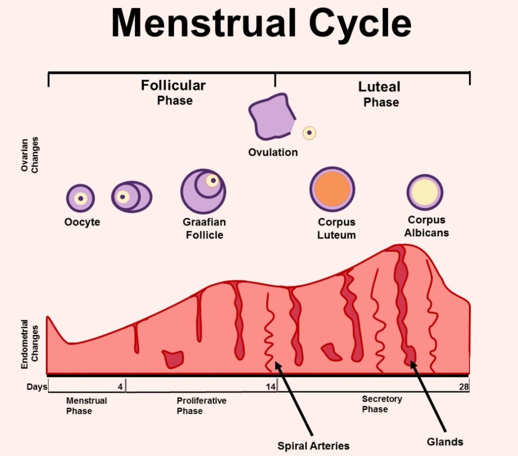
  • Promotes the maturation of ovarian follicles (Graafian follicles), usually one at a time, and triggers estrogen secretion, leading to ovulation.
  • The developing follicle secretes estrogen, causing the uterus to proliferate and the breasts to enlarge, preparing for breastfeeding.
  • About 24-36 hours before ovulation, the anterior pituitary gland secretes luteinizing hormone (LH), causing the Graafian follicle to rupture, leading to ovulation and the formation of the corpus luteum.
  • The corpus luteum secretes progesterone, which thickens the endometrium and maintains pregnancy.

Note:

  1. If fertilization doesn’t occur, progesterone withdrawal happens.
  2. The corpus luteum degenerates into the corpus albicans (a white body) and finally becomes fibrous.
  3. The cycle then begins again due to stimulation by the hypothalamus.

(b) Ovarian Hormones.

1. Oestrogen:

  • Produced by growing follicles (granulosa cells and theca).
  • Responsible for the development of female secondary sexual characteristics (e.g., breast growth).
  • Causes proliferation of the endometrium, prepares the vagina, and promotes the production of cervical mucus.

2. Progesterone:

  • After ovulation, LH stimulates the corpus luteum to produce high levels of progesterone and low levels of estrogen.
  • Progesterone causes the endometrium to become more tortuous, raises basal body temperature, and induces sensations of fullness in the breasts before menstruation.
  • The rise in ovarian hormones decreases the flow of GnRH, leading to reduced production of FSH and LH. This is a negative feedback loop.
  • A positive feedback mechanism occurs when blood estrogen levels rise, stimulating the hypothalamus to secrete more luteinizing hormone-releasing factor.
Uterine Phases Events of the Menstrual or Endometrial Cycle midwives revision

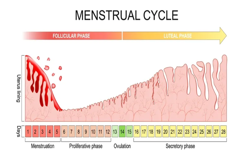
Uterine Phases/Events of the Menstrual or Endometrial Cycle

1. Menstrual Phase.

  • If the ovum is not fertilized, the corpus luteum degenerates, causing progesterone and estrogen levels to fall.
  • The functional layer of the endometrium, which relies on high levels of ovarian hormones, is shed during menstruation.
  • High circulating levels of progesterone and estrogen inhibit FSH and LH production in the anterior pituitary gland. If pregnancy occurs, these hormones prevent the release of another ovum.
  • When the corpus luteum degenerates, falling hormone levels cause the anterior pituitary to resume FSH secretion, initiating the next cycle.

2. Proliferative Phase

  • This phase begins immediately after the menstrual phase and lasts until ovulation. It coincides with the follicular phase.
  • The endometrium regenerates, forming a new layer.
  • This phase typically lasts 10 days in a 28-day cycle:
  1. Early proliferative phase: Occurs 4-7 days after menstruation.
  2. Mid proliferative phase: Occurs 8-10 days after menstruation.
  3. Late proliferative phase: Occurs 11-14 days after menstruation.
  • FSH stimulates the growth and maturation of Graafian follicles, which produce estrogen to repair the endometrium.
  • The endometrium thickens, consisting of three layers:
  1. Basal layer: Next to the myometrium, not shed during menstruation, as it contains the necessary structures to rebuild the endometrium.
  2. Functional layer: Spongy, tubular glands, about 2.5 cm thick.
  3. Compact layer: Cuboidal ciliated epithelium.

Note: The functional and compact layers (b and c) are shed during menstruation.

3. Secretory Phase

  • This phase follows the proliferative phase and is regulated by progesterone.
  • The endometrium becomes edematous (thickens and swells) and develops a spongy appearance.
  • Secretory glands produce increased amounts of glycogen to nourish a potential fertilized ovum.
  • This phase lasts for about 14 days.

Note:

  • If the ovum is not fertilized, menstruation begins, marking the start of a new cycle.
  • If the ovum is fertilized, the zygote travels to the uterus and becomes embedded in the endometrium, producing human chorionic gonadotropin (HCG). This hormone supports the corpus luteum, ensuring continuous secretion of progesterone and estrogen to maintain pregnancy.
  • After about 12 weeks, the placenta forms and takes over hormone production (estrogen, progesterone, and gonadotropins) to sustain the pregnancy.

Gametogenesis 

Gametogenesis is the developmental process that leads to the creation of reproductive cells.

Before fertilisation, gametes have to be formed in the process of gametogenesis which takes place on the principles of meiosis. 

Production of a Mature Ovum (Oogenesis)

Oogenesis is accomplished through meiosis, a process beginning during embryonic development

A primary oocyte produces one secondary oocyte and a polar body.
The secondary oocyte and the first polar body then give rise to a mature ovum and three polar bodies, which degenerate. The ovum is the largest cell in the body.

Gametogenesis midwives revision

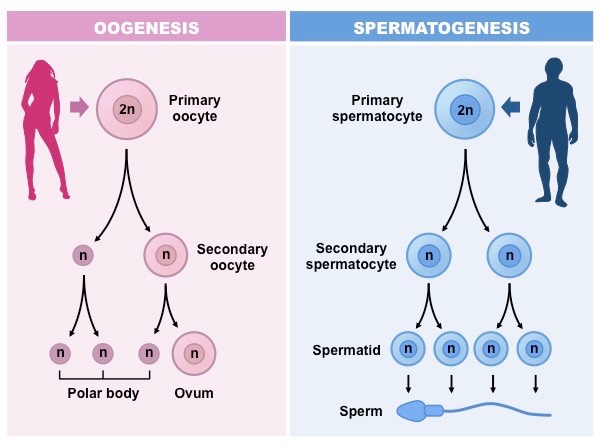
Spermatogenesis (Production of Sperm)

Spermatogenesis, the formation of spermatozoa, begins at puberty (around 14-16 years). Sperms are produced in the basal layer of the germinal epithelium of the testes under the influence of Follicle-Stimulating Hormone (FSH). This occurs through meiotic cell division.

  • Primitive structures (spermatogonia) in the testes are nourished by Sertoli cells, developing into primary spermatocytes. These primary spermatocytes, possessing a diploid number of chromosomes, undergo meiosis to form two daughter cells: secondary spermatocytes.
  • The Luteinizing Hormone (LH) acts on the secondary spermatocytes, initiating the second meiotic division and forming spermatids. The larger portion of the spermatid develops into a spermatozoon (sperm). This transformation of spermatids into spermatozoa is called spermiogenesis.

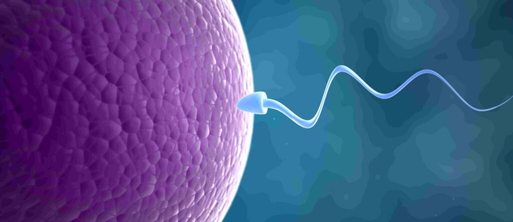
The spermatozoon has three main parts:

  • Head: Contains the acrosome (with the hyaluronidase enzyme, which breaks down the ovum’s outer layer for sperm entry) and the nucleus (containing chromosomes and genetic material).
  • Body (Midpiece): Provides nutrients for the sperm.
  • Tail (Flagellum): Propels the sperm after ejaculation.

Male Hormones:

  1. The hypothalamus produces gonadotropin-releasing factor (GnRH), which stimulates the anterior pituitary gland.
  2. FSH acts on the seminiferous tubules to stimulate spermatozoa production.
  3. LH stimulates interstitial cells to produce testosterone, the primary male sex hormone responsible for the development of secondary sexual characteristics in boys at puberty, including:
  • Increased muscle growth and height/weight gain.
  • Enlargement of the larynx (deeper voice).
  • Growth of hair on the face, armpits, chest, abdomen, and pubic area.
  • Enlargement of the penis, scrotum, and prostate gland

Revision Questions:

  1. Define meiosis.
  2. Describe the formation of a mature ovum.
  3. State the differences between a primary and a secondary oocyte.
  4. What is spermiogenesis?
  5. State two functions of the testes.
  6. Describe the male reproductive system.
  7. List three hormones that influence spermatogenesis.
  8. List three hormones involved in male reproduction.

Menstruation Cycle Read More »

FEMALE BREAST (MAMMARY GLAND)

THE FEMALE BREAST (MAMMARY GLAND) 

Breasts are two accessory glands of the female reproductive system.

Situation: They are situated on the anterior chest wall over the pectoralis major muscles between the 2nd and 6th rib and each extend from the sternum to the axilla forming the axillary tail. It’s stabilized by the suspensory ligament.

Shape:

  • In a prime gravida, the shape is hemispherical and has a tail tissue extending towards the axilla forming the axillary tail.
  • It is flat and pendulous or pawpaw shaped in the multiparous women.
  • This varies with each individual and the stage of development as well as age.

Development: The nipples are present at birth but no further development takes place till puberty when the breast increases in size. Further development takes place during pregnancy, but it reaches its full maturity during lactation.

Gross Structure of breat midwives revision

Gross Structure:

The breast has the following parts:

  1. Axillary Tail: This is a tail of tissue called the axillary tail of Spence, extending from the breast towards the axilla.
  2. Areola: It extends 2.5cm around the nipple and it contains sebaceous glands which become more visible in pregnancy and they are known as Montgomery tubercles. These lubricate the nipple.
  3. The Nipple: It lies in the centre of the areolar at the level of the fourth rib. It is a protuberance about 6mm in length, composed of erectile tissue and is covered with epithelium, contains plain muscle fibres which have a sphincter-like action controlling the flow of milk. The surface of the nipple is perforated by small orifices which open in the lactiferous ducts.

Microscopic Structure:

It is composed largely of glandular tissue but also of some fatty tissue and is covered with skin. The glandular tissue is covered with connective tissue. They are 18-20 in number.

Microscopic Structure of breast midwives revision

The internal structure is said to be composed of the following:

  • Nipple: This is located at the apex of the breast and projects up to 1 cm. 
  • Areola: This is a roughly circular area of skin that surrounds the nipple. Its colour darkens during pregnancy due to the deposition of melanin. The areolar skin contains Montgomery glands which secrete a protective oily lubricant.
  • The Lobes: These separate the different batches of alveoli.
  • The Alveoli: Each gland is called alveolus and is the milk-secreting unit lined by milk-making cells called acini cells. These are covered by myo-epithelium which contract to expel milk.
  • The Lactiferous Tubule: Is also known as small ducts, they extend from the alveoli running into one another uniting to form bigger ducts which run into lactiferous ducts.
  • The Lactiferous Duct: This is the central duct into which small ducts or tubules run.
  • The Ampulla or Lactiferous Sinus: This is a widened out portion underneath the areola. It is a continuation of the lactiferous duct towards the nipple and terminates as minute openings on its surface. The ampulla is a milk reservoir during breastfeeding.

Blood Supply:

  • By internal and external mammary arteries.
  • Intercostal arteries which originate from the aorta.

Venous Return: From a circular network around the nipple and drain into internal mammary and axillary veins.

Lymphatic Drainage: Lymph drains freely between the breasts and into lymph nodes in the axilla and the medial sternum.

Nerve Supply: There is poor nervous supply. The skin is supplied by branches of the 4th, 5th and 6th thoracic nerves. The functions of the breasts are controlled by hormones i.e., oestrogen, progesterone, and prolactin. The breast is supported by the suspensory ligaments.

Functions of the Breast:

  • To supply milk to the baby.
  • To give shape to the female figure.
  • It is a secondary sex organ.

Physiology of the Breast

1. Before Pregnancy

Breasts are present at birth. They develop at puberty due to the effect of oestrogen, which passes from the growing ovarian follicle through the bloodstream to the breast.

Effects of Oestrogen on the Breast:

  • The breasts enlarge and assume the adult female size and shape.
  • It causes further growth of the nipple and areola.
  • It promotes growth and development of the lactiferous tubules and ducts. Therefore, breast enlargement is due to the enlargement of the ducts.

Before menstruation, breast fullness and tingling occur due to progesterone stimulation from the corpus luteum.

2. During Pregnancy

  • Further breast development and enlargement occur due to alveoli hypertrophy. This is due to progesterone stimulation in preparation for milk production. 
  • Progesterone and oestrogen play an important part in the development of glandular tissue and its ducts.

3. After Delivery

  • Prolactin is responsible for milk production, beginning around the third day postpartum. This occurs after oestrogen is fully withdrawn, and the breasts reach their full development.

Factors Affecting Lactation:

  1. Hormonal Control: Placental separation and expulsion alter the oestrogen-progesterone balance, resulting in prolactin release from the anterior pituitary gland.
  2. Physical Factors: The neuro-hormonal reflex, involving oxytocin, causes milk letdown when the baby suckles.
  3. Emotional Factors: Maternal willingness to breastfeed and the baby’s ability to breastfeed facilitate milk production.
  4. Nutritional Factors: Well-nourished lactating women experience more successful breastfeeding.
Physiology of Lactation midwives revision (1)

Physiology of Lactation

1. Hormonal Control:

  • An alteration in the progesterone-oestrogen balance results in prolactin release from the lactotrophs of the anterior pituitary gland.
  • Clinical Note: To suppress lactation (e.g., after baby loss), oestrogen may be administered via drugs like doxinex, cabergoline, etc., which inhibit prolactin.

2. Milk Production:

  • Essential substances are extracted from the increased blood supply to the breast for milk formation. Fatty globules and protein molecules form at the base of acini cells, move into the alveoli, and travel through the lactiferous tubules. Lactation depends not only on hormones but also on breast blood supply.

3. Passage of Milk:

Milk transit from secretory cells to the nipple is aided by:

  • Back Pressure: Newly formed globules push preceding ones into the lactiferous tubules and ducts.
  • Neuro-Hormonal Reflex: Suckling empties the ampulla, causing large lactiferous ducts to contract and force milk towards the nipple. Nipple stimulation triggers oxytocin release from the posterior pituitary, further contracting lactiferous tubules and increasing milk flow.
  • Maintenance of Milk Supply: Supply responds to demand. Frequent breastfeeding maintains supply; infrequent breastfeeding reduces it.

Factors Essential for Maintenance:

  1. Stimulus: Infant suckling creates the neuro-hormonal reflex. If the infant cannot breastfeed, the breast should be emptied by other means.
  2. Complete Emptying: Complete breast emptying ensures milk flow and stimulates new supply.

Important:

  • Infants should breastfeed 5 times a day and 3 times at night (or more, 8 to 12 times in 24 hours).
  • Correct positioning and attachment ensure effective suckling and adequate milk production and prevent sore/cracked nipples.
attachment breast midwives revision

Positioning of the Mother During Breastfeeding:

The mother should sit upright, holding the breast with fingers in a “C” shape (thumb above the areola, fingers below). Avoid the “scissor hold,” which can remove the nipple from the infant’s mouth.

Assessment of Baby Positioning:

  • Baby’s tummy faces the mother’s tummy.
  • Head and body are aligned and supported.
  • Nose faces the nipple, and the baby can look up at the mother’s face. The cradle position is common.

Assessment of Breast Attachment:

  • Chin touches the breast.
  • Mouth is wide open, and most of the areola is in the baby’s mouth.
  • Cheeks are rounded.
  • Lower lip is turned out.
  • More areola is above than below the mouth.

Breastfeeding should be initiated within 30 minutes to 1 hour of life and maintained exclusively for six months. Babies should be breastfed on demand, which includes when the breast is full, the baby cries, or the mother desires. Effective breastfeeding involves the baby sucking and pausing to swallow.

Clinical Procedure: Breast Examination for Antenatal Mother

Objective: To examine the breasts to rule out abnormalities and prepare the mother for successful lactation.

Requirements: A chair, a draw sheet, and a hand towel or handkerchief.

Table

Step

Action

Rationale

1.

Apply soft skills while explaining procedure to the woman.

To facilitate cooperation.

2.

Instruct the mother to obtain a sitting up position and moves her hands from the lateral sides upwards and then places them down.

To expose the breasts and observe any lamps if available.

3.

Inspect the breasts for appearance, colour note symmetry, size, shape and texture, any dry scarring, ulceration or bleeding.

To detect abnormalities.

4.

Lift the two breasts upwards gently to inspect the sub mammary area.

To observe skin infections and hygiene.

5.

Palpate the Axilla and axillary tail of Spence of the left breast and then the right.

To rule out tenderness which signifies signs of infections and for enlarged lymph nodes.

6.

Systematically palpate the whole breast round.

To feel for hard lamps.

7.

Support the chest with the left hand and do the protraction test.

To rule out abnormalities of the nipples.

8.

Do the same to the second breast.


9.

Explain and educate the mother according to the findings.


10.

Make the mother comfortable, clear away and wash hands.


Revision Questions:

  1. What is a breast?
  2. Explain breast development.
  3. Describe the microscopic structure of the breast.
  4. State two functions of the breasts.
  5. Describe the physiology of lactation.
  6. State the factors that affect lactation.

FEMALE BREAST (MAMMARY GLAND) Read More »

Pelvic Floor Muscles midwives revision

The Perineal Body

The perineal body is a small, pyramid-shaped mass made of muscles and fibrous tissue, located between the lower end of the rectum and the vagina.

Size: It measures about 4 cm but can stretch significantly during the second stage of labor.

Shape: It has a triangular shape, with the base being the skin and the apex pointing upwards.

Location: It lies between the vagina and the rectum.

Structure: The perineal body is composed of three layers of tissue:

  1. Outer covering: The skin.
  2. Superficial pelvic floor muscles: These include muscles like the Bulbocavernosus and Transverse Perineal muscles.
  3. Deep pelvic floor muscles: These provide additional support.

Blood Supply

  • Arterial Supply: Pudendal arteries, which are branches of the internal iliac arteries.
  • Venous Drainage: Blood is drained through veins corresponding to the pudendal arteries.

Lymphatic Drainage:  Lymph drains into the inguinal lymph nodes.

Innervation/Nerve Supply: The perineal body is innervated by the perineal branch of the pudendal nerves.

Functions

  • Childbirth: The perineal body stretches to allow the baby to pass through during childbirth.
  • Defecation: It plays a role in the process of defecation.
  • Surgical Importance: It is the area where an episiotomy may be performed during childbirth. Trauma to this area can lead to complications such as urinary or fecal incontinence and prolapse of pelvic organs.
The Perineal Body midwives revision

The Pelvic Floor Muscles

The pelvic floor muscles form a muscular diaphragm that fills the pelvic cavity, supporting the organs within.

Shape: The pelvic floor is gutter-shaped, which influences the rotation of the baby’s presenting part during childbirth.

Functions

  • Support: The pelvic floor muscles support pelvic and abdominal organs, preventing them from prolapsing.
  • Pressure Maintenance: They help maintain pressure during activities like coughing and sneezing.
  • Bowel and Urinary Function: These muscles are involved in bowel movements and urination.
  • Labour: During the second stage of labor, the pelvic floor provides necessary resistance to help push the baby through the birth canal.

Gross Structure: The pelvic floor is pierced by three openings:

  1. Urethral Orifice

  2. Vagina

  3. Rectum

Layers of the Pelvic Floor

From inside to outside, the pelvic floor is composed of:

  1. Peritoneum
  2. Fascia Layer
  3. Deep Muscle Layer
  4. Subcutaneous Layer
  5. Skin

Superficial Pelvic Floor Muscles 

These muscles include:

  1. Urethral Sphincter: Controls the passage of urine.
  2. Ischiocavernosus: Arises from the ischial tuberosities and runs along the pubic arch to the clitoris. (Ischium to the genitals)
  3. Bulbocavernosus (Bulbospongiosus): Encircles and strengthens the vaginal orifice, extending from the perineal body to the pubic bone.
  4. Transverse Perineal Muscle: Runs from the ischial tuberosities to the centre of the perineum. (surface, side to side)
  5. External Anal Sphincter: Surrounds the anus and attaches to the coccyx.

Deep Pelvic Floor Muscles

The deep muscle layer consists of three paired muscles collectively known as the levator ani. They are the most significant muscles of the pelvic floor. They are the floor of the pelvis. Keeps organs from falling out of the pelvis.

  • Attachment Points:
  1. Front: Lateral part of the symphysis pubis.
  2. Back: Ischial spines and pubis.
  3. Sides: Obturator foramen.
  • Muscles:

  1. Pubococcygeus: Arises from the pubic bone, surrounds the urethra, vagina, and anus, and inserts into the coccyx.
  2. Iliococcygeus: Arises from the ilium at the white line of fascia and inserts into the coccyx.

Ischiococcygeus: Arises from the ischial spines and passes backward to insert into the coccyx.

Clinical Notes

  • During Labour: The anterior pelvic wall moves upwards and forwards during the second stage of labour, pulling the bladder and urethra upwards, making them abdominal organs. This can cause urinary retention, so emptying the bladder before the second stage is crucial.
  • Post-Delivery: After childbirth, the pelvic floor, bladder, and urethra return to their pre-delivery positions, a process known as the “swing door action” of the pelvic floor muscles.

Functions of the Pelvic Floor Muscles:

  • Support: The pelvic floor muscles provide essential support for the pelvic organs (bladder, uterus, rectum) and prevent prolapse (the downward displacement of these organs).
  • Continence: The coordinated action of the urethral and anal sphincters and the levator ani muscles is vital for maintaining urinary and fecal continence. Weakness in these muscles can lead to incontinence.
  • Sexual Function: The pelvic floor muscles play a role in sexual function in both men and women. Their strength and tone influence sensations and orgasmic response.
  • Pressure Regulation: These muscles help regulate intra-abdominal pressure, preventing the prolapse of pelvic organs during activities like coughing, sneezing, and lifting heavy objects. Weakness in these muscles can exacerbate prolapse.
  • Childbirth: During the second stage of labor, the pelvic floor muscles provide controlled resistance to facilitate the descent of the baby through the birth canal. The interplay between the muscles’ strength and elasticity influences the risk of perineal tearing.

Superficial Pelvic Floor Muscles:

  • Urethral Sphincter (Internal and External): The internal urethral sphincter is involuntary, while the external urethral sphincter is under voluntary control and contributes to urinary continence.
  • Ischiocavernosus: These muscles contribute to erection and clitoral engorgement in females and penile erection in males.
  • Bulbocavernosus (Bulbospongiosus): This muscle contributes to vaginal constriction, clitoral erection, and ejaculation in females and penile erection and ejaculation in males.
  • Transverse Perineal Muscle: These muscles contribute to support of the perineal body.
  • External Anal Sphincter: This muscle, along with the internal anal sphincter (involuntary), controls defecation.

Injury to Pelvic Floor Muscles During Labor

Overstretching:

  • During late pregnancy, progesterone softens ligaments in preparation for labour.
  • First Stage: Bearing down prematurely can cause excessive strain on the peri-cervical tissue and transverse cervical ligaments, leading to a retroverted uterus.
  • Second Stage: Prolonged labor can overstretch the fascia supporting the bladder, leading to prolapses like cystocele, rectocele, urethrocele, and uterocele.

Tears and Lacerations:

Tears can affect the levator ani muscles and surrounding tissues. These tears are classified by severity into four degrees:

  1. 1st Degree: Involves the fourchette, vaginal mucosa, and perineal skin, but not muscles.
  2. 2nd Degree: Involves the fourchette, skin, and muscles of the perineum.
  3. 3rd Degree: Extends to the anal sphincter.
  4. 4th Degree: Extends through the rectal lining, potentially leading to pelvic floor dysfunction and fecal incontinence.

Prevention of Pelvic Floor Injuries

During Pregnancy:

  • Antenatal Care: Encourage regular antenatal visits and hospital delivery for high-risk mothers.
  • Weight Management: Maintaining a healthy weight gain during pregnancy minimizes strain on the pelvic floor.
  • Ultrasound Scans: Advise mothers to undergo ultrasounds to detect malpresentations and plan the mode of delivery.
  • Nutrition: Promote a nutritious diet to build healthy skin and correct anemia.
  • Hygiene: Educate mothers on maintaining hygiene to prevent infections.
  • Exercises: Recommend exercises like walking and hill climbing to strengthen the pelvic floor muscles.
  • Constipation Management: Constipation increases intra-abdominal pressure, potentially stressing the pelvic floor. Adequate fiber intake, hydration, and stool softeners can help prevent constipation.
  • Hydration: Adequate hydration helps prevent constipation and maintains tissue elasticity.
  • Labor Preparation: Teach mothers what to do during labor, including avoiding native medicines that may weaken the perineal muscles.

During Labor:

  • Pain Control: Effective pain management techniques, such as epidurals, reduce the urge to push prematurely and allow for more controlled descent of the baby, decreasing the risk of perineal trauma.
  • Positional Changes During Labor: Encouraging different labor positions (e.g., upright, lateral) can facilitate optimal fetal descent and reduce the strain on the pelvic floor.
  • Controlled Pushing: Guided pushing techniques, under the guidance of a skilled birth attendant, help prevent excessive straining and reduce the risk of injury.
  • Skilled Birth Attendant: Ensure that delivery is managed by a skilled birth attendant who can recognize and prevent impending tears.
  • Instrumental Delivery Techniques: Proper use of forceps or vacuum extraction minimizes the risk of injury if necessary, and should only be performed by experienced practitioners.
  • Perineal Massage: Perineal massage during the later stages of pregnancy may help increase perineal elasticity and reduce the risk of tearing during labor. (Evidence is mixed, and this needs to be discussed with the patient).
  • Episiotomy: Perform an episiotomy if necessary to prevent severe tears.

Postpartum Prevention:

  • Postnatal Pelvic Floor Assessment: A post-delivery pelvic floor assessment is useful to identify any damage or weakness.
  • Postnatal Pelvic Floor Muscle Training (PFMT): Early initiation of PFMT post-delivery helps repair and strengthen the pelvic floor, improving recovery and reducing the risk of long-term problems.
  • Physiotherapy: Physiotherapy may be beneficial for women with significant pelvic floor damage or persistent issues, providing specialized exercises and guidance.
  • Lifestyle Modifications: Maintaining a healthy lifestyle, including appropriate diet, exercise, and weight management, is essential for long-term pelvic floor health.
  • Continence Management: If necessary, strategies for managing urinary or fecal incontinence (behavioral modification, medication) can be initiated.

Revision Questions

  1. Name four superficial pelvic floor muscles.
  2. List three parts of the perineal body.
  3. Describe the swing door action of the pelvic floor muscles.
  4. Identi fy three types of pelvic floor injuries during labor.
  5. Explain ten ways to prevent perineal tears.
  6. How would you determine if a tear is a third-degree tear?

Pelvic Floor Muscles Read More »

Want notes in PDF? Join our classes!!

Send us a message on WhatsApp
0726113908

Scroll to Top